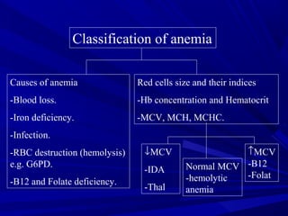
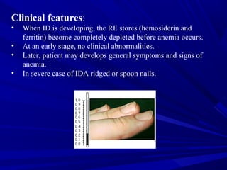
Classification of anemia
Causes of anemia
-Blood loss.
-Iron deficiency.
-Infection.
-RBC destruction (hemolysis)
e.g. G6PD.
-B12 and Folate deficiency.
Red cells size and their indices
-Hb concentration and Hematocrit
-MCV, MCH, MCHC.
↓MCV
-IDA
-Thal
↑MCV
-B12
-Folat
Normal MCV
-hemolytic
anemia
Iron deficiency anemia
Normal iron metabolism:
-The primary function is oxygen transport.
-Iron is absorbed by duodenum and jejunim
-Average total body iron content 3500-4000 mg.
-Approximately 2/3 found in hemoglobin,
-Iron is also stored in RE cells (BM, Spleen and liver)
as hemosiderin and ferratin.
-Also iron found in myglobin and myeloperoxidase
and in certain electron transfer.
-Iron is more stable in ferric state (Fe+++
) than in
ferrous state (Fe++
).
Iron + Hem
Iron Metabolism
Iron Absorption
1-2 mg only
Plasma Fe
Transferrin carriers
4 mg
Body stores
1000 mg (M)
300-500 mg (F)
Myglobin
300 mg
20 mg Fe
Returned to immature RBC
in BM
RBC
2500 mg
R.E. 20 mg
Released daily
Via RE system
90% extra vascular
5-10% intra vascular
Loss (from GI tract)
1-2 mg daily
Daily Fe++
turnover continuous process
Dietary iron:
Iron is present in food as ferric hydroxides (ferric-protein
complexes and hem-protein complexes).
-meat, liver
-vegetables, eggs.
-The average diet contains 10-15mg and only 5-10% is normally
absorbed.
Iron requirements:
It varies depending on sex and age:
Male/female 0.5-1 mg/day
Pregnant female 1-2 mg/day
Children 0.5 mg/day
Clinical features:
• When ID is developing, the RE stores (hemosiderin and
ferritin) become completely depleted before anemia occurs.
• At an early stage, no clinical abnormalities.
• Later, patient may develops general symptoms and signs of
anemia.
• In severe case of IDA ridged or spoon nails.
Causes:
• Chronic blood loss
Fetomaternal Hemorrhage, inherited
bleeding disorders menstrual peroid.
• Maternal iron deficiency (neonate).
• Growth spurts (infants and children).
• Gastrointestinal,
peptic ulcer, aspirin ingestion, carcinoma,
hookworm, colitis, piles etc.
• Pregnancy
• Rarely hematouria,
self-inflicted blood loss, hemoglobinuria.
• Insufficient daily iron intake (poor diet).
• Malabsorption.
Laboratory findings:
•Red cell indices:
Low Hb conc.
MCV, MCH, MCHC* ↓
•Blood film:
Hypochromic microcytic Picture.
Occasional Target cells.
Pencil shaped poikilocytes.
Normal reticulocyte count.
•Bone marrow iron:
Normal to hypercellular.
RBC precursors are increased in number.
Iron stain negative.
•Chemical testing on serum:
Serum iron Decreased
Transferrin/TIBC Normal to High
Serum ferritin Decreased (Very low)
Hypochromic Microcytic picture (IDA)
-ve BM Iron Stain +ve
Reticulocytes

Ida

  • 1.
    Classification of anemia Causesof anemia -Blood loss. -Iron deficiency. -Infection. -RBC destruction (hemolysis) e.g. G6PD. -B12 and Folate deficiency. Red cells size and their indices -Hb concentration and Hematocrit -MCV, MCH, MCHC. ↓MCV -IDA -Thal ↑MCV -B12 -Folat Normal MCV -hemolytic anemia
  • 2.
    Iron deficiency anemia Normaliron metabolism: -The primary function is oxygen transport. -Iron is absorbed by duodenum and jejunim -Average total body iron content 3500-4000 mg. -Approximately 2/3 found in hemoglobin, -Iron is also stored in RE cells (BM, Spleen and liver) as hemosiderin and ferratin. -Also iron found in myglobin and myeloperoxidase and in certain electron transfer. -Iron is more stable in ferric state (Fe+++ ) than in ferrous state (Fe++ ).
  • 4.
    Iron + Hem IronMetabolism
  • 5.
    Iron Absorption 1-2 mgonly Plasma Fe Transferrin carriers 4 mg Body stores 1000 mg (M) 300-500 mg (F) Myglobin 300 mg 20 mg Fe Returned to immature RBC in BM RBC 2500 mg R.E. 20 mg Released daily Via RE system 90% extra vascular 5-10% intra vascular Loss (from GI tract) 1-2 mg daily Daily Fe++ turnover continuous process
  • 6.
    Dietary iron: Iron ispresent in food as ferric hydroxides (ferric-protein complexes and hem-protein complexes). -meat, liver -vegetables, eggs. -The average diet contains 10-15mg and only 5-10% is normally absorbed. Iron requirements: It varies depending on sex and age: Male/female 0.5-1 mg/day Pregnant female 1-2 mg/day Children 0.5 mg/day
  • 7.
    Clinical features: • WhenID is developing, the RE stores (hemosiderin and ferritin) become completely depleted before anemia occurs. • At an early stage, no clinical abnormalities. • Later, patient may develops general symptoms and signs of anemia. • In severe case of IDA ridged or spoon nails.
  • 8.
    Causes: • Chronic bloodloss Fetomaternal Hemorrhage, inherited bleeding disorders menstrual peroid. • Maternal iron deficiency (neonate). • Growth spurts (infants and children). • Gastrointestinal, peptic ulcer, aspirin ingestion, carcinoma, hookworm, colitis, piles etc. • Pregnancy • Rarely hematouria, self-inflicted blood loss, hemoglobinuria. • Insufficient daily iron intake (poor diet). • Malabsorption.
  • 9.
    Laboratory findings: •Red cellindices: Low Hb conc. MCV, MCH, MCHC* ↓ •Blood film: Hypochromic microcytic Picture. Occasional Target cells. Pencil shaped poikilocytes. Normal reticulocyte count. •Bone marrow iron: Normal to hypercellular. RBC precursors are increased in number. Iron stain negative. •Chemical testing on serum: Serum iron Decreased Transferrin/TIBC Normal to High Serum ferritin Decreased (Very low)
  • 10.
    Hypochromic Microcytic picture(IDA) -ve BM Iron Stain +ve
  • 12.