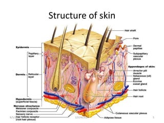
This document provides an overview of the structure and function of skin and its appendages. It discusses the three layers of skin - the epidermis, dermis and hypodermis - and describes the cellular structure and functions of each layer. It also examines skin appendages like hair, nails, sweat and sebaceous glands. The document is intended as a reference for the anatomy of skin and its related tissues.
it describes the microanatomy of skin and its appendages in a concise format. it will give the overview of the integumentary system of our body and largest organ of our body.
Describe the structures, relations, and functions of the adrenal gland.
describe the histological structures and clinical importance of the adrenal gland
basics of skin, review of skin, Integumentary system, the structure of the skin, Functions of skin, skin appendages, Hair, sweat glands, sebaceous glands, Nails, dermis, epidermis,
subcutaneous tissue. anatomy and physiology
Conférence du Pr. Vincent Leroy (Hôpital Universitaire de Grenoble, France), Juin 2014. Prise en charge de l'Hépatite C à l'aide des nouveaux antiviraux Sofosbuvir, Daclatasvir, Siméprévir, en combinaison ou non avec l'Interféron pégylé et la Ribavirine.
The skin is the largest organ of the body, accounting for about 15% of the total body weight in adult humans. It exerts multiple vital protective functions against environmental aggressions, rendered possible thanks to an elaborate structure, associating various tissues of ectodermal and mesodermal origin, arranged in three layers, including (from top to bottom) the epidermis (and its appendages), the dermis and the hypodermis.
The skin : هذا العرض يتحدث عن الجلد الذي يعتبر اكبر عضو بالجسم وشرح الطبقاة المكونة للجلد :
------------------------------------------
https://t.me/GoldenAlzaidy
------------------------------------------------------
youtube::: https://www.youtube.com/watch?v=Orumw-PyNjw
Brief Anatomy of Skin and Skin GraftingRishi Gupta
Brief Anatomy of Skin and Skin Grafting.
Anatomy of Skin
History of skin grafting.
Recent Advances in Skin Grafting.
Dermal Substitutes.
Cell cultures in skin grafting.
this presentation gives the overview of the Gastrointestinal system with detail description of the stomach.
some clinical aspects, like peptic ulcer disease, Virchow's lymph node, gastrostomy are also included.
describes the muscles, nerves and vessels of arm region. it gives an overview to understand to basic anatomical aspect of arm region including cubital fossa.
this presentation describes the detail anatomy of Temporo-mandibular joint with respect to its articulating surfaces, ligaments, muscles and blood and nerve supply.
well describes the development of nervous system from basic to advanced concept including neural tube defects. the concepts are presented in graphical form for easy understanding of concepts.
Unit 8 - Information and Communication Technology (Paper I).pdfThiyagu K
This slides describes the basic concepts of ICT, basics of Email, Emerging Technology and Digital Initiatives in Education. This presentations aligns with the UGC Paper I syllabus.
How to Split Bills in the Odoo 17 POS ModuleCeline George
Bills have a main role in point of sale procedure. It will help to track sales, handling payments and giving receipts to customers. Bill splitting also has an important role in POS. For example, If some friends come together for dinner and if they want to divide the bill then it is possible by POS bill splitting. This slide will show how to split bills in odoo 17 POS.
Model Attribute Check Company Auto PropertyCeline George
In Odoo, the multi-company feature allows you to manage multiple companies within a single Odoo database instance. Each company can have its own configurations while still sharing common resources such as products, customers, and suppliers.
This is a presentation by Dada Robert in a Your Skill Boost masterclass organised by the Excellence Foundation for South Sudan (EFSS) on Saturday, the 25th and Sunday, the 26th of May 2024.
He discussed the concept of quality improvement, emphasizing its applicability to various aspects of life, including personal, project, and program improvements. He defined quality as doing the right thing at the right time in the right way to achieve the best possible results and discussed the concept of the "gap" between what we know and what we do, and how this gap represents the areas we need to improve. He explained the scientific approach to quality improvement, which involves systematic performance analysis, testing and learning, and implementing change ideas. He also highlighted the importance of client focus and a team approach to quality improvement.
Ethnobotany and Ethnopharmacology:
Ethnobotany in herbal drug evaluation,
Impact of Ethnobotany in traditional medicine,
New development in herbals,
Bio-prospecting tools for drug discovery,
Role of Ethnopharmacology in drug evaluation,
Reverse Pharmacology.
Read| The latest issue of The Challenger is here! We are thrilled to announce that our school paper has qualified for the NATIONAL SCHOOLS PRESS CONFERENCE (NSPC) 2024. Thank you for your unwavering support and trust. Dive into the stories that made us stand out!
The Art Pastor's Guide to Sabbath | Steve ThomasonSteve Thomason
What is the purpose of the Sabbath Law in the Torah. It is interesting to compare how the context of the law shifts from Exodus to Deuteronomy. Who gets to rest, and why?
2. Introduction
– our largest organ
– Accounts for 12% of body weight…it weighs twice
as much as your brain!
– Varies in thickness at different parts (< 0.5
mm at eyelids to > 5 mm on middle of upper
back)
– Divided into three distinct layers
• Epidermis
• Dermis
• Hypodermis – lies deep to the dermis
8/5/2012 1212413114 2
4. Functions
• Barrier against loss of body fluid.
• Protection and excretion
• Sensory function
• Metabolic function
• Temperature regulation
• Adaptation during edema and pregnancy
• Legal and anthropological importance
8/5/2012 1212413114 4
6. Epidermis
• Primarily made up of keratinized stratified
squamous epithelium(keratinocytes)
• gives strength to the skin.
• Varies in thickness from thick skin to thin skin
• It does not have any vascularization, so it
relies on the connective tissues deep to it.
• Also contain melanocytes, merkel’s cells and
Langerhans cell
8/5/2012 1212413114 6
7. Layers of epidermis
• Stratum basale (the deepest layer)
• Stratum spinosum
• Stratum granulosum
• Stratum lucidum (only in thick skin)
• Stratum corneum (most superficial layer of
epidermis
8/5/2012 1212413114 7
9. STRATUM BASALE
• Consists of single layer of basophilic
columner or cuboidal cells.
• Along with S. spinosum it is component of
Malpighian layer
• Cells are bound to each other by
desmosomes and to basal lamina by
hemidesmosomes.
• All cells contain intermediate keratin
filaments, number of which increases as cells
progress upward.
8/5/2012 1212413114 9
10. Stratum Spinosum
• Also contain the dividing cells as in basale.
• Cells contain bundles of intermediate
filament ( tonofilaments) projecting into the
processses of cells which give attachment to
the desmosomes, so giving spined
appearance.
• Tonofilaments provide resistant to the
abrasion so this layer is thicker in the areas
prone to abrasion( thick skin)
8/5/2012 1212413114 10
12. Stratum Granulosum
• Consists of polygonal cells , cytoplasm of which is
filled with the basophilic granule , keratohyaline
granules. It is rich in phosphorylated histidine and
cystine.
• Cells contain, lamellated bodies, made up of lipid.
It fuses with the cell membrane and it come out of
cells and function as a intercellular cement or
sealing agent.
• This sealing effect is first evolutionary adaptation
to terrestrial life.
8/5/2012 1212413114 12
13. Stratum Lucidum
• more prominent in thick skin . Cellular
organells and nuclei are not prominent.
• It is composed of clear non-nucleated cells.
• In the palms and soles, the stratum lucidum is
present. The tan colored protein blocks the
underlying melanocytes from view
8/5/2012 1212413114 13
14. Stratum corneum
• The main difference between thick skin and thin
skin relates to the thickness of the Stratum
corneum.
• These are the dead cells, flaking off. The cells lose
their nucleus and fuse to form squamous
sheets, which are eventually shed from the surface
(desquamation).
• It takes about 40 50 days days for a cell to move
from the stratum basale to the distal edge of the
stratum corneum and shed.
8/5/2012 1212413114 14
15. Keratin
• Keratin is a waxy protein substance found in the
epidermis. It also makes up the nails and hairs.
• It can absorb water, so keratin swells when
soaking wet.
• It makes the skin look wrinkled when you are in
the tub too long. The water evaporates when you
dry off, and pulls more water out of your body, so
soaking in the tub will dry your skin out…unless
you put lotion on right away to keep the water in
the epidermal and dermal layers. Lotions will not
penetrate to the dermis, just water.
8/5/2012 1212413114 15
16. • Icthyosis – excessive keratinisation of the skin
with autosomal recessive inheritance pattern.
• Grotesque appearance due to icthyosis in
fetus- harlequin fetus.
8/5/2012 1212413114 16
17. Melanocytes- neural crest cells
•During the first 3 months
of development, the
epidermis is invaded by
cells arising from the
neural crest.
• These cells synthesize
melanin pigment, which
can be transferred to
other cells of the
epidermis by way of
dendritic processes.
8/5/2012 1212413114 17
18. Melanin synthesis
• Stage 1 – tyrosinase
activity in vesicle
• Stage 2- melanosome
with tyrosinase and
filaments of melanin
• Stage 3- more filaments
of melanin
• Stage 4- melanin
completely filled the
vesicles.
8/5/2012 1212413114 18
19. • Addison disease – adrenal cortex hypo
function resulting increased in ACTH
production---- increased melanin synthesis.
• Albinism – hereditary condition of inability of
melanocytes to produce melanin.
• Vitiligo- degeneration of entire pool of
melanocytes.
• Nevus and Malignant melanoma
8/5/2012 1212413114 19
20. Melanin
• Tyrosine by the action of tyrosinase converted
into the DOPA, which undergo polymerization
to form the melanin.
• it is located in supranuclear position inside the
keratonocytes so that it can prevent the
nuclear damage by the UV radiation coming
from above.
8/5/2012 1212413114 20
21. Langerhans cells
• These are star shaped bone marrow derived
cells. Migrated to the skin via blood.
• Present in the S. spinosum of the skin
• These cells are antigen presenting cells so
participate in immune related reaction.
• These are component of SALT.
8/5/2012 1212413114 21
22. Merkel’s cell
• Neural crest in origin
• Found in the basal layer of the thick skin . Has
sensory function.
• Expanded terminal disc of free nerve endings
are found just beneath the merkel’s cell.
• These cells also acts as the neuroendocrine
function.
8/5/2012 1212413114 22
23. Dermis
• It is connective tissue that support the epidermis
and attaches the epidermis to the hypodermis.
• Its surface consists of many ridges ( dermal
papillae) which interdigitate with epidermal
ridges.
• Dermal papillae are more numerous in the skin
that is more prone to pressure and friction.
• Has 2 layer - Papillary layer and reticular layer
8/5/2012 1212413114 23
24. Dermoepidermal junction
• Dermis is consists of the collagen fibers and
elastic fibers which are thicker in reticular
layer and thinner in papillary layer.
• Special collagen fibers extend from the dermal
papillae and inserted in the basal lamina
forming the dermoepidermal junction.
• Loss of this junction lead to pemphigoid
• Loss of intercellular junction lead to
pemphigus.
8/5/2012 1212413114 24
25. Papillary layer
• Made up of loose connective tissue having
thin fibers and many cellular structures.
• Has ridges to increase surface area for contact
with the epidermis.
• Along the ridges sweat gland open at regular
interval.
• The papillary layer in the dermis is what forms
our fingerprints.
8/5/2012 1212413114 25
26. Reticular layer
• Dense irregular Connective Tissue
• Has bundles of Collagen and Elastic fibers and
less number of cells.
• Arrangement of bundle in the direction of
mechanical force give rise to the cleavage lines of
Langer.
• strongest layer of the Dermis. Gives the area
strength.
• Leather is made of this layer.
• EDS
8/5/2012 1212413114 26
27. • The dermis is also the area where all the
glands of the body are located.
• A transdermal patch (nicotine patch, etc) must
diffuse all the way from the epidermis into the
dermis to reach the blood vessels there.
• The blood vessels in the dermis are what give
a pink color to Caucasian people.
8/5/2012 1212413114 27
28. Hypodermis
• Consists of loose connective tissue which
helps in sliding the skin over the deep
structure.
• Consists of layer of fat according to the
nutritional status of the person.
• Also called as superficial fascia or panniculus
adiposus
8/5/2012 1212413114 28
29. Vessels in skin
• Arteries form the 2 plexuses. One at the
junction of papillary and reticular layer( sub-
papillary plexus) and another at junction of
dermis and hypodermis (cutaneous plexus).
• Veins form the three plexuses – 2 in same
position as for arterial and another in the
middle of the dermis.
8/5/2012 1212413114 29
33. Skin color
1) MELANIN: (dark brown pigment).
2) CAROTENE: (a yellowish/orange pigment found only
in plants). Accumulates more in the skins of Asians
and Native Americans.
3) SKIN THICKNESS: blood vessels seen, looks pinker
4) HEMOGLOBIN: The DERMIS contains the blood
vessels that give Caucasians the pink color to the
skin. Even veins are red because blood is red. But
when you look at veins through the adipose layer
(the hypodermis), they look blue.
8/5/2012 1212413114 33
34. Condition of skin
• If you rub the skin, it stimulates cell division = callous
• If you rub the skin too hard, the stratum basale tears
away from the basement membrane, and causes a
gap, which fills with fluid: BLISTER.
• The epidermis then dies because it’s too far away
from nutrients. That’s why the top of a blister dries
up
• If the blisters are small (less than 5 mm in
diameter), they are known as vesicles; if they are
larger (greater than 5 mm in diameter), they are
termed bullae
8/5/2012 1212413114 34
35. • Psoriasis – decrease in cell turnover time and
increased proliferation of basal layer cells.
Results in greater epidermal thickness and
rapid renewal of epidermis.
• Darkening of the skin to solar radiation-
darkens the preexisting melanin and also
increase the synthesis of melanin.
8/5/2012 1212413114 35
36. Skin cancer
Major types
• Squamous cell carcinoma
• Basal cell carcinoma
• Malignant melanoma
8/5/2012 1212413114 36
37. Appendages of the skin
• Hairs
• Sebaceous gland
• Sweat gland
• Nails
8/5/2012 1212413114 37
38. Hair
• It is keratinized elongated structure derived
from the invagination of epidermis(hair
follicle) .
• By 3rd month first hairs appear over eyebrow
and upper lip.
• It acts for thermoregulation, sensory function.
• Distribution of hair after puberty possesses
distinctive sex differences.
• Made up of shaft and root
8/5/2012 1212413114 38
39. Hair
• The hair follicle is a tubular structure
consisting of five concentric layers of epithelial
cells.
• At the base, there is a bulbous expansion, the
hair bulb, enclosing the hair papilla .
• During hair formation inner 3 layer undergo
keratinisation and outer 2 layer form epithelial
sheath.
8/5/2012 1212413114 39
41. Stages of Hair growth
• Anangen phase- fast growing phase
• Catangen phase- involution phase
• Telogen phase- rest phase
Types of hairs
• Laungo hairs- foetal hair
• Vellus hairs- infant hair and fine hairs of body
• Terminal hairs- coarse hair
8/5/2012 1212413114 41
42. Arrector pili muscle
• extends from the
dermal coat of isthmus
of the hair follicle to the
papillary layer of the
dermis.
• Gives gooseflesh
appearance.
• Cholinergic sympathetic
supply
8/5/2012 1212413114 42
43. Nail
• Nails are plates of keratinized epithelial cells
on the dorsal surface of each distal phalanx.
• The nearly transparent nail plate and the thin
epithelium of the nail bed provide a useful
window on the amount of oxygen in the blood
by showing the color of blood in the dermal
vessels.
• Growth rate 1-3mm/month
8/5/2012 1212413114 43
44. • Nails develop from thickened areas of
epidermis at the tips of each digit called nail
fields.
• Later these nail fields migrate onto the dorsal
surface surrounded laterally and proximally by
folds of epidermis called nail folds.
8/5/2012 1212413114 44
47. Beau’s line
• Transverse depressions
due to disruption of
proximal matrix mitotic
activity.
• Common in coronary
artery
occlusion, hypocalcemia
, malnutrition and
trauma.
8/5/2012 1212413114 47
48. Onychomadesis/ nail shedding
• Proximal detachment
of the nail plate from
the proximal nail fold.
8/5/2012 1212413114 48
49. Nail pitting
• Punctate depressions
of the nail plate surface.
• Common in psoriasis
8/5/2012 1212413114 49
50. Onychorrhexis
• Longitudinal ridging
and fissuring of the
Plate.
• Common in lichen
planus .
• Normal in aging
process.
8/5/2012 1212413114 50
51. Leukonychia
• Because of
parakeratotic cells with
in ventral portion of nail
plate, nail appears
whitish.
• May be hereditary or
associated with
congenital disorders.
8/5/2012 1212413114 51
52. Koilonychia / spoon shaped nail
• Concave nail due to
upward eversion of
lateral nail edge.
• Common in iron
deficiency anemia, PVS
8/5/2012 1212413114 52
53. Splinter hemorrhage
• One of the diagnostic
sign of infective
endocarditis.
• Also common in
vasculitis.
8/5/2012 1212413114 53
54. Nail clubbing
• Schamroth's window
test is the test to define
clubbing.
• Lovibond’s angle > 165
degree
• Common in heart
disease, lung disease
and GI disease.
8/5/2012 1212413114 54
55. Sebaceous gland
• Holocrine type of branched acinar gland.
• Acinus consists of rounded cells filled with lipid.
• Cell lost by holocrine secretion are replaced by
the proliferation of basal cell of acinus.
• Open either with in hair follicle(common) or on
the skin surface( rare).
• These are not under nervous control.
• Pilo-sebaceous apparatus.
8/5/2012 1212413114 55
56. • Gland lies within fibrous
sheath of hair follicle.
• Glandular epithelium
represents the external
root sheath.
• Discharge their
secretion to hair shaft
and then to skin
surface.
8/5/2012 1212413114 56
57. Eccrine sweat gland
• Found everywhere in the skin
• Secrets watery secretion –sweat which helps
in thermoregulation.
• It has 2 component- secretory glandular tissue
and duct system . Both are coiled structures.
• Secretory part has 3 types of cells- clear
cells, dark cells and myoepithelial cells.
8/5/2012 1212413114 57
59. Apocrine gland
• Apocrine glands are mainly confined to the
areolae of the breasts, axillae and genital
regions.
• produce a viscid, milky secretion which
becomes malodorous after the action of skin
commensal bacteria.
• Open into the hair follicle.
• Matured only after the puberty
8/5/2012 1212413114 59
60. Apocrine gland ….
• These are much larger in shape than eccrine
glands.
• Innervated by adrenergic nerve ending not by
cholinergic as in eccrine glands.
• Glands of moll of eyelid and ceruminous gland
of ear are modified sweat gland.
8/5/2012 1212413114 60
61. References
• Basic histology ( 11th edition) by Luiz Carlos
Junqueira and Jose Carneiro.
• WHEATER’S functional histology(5th edition)
• General anatomy – by AK Dutta
• Langman Medical Embryology (9th edition)
• Various websites.
8/5/2012 1212413114 61