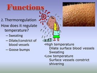
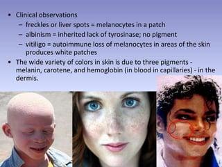
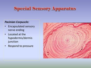
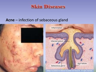
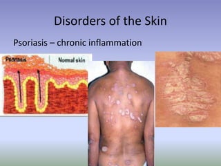
Skin (cutaneous membrane)
•largest organ of the human body with a
surface area of 1.5-2.0 square meters in
an average adult.
•Each 5 square cm may have up to:
600 sweat glands
20 blood vessels
60,000 melanocytes and over a
thousand nerve endings.
“A waterproof, stretchable, washable and permanent-pressed coat
of the human body that is guaranteed to last a lifetime with
reasonable care”
1. Protection Integument
(“covering”)
– tight cell junctions prevent
bacterial invasion
– lipids released retard
evaporation
– pigment protects somewhat
against UV light
– Langerhans cells alert
immune system
2. Thermoregulation
How does it regulate
temperature?
– Sweating
– Dilate/constrict of
blood vessels
– Goose bumps
•High temperature
Dilate surface blood vessels
Sweating
•Low temperature
Surface vessels constrict
shivering
• Shivering and constriction of surface vessels
– raise internal body temperature as needed
• Exercise
– in moderate exercise, more blood brought to
surface helps lower temperature
– with extreme exercise, blood is shunted to
muscles and body temperature rises
3. Excretes Waste
The skin removes the
excess of water,
pigments and salts
from the blood
through the sweat
glands.
subcutaneous
4. Reduces water loss
The uppermost layer of the skin, a thick
impermeable membrane, is full of Keratin and
is “cornified” or hardened
5. Houses sensory receptors
– touch, pressure,
vibration, tickle, heat,
cold, and pain arise in
the skin
6.Synthesis of Vit. D
– activation of a precursor
molecule in the skin by UV
light
– enzymes in the liver and
kidneys modify the activated
molecule to produce
calcitriol, the most active
form of vitamin D.
– necessary vitamin for
absorption of calcium from
food in the gastrointestinal
tract
Cutaneous membrane
• Protects tissues from
physical trauma,
biological pathogens,
and chemical trauma
• Provides sensations
Accessory Structures
• Provides sensations
• Produces secretions
• Protects epidermal
surfaces
Keratinized stratified squamous epithelium
Stratum Basale:
• Mitotic layer, continuously
replaces epidermal cells,
turnover rate between 25 to
50 days
Stratum Spinosum:
• Cells are pushed upward
and flatten out
• Stratum Granulosum:
Cells contain granules of
Keratin
Stratum Lucidum:
• Observed only in non-hairy
or thick skin. Several layers of
dead cells with indistinct
boundaries
Stratum Corneum:
• Composed of 25 or more
layers of dead squamous
cells still joined by
desmosomes. Eventually
desmosomes break and cells
flake off in a process called
desquamation
Keratinocytes:
Most common cells of the
epidermis. Provides
protection and
waterproofing sealant
Melanocytes:
Produces and transfer the
protein melanin to
Keratinocytes. Melanin is a
brown/black pigment that
absorbs UV-light.
Langerhans cells:
Arise from red bone marrow
and migrate to the
epidermis. They participate
in immune responses against
bacteria and viruses. Easily
damaged by UV
Merkel Cells:
Found in the Stratum Basale,
they contact the flattened
process of sensory neuron.
They respond to touch
sensations
• Melanin produced in epidermis by melanocytes
– melanocytes convert tyrosine to melanin
• UV in sunlight increases melanin production
– same number of melanocytes in everyone, but differing
amounts of pigment produced
– results vary from yellow to tan to black color
• Clinical observations
– freckles or liver spots = melanocytes in a patch
– albinism = inherited lack of tyrosinase; no pigment
– vitiligo = autoimmune loss of melanocytes in areas of the skin
produces white patches
• The wide variety of colors in skin is due to three pigments -
melanin, carotene, and hemoglobin (in blood in capillaries) - in the
dermis.
Carotene in dermis:
yellow-orange pigment (precursor of vitamin A)
found in stratum corneum & dermis
Hemoglobin in dermis:
red, oxygen-carrying pigment in blood cells
if other pigments are not present, epidermis is
translucent so pinkness will be evident
Deeper layers of skin
10-20 times thicker
than epidermis.
Top layer arranged
In ridges.
Why are there
ridges?
Ridges help the epidermis bind
to the dermis. The uneven
ridges create fingerprints
Dermis
• Reticular region:
consists of dense irregular CT
Contain most of the accessory
Structures
3. Papillary Region:
Consist of areolar Tissue
4. Dermal Papilla:
Fingerlike projections that
greatly increase surface area
1
2
3
4
Apocrine sweat glands:
• Found in the Axillae, nipples,
labia, and glans penis.
• Begin to function at puberty
and are affected by
hormones
• Produce odorous thick
secretion
• Possible pheromone function
Sudoriferous (eccrine)
Sweat Gland:
• Widespread (3,000/sq. in.)
• Produce thin watery
secretion
• Controlled by nervous
system
• Thermoregulation
• Excretion of urea
• Antibacterial action
Sebaceous “Oil” gland:
• Secrete sebum
• Coats hair shaft and lubricates
the epidermis
• Secreted to hair follicles
• Not associated with hair on
the labia, glans penis, and lips
• Activity controlled by sex-
hormones
• Modified in external ear canal
to produce cerumen or ear
wax (ceruminous glands)
Mammary Glands:
• Anatomically related to
apocrine sweat glands
• Development controls by
sex hormones and
pituitary hormones
• Produce milk
Ceruminous Glands:
• Modified sweat glands
found in the external
auditory canal
• Produce cerumen or ear
wax
• Helps trap foreign
particles from reaching
the eardrum
Hair:
• Present on most skin
except palmer
surface, and plantar
surface
• Growth controlled by
genetics and sex
hormones
• Provides protection
and sensory input
• Each hair has an
arrector pili muscle
Split ends
Nails – protective
covers of ends of
fingers and toes.
Meissner’s Corpuscles:
• Present in dermal papilla
• Specialized sensory neuron nerve endings
• Respond to touch ( tactile)
• Most numerous in thick or non-hairy skin of the palmar and
plantar surfaces
Pacinian Corpuscle:
• Encapsulated sensory
nerve ending
• Located at the
hypodermis/dermis
junction
• Respond to pressure
• The hair growth cycle consists of a growing stage and a resting stage.
– Growth cycle = growth stage & resting stage
• Growth stage
– lasts for 2 to 6 years
– matrix cells at base of hair root producing length
• Resting stage
– lasts for 3 months
– matrix cells inactive & follicle atrophies
– Old hair falls out as growth stage begins again
• normal hair loss is 70 to 100 hairs per day
• Both rate of growth and the replacement cycle can be altered by
illness, diet, high fever, surgery, blood loss, severe emotional stress,
and gender.
• Chemotherapeutic agents affect the rapidly dividing matrix hair cells
resulting in hair loss.
• Hair color is due primarily to the amount and type of melanin.
• Graying of hair occurs because of a progressive decline in
tyrosinase.
– Dark hair contains true melanin
– Blond and red hair contain melanin with iron and sulfur added
– Graying hair is result of decline in melanin production
– White hair has air bubbles in the medullary shaft
• Hormones influence the growth and loss of hair (Clinical
applications).
Acne – infection of sebaceous gland
Whitehead-sebum lodges in ducts bec of improper discharging
blackhead- whitehead blackened by oxidation
Furuncles and carbuncles
Benign tumor – fleshy growths on neck, armpits
and body.
Harmless!
Tumor
Vascular Birthmarks – Blood vessel abnormality
affecting .5% of population – darkens skin
Dermatitis – dry, sensitive skin
Athlete’s Foot
Ring WormFungal Infection
Impetigo – bacterial infection
Disorders of the Skin
Psoriasis – chronic inflammation
SkinCancer: three common types
Melanoma
Squamous cell Carcinoma
Basal cell
carcinoma
ABCD Rule for Detection
Warts- viral infection
Plane warts
Common wart
Plantar wart
VAMPIRES AND WEREWOLVES
• Porphyria- rare skin condition; genetic
- can not make the iron-containing heme part of
hemoglobin
-accumulation of porphyrins; lesions appear
when exposed to the sun
-fingers, toes, nose become mutilated, degeneration of
gums, rampant hair growth
-symptoms become aggravated by exposure to some
chemicals including garlic
Characteristics of Porphyria ( 5Ps)
a. Puberty
b. Psychiatric abnormality
c. Pain
d. Polyneuropathy
e. Photosensitivity
Treatments:
• Injection of heme molecules from healthy
rbcs
• Middle ages ( no injections)- drank blood
Touch me Nots- Epidermolysis bullosa
• Hereditary
• Lack of keratin in basal cells of epidermis
• A simple touch can cause layers to separate and
blister
SKIN SIGNS
1. Red skin- HBP, polycythemia, allergy, blushing
2. Pale ( pallor)- anemia, LBP, fear and anger
3. Purplish/bluish- severe heart disease, pneumonia
4. Yellow-jaundice
5. Bronze – addison’s disease
6. Moist/hot- hyperthyroidism
7. Rashes- measles, chicken pox and infectious dss
8. Vitamin deficiency- rough, cracked scaly around
corners of mouth
9. Black/bluish marks- hematoma (clots)
• When an injury extends to tissues deep to the epidermis, the repair
process is more complex than epidermal healing, and scar formation
results.
• Healing occurs in 4 phases
– inflammatory phase has clot unite wound edges and WBCs arrive from
dilated and more permeable blood vessels
– migratory phase begins the regrowth of epithelial cells and the
formation of scar tissue by the fibroblasts
– proliferative phase is a completion of tissue formation
– maturation phase sees the scab fall off
• Scar formation
– hypertrophic scar remains within the boundaries of the original wound
– keloid scar extends into previously normal tissue
• collagen fibers are very dense and fewer blood vessels are present
so the tissue is lighter in color
1. Inflammatory stage:
a. Blood clot forms
b. Inflammation is the result of vasodilation which
helps deliver WBC’s to help eliminate
microbes
2. Migratory Phase:
a. Clot becomes a scab
b. Epithelial cells migrate beneath
scab to repair epithelium
c. Fibroblast migrate into the area to repair
connective tissue
d. New tissue is called granulation
tissue
3. Proliferative phase:
a. Extensive growth of epithelial cells
b. Deposition of collagen fibers by
fibroblasts
c. Continued growth of vascular network
4. Maturation phase:
a. Scab sloughs off
b. Collagen fibers become more
organized
c. Fibroblasts decrease in number
• Collagen fibers decrease in number & stiffen
• Elastic fibers become less elastic
• Fibroblasts decrease in number
• decrease in number of melanocytes (gray hair, blotching)
• decrease in Langerhans cells (decreased immune
responsiveness)
• reduced number and less-efficient phagocytes
• Your body is composed of approximately 100 Trillion
cells
• About 16% of your body weight is skin
• The skin is completely renewed every 27 days
• You will make almost 1000 new skins in a lifetime
• If all the layers of your skin were laid out on the
ground, it would cover about 20 m2 or 2 parking
spaces
• A fingernail or toenail takes about 6 months to
grow from base to tip
• Fingernails grow faster than toenails
• An average human scalp has 100,000 hairs
• We lose between 40 and 100 hairs per day
• Blondes have more hair than brunettes
• Fingerprints provide traction for grasping objects
• Even identical twins have different fingerprints
• Every square inch of dermis contains twenty feet of
blood vessels
• Skin on our hands and feet is thicker. When we
bathe, skin takes on water and swells slightly.
• In the thicker areas, increased surface area creates
crowding. The skin must wrinkle to accommodate
the changes
• Friction of the epidermis causes cell division to
increase.
• This outward thickening is called a callus.
• Sometimes growth is inward, creating a corn.
• Humans shed about 600,000 particles of skin per
hour – about 1.5 pounds per year.
• At age 70, you will have lost about 105 lbs. of skin.

3. integumentary system

  • 2.
    Skin (cutaneous membrane) •largestorgan of the human body with a surface area of 1.5-2.0 square meters in an average adult. •Each 5 square cm may have up to: 600 sweat glands 20 blood vessels 60,000 melanocytes and over a thousand nerve endings. “A waterproof, stretchable, washable and permanent-pressed coat of the human body that is guaranteed to last a lifetime with reasonable care”
  • 3.
    1. Protection Integument (“covering”) –tight cell junctions prevent bacterial invasion – lipids released retard evaporation – pigment protects somewhat against UV light – Langerhans cells alert immune system
  • 4.
    2. Thermoregulation How doesit regulate temperature? – Sweating – Dilate/constrict of blood vessels – Goose bumps •High temperature Dilate surface blood vessels Sweating •Low temperature Surface vessels constrict shivering
  • 5.
    • Shivering andconstriction of surface vessels – raise internal body temperature as needed • Exercise – in moderate exercise, more blood brought to surface helps lower temperature – with extreme exercise, blood is shunted to muscles and body temperature rises
  • 7.
    3. Excretes Waste Theskin removes the excess of water, pigments and salts from the blood through the sweat glands. subcutaneous
  • 8.
    4. Reduces waterloss The uppermost layer of the skin, a thick impermeable membrane, is full of Keratin and is “cornified” or hardened
  • 9.
    5. Houses sensoryreceptors – touch, pressure, vibration, tickle, heat, cold, and pain arise in the skin
  • 10.
    6.Synthesis of Vit.D – activation of a precursor molecule in the skin by UV light – enzymes in the liver and kidneys modify the activated molecule to produce calcitriol, the most active form of vitamin D. – necessary vitamin for absorption of calcium from food in the gastrointestinal tract
  • 13.
    Cutaneous membrane • Protectstissues from physical trauma, biological pathogens, and chemical trauma • Provides sensations Accessory Structures • Provides sensations • Produces secretions • Protects epidermal surfaces
  • 14.
  • 16.
    Stratum Basale: • Mitoticlayer, continuously replaces epidermal cells, turnover rate between 25 to 50 days Stratum Spinosum: • Cells are pushed upward and flatten out • Stratum Granulosum: Cells contain granules of Keratin
  • 17.
    Stratum Lucidum: • Observedonly in non-hairy or thick skin. Several layers of dead cells with indistinct boundaries Stratum Corneum: • Composed of 25 or more layers of dead squamous cells still joined by desmosomes. Eventually desmosomes break and cells flake off in a process called desquamation
  • 18.
    Keratinocytes: Most common cellsof the epidermis. Provides protection and waterproofing sealant Melanocytes: Produces and transfer the protein melanin to Keratinocytes. Melanin is a brown/black pigment that absorbs UV-light.
  • 19.
    Langerhans cells: Arise fromred bone marrow and migrate to the epidermis. They participate in immune responses against bacteria and viruses. Easily damaged by UV Merkel Cells: Found in the Stratum Basale, they contact the flattened process of sensory neuron. They respond to touch sensations
  • 20.
    • Melanin producedin epidermis by melanocytes – melanocytes convert tyrosine to melanin • UV in sunlight increases melanin production – same number of melanocytes in everyone, but differing amounts of pigment produced – results vary from yellow to tan to black color
  • 21.
    • Clinical observations –freckles or liver spots = melanocytes in a patch – albinism = inherited lack of tyrosinase; no pigment – vitiligo = autoimmune loss of melanocytes in areas of the skin produces white patches • The wide variety of colors in skin is due to three pigments - melanin, carotene, and hemoglobin (in blood in capillaries) - in the dermis.
  • 22.
    Carotene in dermis: yellow-orangepigment (precursor of vitamin A) found in stratum corneum & dermis Hemoglobin in dermis: red, oxygen-carrying pigment in blood cells if other pigments are not present, epidermis is translucent so pinkness will be evident
  • 23.
    Deeper layers ofskin 10-20 times thicker than epidermis. Top layer arranged In ridges. Why are there ridges?
  • 24.
    Ridges help theepidermis bind to the dermis. The uneven ridges create fingerprints
  • 25.
    Dermis • Reticular region: consistsof dense irregular CT Contain most of the accessory Structures 3. Papillary Region: Consist of areolar Tissue 4. Dermal Papilla: Fingerlike projections that greatly increase surface area 1 2 3 4
  • 27.
    Apocrine sweat glands: •Found in the Axillae, nipples, labia, and glans penis. • Begin to function at puberty and are affected by hormones • Produce odorous thick secretion • Possible pheromone function Sudoriferous (eccrine) Sweat Gland: • Widespread (3,000/sq. in.) • Produce thin watery secretion • Controlled by nervous system • Thermoregulation • Excretion of urea • Antibacterial action
  • 28.
    Sebaceous “Oil” gland: •Secrete sebum • Coats hair shaft and lubricates the epidermis • Secreted to hair follicles • Not associated with hair on the labia, glans penis, and lips • Activity controlled by sex- hormones • Modified in external ear canal to produce cerumen or ear wax (ceruminous glands)
  • 29.
    Mammary Glands: • Anatomicallyrelated to apocrine sweat glands • Development controls by sex hormones and pituitary hormones • Produce milk
  • 30.
    Ceruminous Glands: • Modifiedsweat glands found in the external auditory canal • Produce cerumen or ear wax • Helps trap foreign particles from reaching the eardrum
  • 31.
    Hair: • Present onmost skin except palmer surface, and plantar surface • Growth controlled by genetics and sex hormones • Provides protection and sensory input • Each hair has an arrector pili muscle
  • 32.
  • 33.
    Nails – protective coversof ends of fingers and toes.
  • 34.
    Meissner’s Corpuscles: • Presentin dermal papilla • Specialized sensory neuron nerve endings • Respond to touch ( tactile) • Most numerous in thick or non-hairy skin of the palmar and plantar surfaces
  • 35.
    Pacinian Corpuscle: • Encapsulatedsensory nerve ending • Located at the hypodermis/dermis junction • Respond to pressure
  • 36.
    • The hairgrowth cycle consists of a growing stage and a resting stage. – Growth cycle = growth stage & resting stage • Growth stage – lasts for 2 to 6 years – matrix cells at base of hair root producing length • Resting stage – lasts for 3 months – matrix cells inactive & follicle atrophies – Old hair falls out as growth stage begins again • normal hair loss is 70 to 100 hairs per day • Both rate of growth and the replacement cycle can be altered by illness, diet, high fever, surgery, blood loss, severe emotional stress, and gender. • Chemotherapeutic agents affect the rapidly dividing matrix hair cells resulting in hair loss.
  • 37.
    • Hair coloris due primarily to the amount and type of melanin. • Graying of hair occurs because of a progressive decline in tyrosinase. – Dark hair contains true melanin – Blond and red hair contain melanin with iron and sulfur added – Graying hair is result of decline in melanin production – White hair has air bubbles in the medullary shaft • Hormones influence the growth and loss of hair (Clinical applications).
  • 38.
    Acne – infectionof sebaceous gland
  • 39.
    Whitehead-sebum lodges inducts bec of improper discharging blackhead- whitehead blackened by oxidation
  • 40.
  • 41.
    Benign tumor –fleshy growths on neck, armpits and body. Harmless! Tumor
  • 42.
    Vascular Birthmarks –Blood vessel abnormality affecting .5% of population – darkens skin
  • 43.
    Dermatitis – dry,sensitive skin
  • 44.
  • 45.
  • 46.
    Disorders of theSkin Psoriasis – chronic inflammation
  • 47.
    SkinCancer: three commontypes Melanoma Squamous cell Carcinoma Basal cell carcinoma
  • 48.
    ABCD Rule forDetection
  • 49.
    Warts- viral infection Planewarts Common wart Plantar wart
  • 50.
    VAMPIRES AND WEREWOLVES •Porphyria- rare skin condition; genetic - can not make the iron-containing heme part of hemoglobin -accumulation of porphyrins; lesions appear when exposed to the sun -fingers, toes, nose become mutilated, degeneration of gums, rampant hair growth -symptoms become aggravated by exposure to some chemicals including garlic
  • 51.
    Characteristics of Porphyria( 5Ps) a. Puberty b. Psychiatric abnormality c. Pain d. Polyneuropathy e. Photosensitivity Treatments: • Injection of heme molecules from healthy rbcs • Middle ages ( no injections)- drank blood
  • 52.
    Touch me Nots-Epidermolysis bullosa • Hereditary • Lack of keratin in basal cells of epidermis • A simple touch can cause layers to separate and blister
  • 53.
    SKIN SIGNS 1. Redskin- HBP, polycythemia, allergy, blushing 2. Pale ( pallor)- anemia, LBP, fear and anger 3. Purplish/bluish- severe heart disease, pneumonia 4. Yellow-jaundice 5. Bronze – addison’s disease 6. Moist/hot- hyperthyroidism 7. Rashes- measles, chicken pox and infectious dss 8. Vitamin deficiency- rough, cracked scaly around corners of mouth 9. Black/bluish marks- hematoma (clots)
  • 55.
    • When aninjury extends to tissues deep to the epidermis, the repair process is more complex than epidermal healing, and scar formation results. • Healing occurs in 4 phases – inflammatory phase has clot unite wound edges and WBCs arrive from dilated and more permeable blood vessels – migratory phase begins the regrowth of epithelial cells and the formation of scar tissue by the fibroblasts – proliferative phase is a completion of tissue formation – maturation phase sees the scab fall off • Scar formation – hypertrophic scar remains within the boundaries of the original wound – keloid scar extends into previously normal tissue • collagen fibers are very dense and fewer blood vessels are present so the tissue is lighter in color
  • 56.
    1. Inflammatory stage: a.Blood clot forms b. Inflammation is the result of vasodilation which helps deliver WBC’s to help eliminate microbes 2. Migratory Phase: a. Clot becomes a scab b. Epithelial cells migrate beneath scab to repair epithelium c. Fibroblast migrate into the area to repair connective tissue d. New tissue is called granulation tissue
  • 57.
    3. Proliferative phase: a.Extensive growth of epithelial cells b. Deposition of collagen fibers by fibroblasts c. Continued growth of vascular network 4. Maturation phase: a. Scab sloughs off b. Collagen fibers become more organized c. Fibroblasts decrease in number
  • 59.
    • Collagen fibersdecrease in number & stiffen • Elastic fibers become less elastic • Fibroblasts decrease in number • decrease in number of melanocytes (gray hair, blotching) • decrease in Langerhans cells (decreased immune responsiveness) • reduced number and less-efficient phagocytes
  • 60.
    • Your bodyis composed of approximately 100 Trillion cells • About 16% of your body weight is skin • The skin is completely renewed every 27 days • You will make almost 1000 new skins in a lifetime • If all the layers of your skin were laid out on the ground, it would cover about 20 m2 or 2 parking spaces
  • 61.
    • A fingernailor toenail takes about 6 months to grow from base to tip • Fingernails grow faster than toenails • An average human scalp has 100,000 hairs • We lose between 40 and 100 hairs per day • Blondes have more hair than brunettes
  • 62.
    • Fingerprints providetraction for grasping objects • Even identical twins have different fingerprints • Every square inch of dermis contains twenty feet of blood vessels • Skin on our hands and feet is thicker. When we bathe, skin takes on water and swells slightly. • In the thicker areas, increased surface area creates crowding. The skin must wrinkle to accommodate the changes
  • 63.
    • Friction ofthe epidermis causes cell division to increase. • This outward thickening is called a callus. • Sometimes growth is inward, creating a corn. • Humans shed about 600,000 particles of skin per hour – about 1.5 pounds per year. • At age 70, you will have lost about 105 lbs. of skin.