Download to read offline
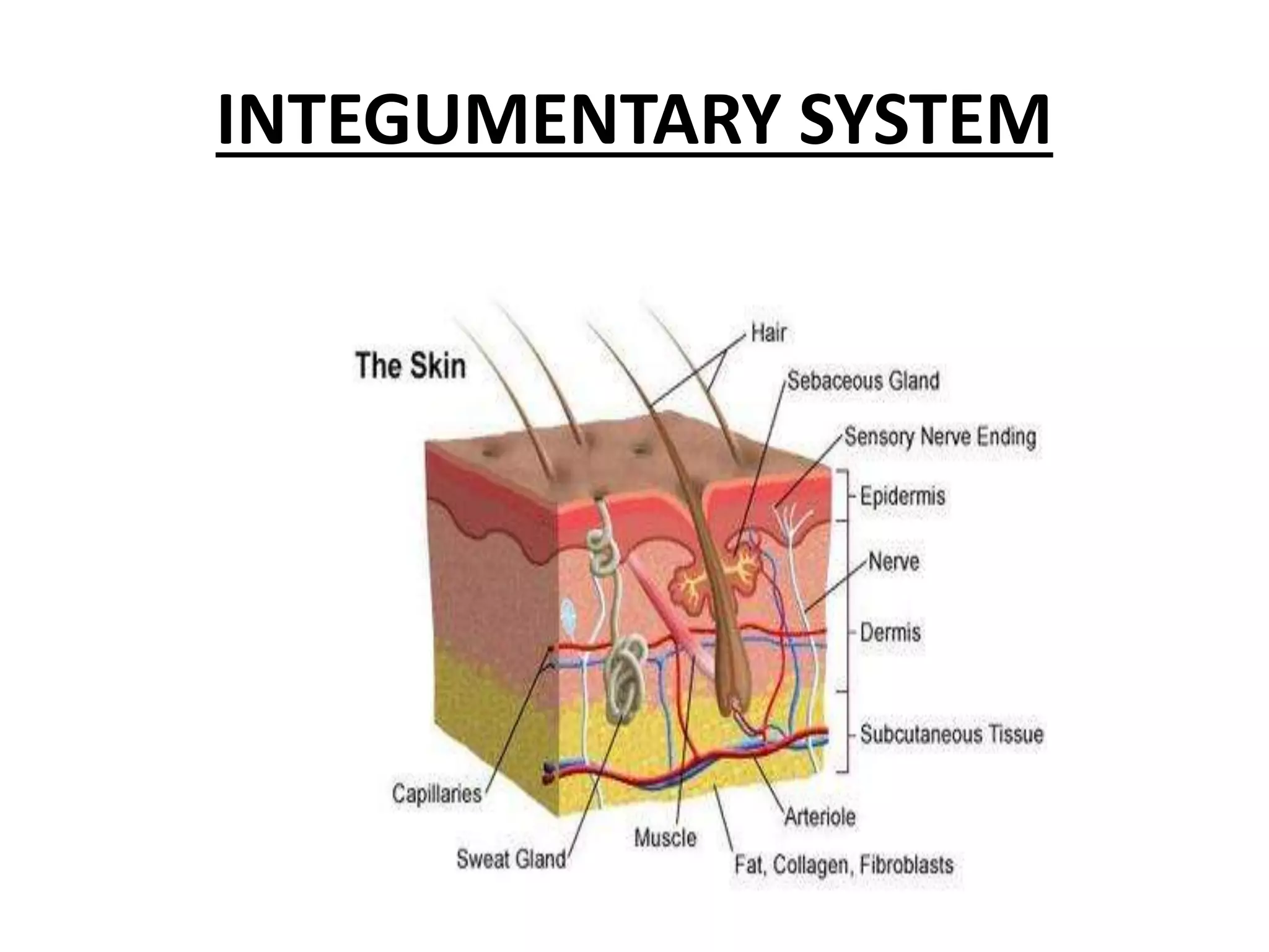


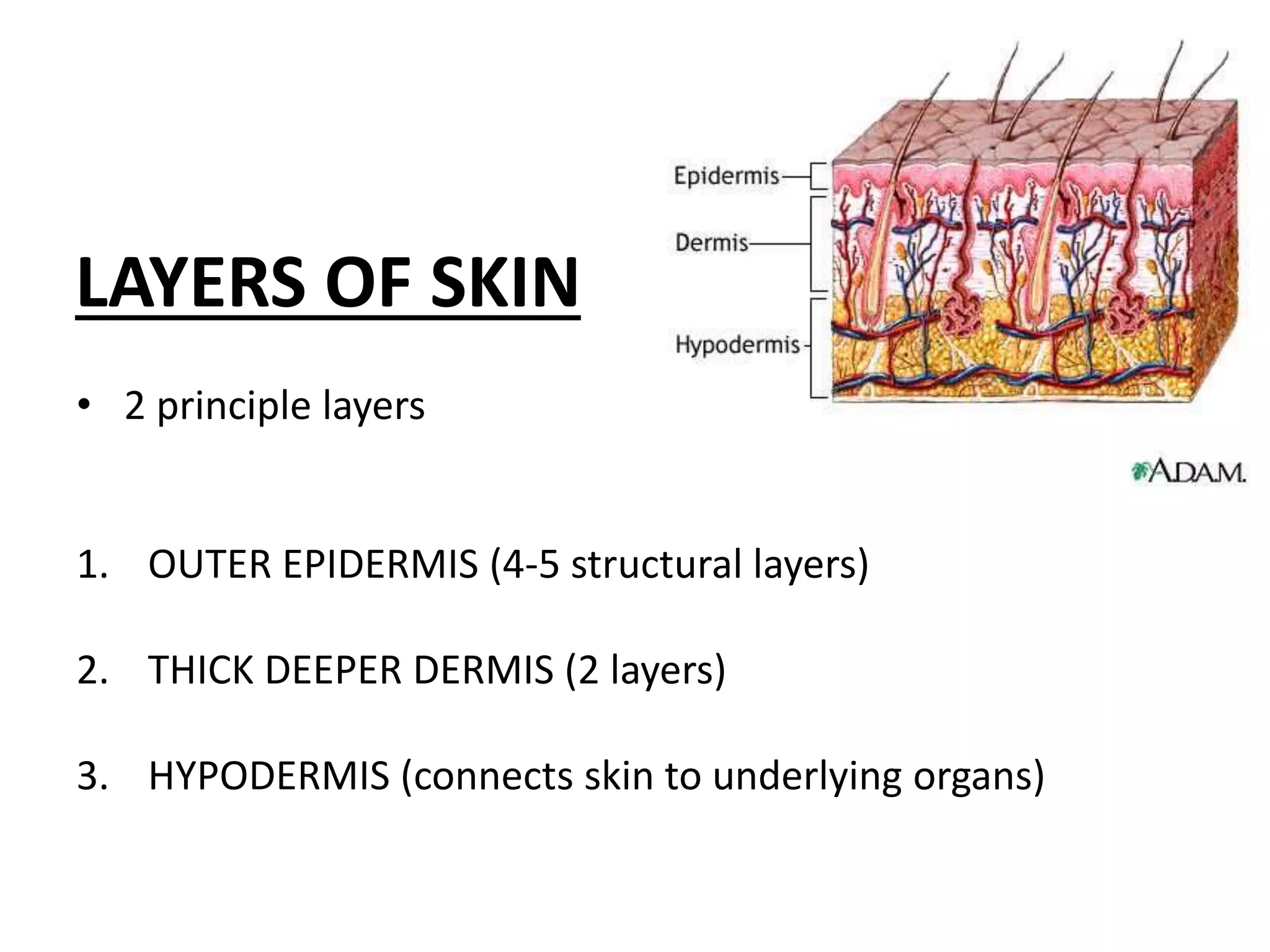


The integumentary system consists of the skin, hair, nails, and glands. The skin is the largest organ of the body and has three main layers - the epidermis, dermis, and hypodermis. The epidermis is made up of stratified squamous epithelium in multiple layers that acts as a protective barrier. Below this is the dermis, which contains hair follicles, sweat and oil glands, nerves, and supplies the epidermis with nutrients. The deepest layer, the hypodermis, connects the skin to underlying tissue. Together these layers allow the skin to regulate temperature, protect the body, and sense the environment.


