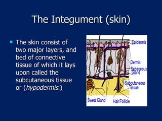
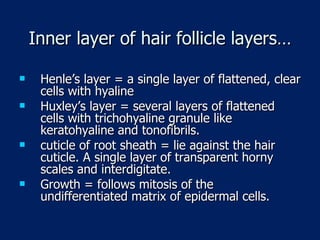
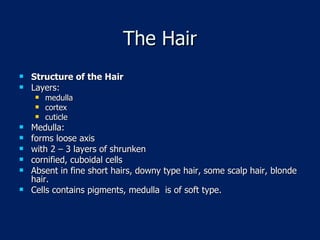
www.Examville.com Online practice tests, live classes, tutoring, study guides Q&A, premium content and more .
The Integumentary system
The Integument The Integumentary System (  the integument ) or simply the skin system is one of the largest and heaviest organ systems of the human body. The skin covers the outer surface of the body and is continuous with the mucous membranes found insides the eyelids, nostrils, and other openings of the human body.
The Integument (skin) The skin consist of two major layers, and bed of connective tissue of which it lays upon called the subcutaneous tissue or ( hypodermis .)
The skin The two major layers are: -(1) The outer  epidermis , derived from the embryonic ectoderm, and -(2) the inner  dermis , which develops from the mesode  Beneath the dermis is the  hypodermis  (sometimes called the subcutaneous tissue layer or the superficial fascia)
The skin and its appendages  hair,  nails,  sebaceous and  sweat glands
The skin and its appendages make up a very complex set of organs that accomplishes several functions for the human body, namely: 1. Protection for internal organs from the outside environment, 2. Protection from the loss of internal fluids or dehydration, 3. Maintenanc of homeostasis, interms of body overall temperature regulation,
The skin and its appendages…. 4. Excretion of some waste products via the  sebaceous  and  sweat  glands,  5. Maintenance of the sites for reception of external cutaneous sensations, 6. Serves as a blood reservoir, and finally, 7. The skin does some metabolic functions.
The Skin The skin layers: Epidermis – specialized epithelium, derived from the ectoderm. Dermis ( corium) – characteristics:  vascular dense connective tissue from the mesoderm.  Corresponds to the Lamina propria of the mucous membrane. Connected to the underlying hypodermis by connective tissue fibers . Hypodermis – lower layer, not part of the skin.
The skin….. Dermal ridges – patterns of ridges over the  skin surface consists of whorls, loops, arches and determined by hereditary traits. Formed during 3rd and 4th months of fetal life   Classification: thick skin = over the palms of hands and feet thin skin = over the rest of the body.
The Epidermis. Cell types: Keratinocyte = differentiate to keratin. Melanocyte Langerhans cell Merkel cell Basal cell layer mitotic activity   cells displaced to higher level    elaborate keratin    keratin replaces the cytoplasm    cell dies    shedding.
Epidermis of the palms and sole 1.  Stratum germinativum: The stratum germinatum ( SG ) provides the germinal cells necessary for the regeneration of the layers of the epidermis. These germinal cells are separated from the  dermis  by a thin layer of basement membrane. After a mitotic division a newly formed cell will undergo a progressive maturation called keratinization as it migrates to the surface. Tonofibrils  = aggregate   bundles of fine filaments, where mitotic activities are inititated.
Epidermis of the palms and sole 2.  Stratum Spinosum The cells that divide in the statum germinativum soon begin to accumulate many desmosomes on their outer surface which provide the characteristic “prickles” of the stratum spinosum ( SS ), which is often called the prickle-cell layer. Composed of irregular, polyhedral cells, separate and flattened at the surface. Cytoplasm is basophilic, intercellular b ridges with short process to adjacent cells MALPHIGIAN layer = stra. Germinativum & stra. Spinosum: a.  responsible for proliferation & initiation of keratinization process. b. contains  Melanocytes  melanin, for pigmentation. c. with scattered  langerhans and Merkel cells.
Epidermis of the palms and sole 3.  Stratum granulosum The progressive maturation of a keratinocyte is charcterized by the accumulation of keratin, called keratinization. The cells of the stratum granulosum ( SGR ) accumulae dense basophilic keratohyalin granules (seen on the close-up view). These granules contain lipids, which along with the desmosomal connections, help to form a  waterproof barrier  that functions to prevent fluid loss from the body-  keratinosomes . Consists of 3 to 5 layers of flattened cells whose long axis is parallel to the skin surface, contains  Keratohyalin  granules, cells become pale, eventually dies.
Epidermis of the palms and sole 4.  Stratum Lucidum Epidermis varies in thickness throughout the body depending mainly on frictional forces and is thickest on the palms of the hands and soles of the feet. The stratum lucidum is normally only well seen in thick epidermis and represents a transition from the stratum granulosum to the stratum corneum.  Clear translucent layer 3 – 5 cell layers, flattened  and closely packed, nuclei are indistinct or absent. Cytoplasm has keratohyalin and distributed among the tonofibrils.
Epidermis of the palms and sole 5.  Stratum Corneum As a cell accumulates keratinohyalin granules, it is thought that rupture of lysosomal membranes release lysosomal enzymes that eventually cause cell death. The dead and dying cells filled with mature keratin form the stratum corneum ( SC ). The deeper cells of the stratum corneum retain their desmosomal junctions, but as they are pushed to the surface by newly forming cells of the stratum germinativum ( SG ), the dead cells gradually break apart and are lost, a process called  desquamation .
Epidermis of the palms and sole Stratum Corneum Nucleus is absent, cytoplasm is replaced with Keratin from the tonofibrils and deeper epidermal layer ( soft keratin) low in sulfur content. Stains pink in Eosin and often shredded during preparation. Stratum disjunctum = most superficial layer , with flat horny plates and desquamated constantly.
Pigmentation of skin: Carotene = yellow color. Blood = reddish hue. melanin pigments = shades of brown melanin  = produced by melanocytes within the melanosomes melanocytes  between keratinocytes of stra. Germinativum and spinosum within hair follicles dermal connective tissue.
Pigmentation of skin….. Tyrosinase  ( enzyme from the ribosomes)    transferred to endoplasmic reticulum    Golgi zone    packaged into vesicles   fused into premelanosomes    mature into 4 stages with melanin packed melanosomes    transferred to keratinocytes of stra. Germinativum and spinosum via phagocytosis of melanosome containing processes of  melanocytes    pigments are injected into the keratinocytes.
Pigmentation of skin….. MSH  – melanocyte stimulating hormones stimulates migration of melanosomes in dendritic process and the transfer of keratinocytes Ultraviolet rays increases the melanocytes enzymatic activity   Tanning
Langerhans cells the 3rd cellular population within the epidermis. Star- shaped, with numerous dendritic process  Found principally within the stra. Spinosum. Berbick granules = rod-like  inclusions in cytoplasm, strat epith. In oral mucosa, esophagus, vagina, hair follicles, sebaceous and apocrine glands, thymus and lymph nodes. Functions by fixing and processes exogenous antigens.  Associated with B and T lymphos .
Merkel cells the 4th cellular population within the epidermis. Wide epidermal distribution, found in stra. Germinativum With irregular shaped nuclei, cytoplasm less dense, concentrated at the basal region of cells. Attached to neighboring keratinocytes by  numerous desmosomes. Functions as mechanoreceptors. Has APUD ( amino precursor uptake and decarboxylation) like activity.
The Dermis 1.  The Papillary Dermis The papillary dermis ( PD ) contains vascular networks that have two important functions. The first being to support the avascular epidermis with vital nutrients and secondly to provide a network for thermoregulation. The vasculature is organized so that by increasing or decreasing blood flow, heat can either be conserved or dissipated. The vasculature interdigitates in areas called dermal papillae ( DP ). The papillary dermis also contains the free sensory nerve endings and structures called Meissner’s corpuscles in highly sensitive areas.
The Dermis…. 2.  The Reticular Dermis The main fibrous bed of the dermis. The reticular layer of the dermis ( RD )  consists of dense irregular connective tissue, which differs from the papillary layer ( PD ), which is made up of mainly loose connective tissue. The reticular layer of the dermis is important in giving the skin it overall strength and elasticity, as well as housing other important epithelial derived structures such as glands and hair follicles. Langer lines =  direction of fiber lines of skin tension, of surgical importance, incisions heal with less gap and scar tissues. Ground substance is of amorphous matrix.
Skin…… 3 principal glycosaminoglycans of the skin  a. hyaluronic acid,  b. dermatan sulfates, c. chondroitin sulfates.
Cellular elements: fibrblasts macrophages fat cells chromatophores connective tissue cells true dermal melanocytes is rare dopa positive accumulate in the sacral region = Mongolian spots accumulate in dermal tumors = blue nevi generally papillary layer has more cells and smaller and finer connective  tissue fibers and reticular fibers.
Muscle fibers maybe found in the dermis arrectores pilorum muscles. Scattered in the dermis = nipple, penis, scrotum, parts of the perineum, contraction gives a wrinkled appearance.
Hypodermis  the subcutaneous layer( superficial) not part of the skin deep extension of the dermis density and arrangement determines the mobility of the skin panniculus adiposus  = a fat pad due to a continuous lobules of fat present up to 3 cm in the abdomen. Devoid of fats = eyelids, penis and scrotum. Superficial zone contains hair follicles and sweat glands.
Skin Appendages Nails Horny plates of epidermis that forms a protective covering on the dorsal plates of the terminal phalanges, do  not desquamate. The body is translucent, pink color due to vascularity in the nail bed. The root is more opaque and becomes continuous with the body of the nail.  3rd month, the begin to form as nail groove    differentiate into soft matrix nail plates    advance into the dorsal surface of the digits  top form nail beds. It lacks sweat glands and hair follicles. Hyponychium  = thickened epidermis at the junction of stra. Corneum. Lonule  = a crescentric margin at the nail bed. Nail groove  is lined by modified epidermis Eponychium  =  cuticle , extension of the stra. Corneum from nail bed to free surface of the nail plate. Growth rates is  0.5 mm per week Faster growth  in the fingernail than in toenails.
Hair Follicle Hair Follicles  elastic keratinized threads that developed from the epidermis. Distributed thru out the entire skin surface except: palms sole dorsal surface of distal phalanges anal apperture urogenital apperture has shaft and root embedded in the skin hair follicle consists of epidermal and dermal tissues
Hair Follicle Structure = dermal root sheath derive from external connective sheath.  With 3 layers:  outer poorly defined, coarse bundle of collagen fibers ( corresponds to reticular layer) midlayer = thicker, corresponds to papillary layer, cellular and with fine connective tissue layer. Inner layer, glassy membrane, corresponds to basal lamina, consists of amorphous ground substance.
Hair Follicle epithermal (epithelial root sheath) =  from the epidermis outer epithelial root sheath = posses a single row of tall cells and an inner polygonal stratum. Inner sheath = keratinized root sheath
Inner layer of hair follicle layers: Henle’s layer = a single layer of flattened, clear cells with hyaline Huxley’s layer = several layers of flattened cells with trichohyaline granule like keratohyaline and tonofibrils. cuticle of root sheath = lie against the hair cuticle. A single layer of transparent horny scales and interdigitate.
Inner layer of hair follicle layers… Henle’s layer = a single layer of flattened, clear cells with hyaline Huxley’s layer = several layers of flattened cells with trichohyaline granule like keratohyaline and tonofibrils. cuticle of root sheath = lie against the hair cuticle. A single layer of transparent horny scales and interdigitate. Growth = follows mitosis of the undifferentiated matrix of epidermal cells.
Definite period for growth: Definite period for growth: Head hair = growth is 2 – 4 years Eyelashes = 3 – 4 months. Upon cessation of growth, multiplication at the bases stopped    root detach from matrix    hair falls out or is pulled. After a period of rest, a new germinal matrix develops to grow up the reformed follicle.
The Hair Structure of the Hair Layers: medulla cortex cuticle Medulla:  forms loose axis with 2 – 3 layers of shrunken  cornified, cuboidal cells Absent in fine short hairs, downy type hair, some scalp hair, blonde hair.  Cells contains pigments, medulla  is of soft type.
Cortex make up the bulk of the hair composed of several layers of long flattened spindle shaped cornified cells. Keratin is of the hard type Keratin fibrils are oriented  parallel to the long axis of the hair Pigment granules are found in between cells and are oxidized Air accumulates in the intercellular spaces, modifies hair color.
Cuticle single layer of thin clear cells cornified cells, have lost their nuclei cells overlap with free edges directed upwards x section, round  = Chinese, Eskimos, American Indians. Oval = Caucasians Elliptical or reniform = wooly hair of blacks.
Sweat Glands   Eccrine or merocrine sweat glands  or ordinary sweat glands.  Coiled, tubular, distributed thru out the skin.  Not found in: nail beds margin of lips glans penis eardrum Most numerous in the: palms  sole Apocrine sweat glands  = found in axilla, nipple, areola, labia majora, circumanal region. = has a thicker secretion, opens into the upper part of hair follicles. Less coiling, the lumen is much wider., myo-epithelial cells are much larger and form a complete layer, begins to function only at puberty. Secretes a waxy substance = cerumen from glands of the external audoitory meatus  and the eyelids glands of Moll.
Sebaceous glands   connected with hair follicles several drain into a single follicle others drain into free surface, e.g., glans penis, labia minora, tarsal ( meibomian) glands absent in palms and soles each is encapsulated by a thin layer of connective tissue. alveolar ( saccular) glands which synthesize lipids. Alveoli are completely filled with stratified epithelium
Sebaceous glands Alveoli are completely filled with stratified epithelium, the internal surface is a single row of small cuboidal cells    continuous with basal cells of epidermis    incr. agranular endoplasmic reticulum    becomes active in Lipogenesis   center of alveoli nucleus becomes larger   cytoplasm is distended with droplets, containing cholesterol, phospholipids, triglycerides    nuclei gradually shrinks and disappears    cells breakdown into fatty mass of debris   SEBUM , holocrine type, total destruction of epithelial cells. * discharge of sebum is aided by the arector pili muscle  and pressure within alveolus.
Sebaceous gland….. Vascularity = rete cutaneum , around the junction zone between the dermis and the hypodermis   rete subpapillare ( network between papillary and reticular layer)
It’s FREE to join. http://www.examville.com

The Skin

  • 1.
    www.Examville.com Online practicetests, live classes, tutoring, study guides Q&A, premium content and more .
  • 2.
  • 3.
    The Integument TheIntegumentary System ( the integument ) or simply the skin system is one of the largest and heaviest organ systems of the human body. The skin covers the outer surface of the body and is continuous with the mucous membranes found insides the eyelids, nostrils, and other openings of the human body.
  • 4.
    The Integument (skin)The skin consist of two major layers, and bed of connective tissue of which it lays upon called the subcutaneous tissue or ( hypodermis .)
  • 5.
    The skin Thetwo major layers are: -(1) The outer epidermis , derived from the embryonic ectoderm, and -(2) the inner dermis , which develops from the mesode Beneath the dermis is the hypodermis (sometimes called the subcutaneous tissue layer or the superficial fascia)
  • 6.
    The skin andits appendages hair, nails, sebaceous and sweat glands
  • 7.
    The skin andits appendages make up a very complex set of organs that accomplishes several functions for the human body, namely: 1. Protection for internal organs from the outside environment, 2. Protection from the loss of internal fluids or dehydration, 3. Maintenanc of homeostasis, interms of body overall temperature regulation,
  • 8.
    The skin andits appendages…. 4. Excretion of some waste products via the sebaceous and sweat glands, 5. Maintenance of the sites for reception of external cutaneous sensations, 6. Serves as a blood reservoir, and finally, 7. The skin does some metabolic functions.
  • 9.
    The Skin Theskin layers: Epidermis – specialized epithelium, derived from the ectoderm. Dermis ( corium) – characteristics: vascular dense connective tissue from the mesoderm. Corresponds to the Lamina propria of the mucous membrane. Connected to the underlying hypodermis by connective tissue fibers . Hypodermis – lower layer, not part of the skin.
  • 10.
    The skin….. Dermalridges – patterns of ridges over the skin surface consists of whorls, loops, arches and determined by hereditary traits. Formed during 3rd and 4th months of fetal life Classification: thick skin = over the palms of hands and feet thin skin = over the rest of the body.
  • 11.
    The Epidermis. Celltypes: Keratinocyte = differentiate to keratin. Melanocyte Langerhans cell Merkel cell Basal cell layer mitotic activity  cells displaced to higher level  elaborate keratin  keratin replaces the cytoplasm  cell dies  shedding.
  • 12.
    Epidermis of thepalms and sole 1. Stratum germinativum: The stratum germinatum ( SG ) provides the germinal cells necessary for the regeneration of the layers of the epidermis. These germinal cells are separated from the dermis by a thin layer of basement membrane. After a mitotic division a newly formed cell will undergo a progressive maturation called keratinization as it migrates to the surface. Tonofibrils = aggregate bundles of fine filaments, where mitotic activities are inititated.
  • 13.
    Epidermis of thepalms and sole 2. Stratum Spinosum The cells that divide in the statum germinativum soon begin to accumulate many desmosomes on their outer surface which provide the characteristic “prickles” of the stratum spinosum ( SS ), which is often called the prickle-cell layer. Composed of irregular, polyhedral cells, separate and flattened at the surface. Cytoplasm is basophilic, intercellular b ridges with short process to adjacent cells MALPHIGIAN layer = stra. Germinativum & stra. Spinosum: a. responsible for proliferation & initiation of keratinization process. b. contains Melanocytes  melanin, for pigmentation. c. with scattered langerhans and Merkel cells.
  • 14.
    Epidermis of thepalms and sole 3. Stratum granulosum The progressive maturation of a keratinocyte is charcterized by the accumulation of keratin, called keratinization. The cells of the stratum granulosum ( SGR ) accumulae dense basophilic keratohyalin granules (seen on the close-up view). These granules contain lipids, which along with the desmosomal connections, help to form a waterproof barrier that functions to prevent fluid loss from the body- keratinosomes . Consists of 3 to 5 layers of flattened cells whose long axis is parallel to the skin surface, contains Keratohyalin granules, cells become pale, eventually dies.
  • 15.
    Epidermis of thepalms and sole 4. Stratum Lucidum Epidermis varies in thickness throughout the body depending mainly on frictional forces and is thickest on the palms of the hands and soles of the feet. The stratum lucidum is normally only well seen in thick epidermis and represents a transition from the stratum granulosum to the stratum corneum. Clear translucent layer 3 – 5 cell layers, flattened and closely packed, nuclei are indistinct or absent. Cytoplasm has keratohyalin and distributed among the tonofibrils.
  • 16.
    Epidermis of thepalms and sole 5. Stratum Corneum As a cell accumulates keratinohyalin granules, it is thought that rupture of lysosomal membranes release lysosomal enzymes that eventually cause cell death. The dead and dying cells filled with mature keratin form the stratum corneum ( SC ). The deeper cells of the stratum corneum retain their desmosomal junctions, but as they are pushed to the surface by newly forming cells of the stratum germinativum ( SG ), the dead cells gradually break apart and are lost, a process called desquamation .
  • 17.
    Epidermis of thepalms and sole Stratum Corneum Nucleus is absent, cytoplasm is replaced with Keratin from the tonofibrils and deeper epidermal layer ( soft keratin) low in sulfur content. Stains pink in Eosin and often shredded during preparation. Stratum disjunctum = most superficial layer , with flat horny plates and desquamated constantly.
  • 18.
    Pigmentation of skin:Carotene = yellow color. Blood = reddish hue. melanin pigments = shades of brown melanin = produced by melanocytes within the melanosomes melanocytes between keratinocytes of stra. Germinativum and spinosum within hair follicles dermal connective tissue.
  • 19.
    Pigmentation of skin…..Tyrosinase ( enzyme from the ribosomes)  transferred to endoplasmic reticulum  Golgi zone  packaged into vesicles  fused into premelanosomes  mature into 4 stages with melanin packed melanosomes  transferred to keratinocytes of stra. Germinativum and spinosum via phagocytosis of melanosome containing processes of melanocytes  pigments are injected into the keratinocytes.
  • 20.
    Pigmentation of skin…..MSH – melanocyte stimulating hormones stimulates migration of melanosomes in dendritic process and the transfer of keratinocytes Ultraviolet rays increases the melanocytes enzymatic activity  Tanning
  • 21.
    Langerhans cells the3rd cellular population within the epidermis. Star- shaped, with numerous dendritic process Found principally within the stra. Spinosum. Berbick granules = rod-like inclusions in cytoplasm, strat epith. In oral mucosa, esophagus, vagina, hair follicles, sebaceous and apocrine glands, thymus and lymph nodes. Functions by fixing and processes exogenous antigens. Associated with B and T lymphos .
  • 22.
    Merkel cells the4th cellular population within the epidermis. Wide epidermal distribution, found in stra. Germinativum With irregular shaped nuclei, cytoplasm less dense, concentrated at the basal region of cells. Attached to neighboring keratinocytes by numerous desmosomes. Functions as mechanoreceptors. Has APUD ( amino precursor uptake and decarboxylation) like activity.
  • 23.
    The Dermis 1. The Papillary Dermis The papillary dermis ( PD ) contains vascular networks that have two important functions. The first being to support the avascular epidermis with vital nutrients and secondly to provide a network for thermoregulation. The vasculature is organized so that by increasing or decreasing blood flow, heat can either be conserved or dissipated. The vasculature interdigitates in areas called dermal papillae ( DP ). The papillary dermis also contains the free sensory nerve endings and structures called Meissner’s corpuscles in highly sensitive areas.
  • 24.
    The Dermis…. 2. The Reticular Dermis The main fibrous bed of the dermis. The reticular layer of the dermis ( RD ) consists of dense irregular connective tissue, which differs from the papillary layer ( PD ), which is made up of mainly loose connective tissue. The reticular layer of the dermis is important in giving the skin it overall strength and elasticity, as well as housing other important epithelial derived structures such as glands and hair follicles. Langer lines = direction of fiber lines of skin tension, of surgical importance, incisions heal with less gap and scar tissues. Ground substance is of amorphous matrix.
  • 25.
    Skin…… 3 principalglycosaminoglycans of the skin a. hyaluronic acid, b. dermatan sulfates, c. chondroitin sulfates.
  • 26.
    Cellular elements: fibrblastsmacrophages fat cells chromatophores connective tissue cells true dermal melanocytes is rare dopa positive accumulate in the sacral region = Mongolian spots accumulate in dermal tumors = blue nevi generally papillary layer has more cells and smaller and finer connective tissue fibers and reticular fibers.
  • 27.
    Muscle fibers maybefound in the dermis arrectores pilorum muscles. Scattered in the dermis = nipple, penis, scrotum, parts of the perineum, contraction gives a wrinkled appearance.
  • 28.
    Hypodermis thesubcutaneous layer( superficial) not part of the skin deep extension of the dermis density and arrangement determines the mobility of the skin panniculus adiposus = a fat pad due to a continuous lobules of fat present up to 3 cm in the abdomen. Devoid of fats = eyelids, penis and scrotum. Superficial zone contains hair follicles and sweat glands.
  • 29.
    Skin Appendages NailsHorny plates of epidermis that forms a protective covering on the dorsal plates of the terminal phalanges, do not desquamate. The body is translucent, pink color due to vascularity in the nail bed. The root is more opaque and becomes continuous with the body of the nail. 3rd month, the begin to form as nail groove  differentiate into soft matrix nail plates  advance into the dorsal surface of the digits top form nail beds. It lacks sweat glands and hair follicles. Hyponychium = thickened epidermis at the junction of stra. Corneum. Lonule = a crescentric margin at the nail bed. Nail groove is lined by modified epidermis Eponychium = cuticle , extension of the stra. Corneum from nail bed to free surface of the nail plate. Growth rates is 0.5 mm per week Faster growth in the fingernail than in toenails.
  • 30.
    Hair Follicle HairFollicles elastic keratinized threads that developed from the epidermis. Distributed thru out the entire skin surface except: palms sole dorsal surface of distal phalanges anal apperture urogenital apperture has shaft and root embedded in the skin hair follicle consists of epidermal and dermal tissues
  • 31.
    Hair Follicle Structure= dermal root sheath derive from external connective sheath. With 3 layers: outer poorly defined, coarse bundle of collagen fibers ( corresponds to reticular layer) midlayer = thicker, corresponds to papillary layer, cellular and with fine connective tissue layer. Inner layer, glassy membrane, corresponds to basal lamina, consists of amorphous ground substance.
  • 32.
    Hair Follicle epithermal(epithelial root sheath) = from the epidermis outer epithelial root sheath = posses a single row of tall cells and an inner polygonal stratum. Inner sheath = keratinized root sheath
  • 33.
    Inner layer ofhair follicle layers: Henle’s layer = a single layer of flattened, clear cells with hyaline Huxley’s layer = several layers of flattened cells with trichohyaline granule like keratohyaline and tonofibrils. cuticle of root sheath = lie against the hair cuticle. A single layer of transparent horny scales and interdigitate.
  • 34.
    Inner layer ofhair follicle layers… Henle’s layer = a single layer of flattened, clear cells with hyaline Huxley’s layer = several layers of flattened cells with trichohyaline granule like keratohyaline and tonofibrils. cuticle of root sheath = lie against the hair cuticle. A single layer of transparent horny scales and interdigitate. Growth = follows mitosis of the undifferentiated matrix of epidermal cells.
  • 35.
    Definite period forgrowth: Definite period for growth: Head hair = growth is 2 – 4 years Eyelashes = 3 – 4 months. Upon cessation of growth, multiplication at the bases stopped  root detach from matrix  hair falls out or is pulled. After a period of rest, a new germinal matrix develops to grow up the reformed follicle.
  • 36.
    The Hair Structureof the Hair Layers: medulla cortex cuticle Medulla: forms loose axis with 2 – 3 layers of shrunken cornified, cuboidal cells Absent in fine short hairs, downy type hair, some scalp hair, blonde hair. Cells contains pigments, medulla is of soft type.
  • 37.
    Cortex make upthe bulk of the hair composed of several layers of long flattened spindle shaped cornified cells. Keratin is of the hard type Keratin fibrils are oriented parallel to the long axis of the hair Pigment granules are found in between cells and are oxidized Air accumulates in the intercellular spaces, modifies hair color.
  • 38.
    Cuticle single layerof thin clear cells cornified cells, have lost their nuclei cells overlap with free edges directed upwards x section, round = Chinese, Eskimos, American Indians. Oval = Caucasians Elliptical or reniform = wooly hair of blacks.
  • 39.
    Sweat Glands Eccrine or merocrine sweat glands or ordinary sweat glands. Coiled, tubular, distributed thru out the skin. Not found in: nail beds margin of lips glans penis eardrum Most numerous in the: palms sole Apocrine sweat glands = found in axilla, nipple, areola, labia majora, circumanal region. = has a thicker secretion, opens into the upper part of hair follicles. Less coiling, the lumen is much wider., myo-epithelial cells are much larger and form a complete layer, begins to function only at puberty. Secretes a waxy substance = cerumen from glands of the external audoitory meatus and the eyelids glands of Moll.
  • 40.
    Sebaceous glands connected with hair follicles several drain into a single follicle others drain into free surface, e.g., glans penis, labia minora, tarsal ( meibomian) glands absent in palms and soles each is encapsulated by a thin layer of connective tissue. alveolar ( saccular) glands which synthesize lipids. Alveoli are completely filled with stratified epithelium
  • 41.
    Sebaceous glands Alveoliare completely filled with stratified epithelium, the internal surface is a single row of small cuboidal cells  continuous with basal cells of epidermis  incr. agranular endoplasmic reticulum  becomes active in Lipogenesis  center of alveoli nucleus becomes larger  cytoplasm is distended with droplets, containing cholesterol, phospholipids, triglycerides  nuclei gradually shrinks and disappears  cells breakdown into fatty mass of debris  SEBUM , holocrine type, total destruction of epithelial cells. * discharge of sebum is aided by the arector pili muscle and pressure within alveolus.
  • 42.
    Sebaceous gland….. Vascularity= rete cutaneum , around the junction zone between the dermis and the hypodermis  rete subpapillare ( network between papillary and reticular layer)
  • 43.
    It’s FREE tojoin. http://www.examville.com