Downloaded 183 times


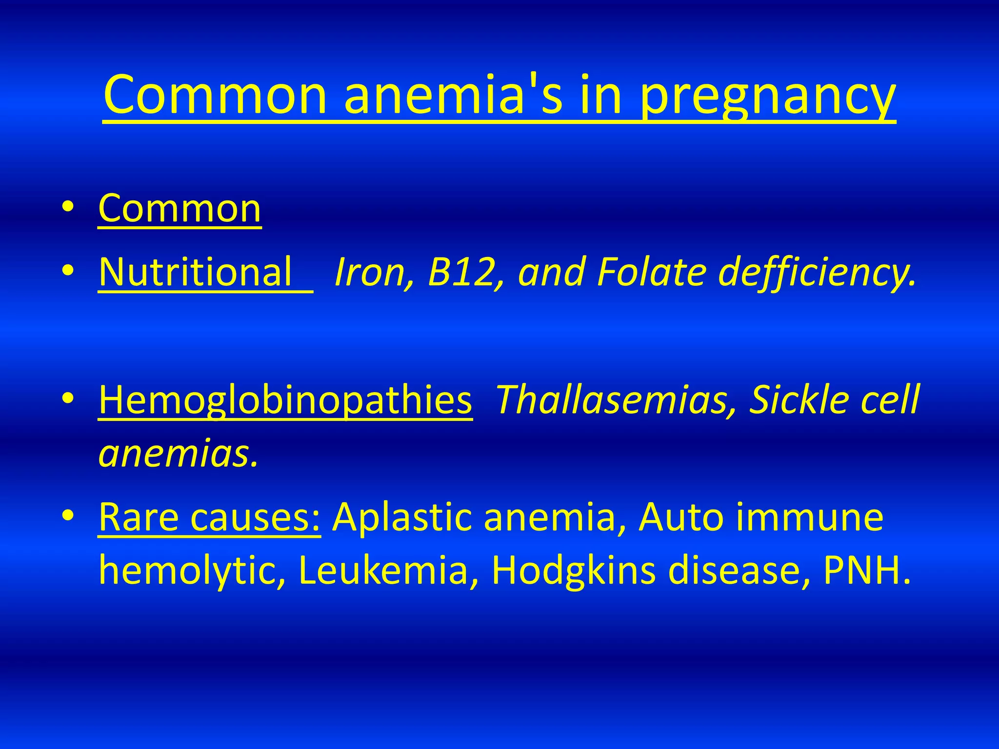

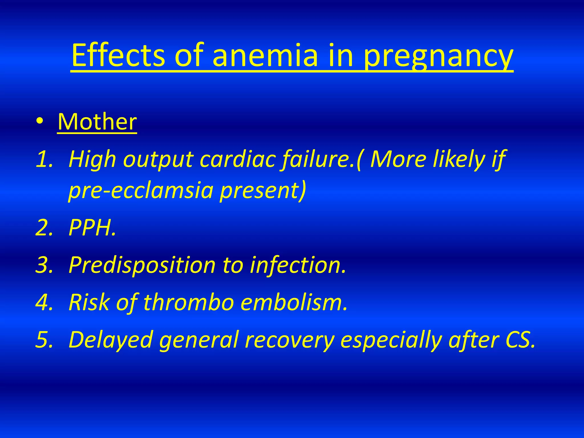


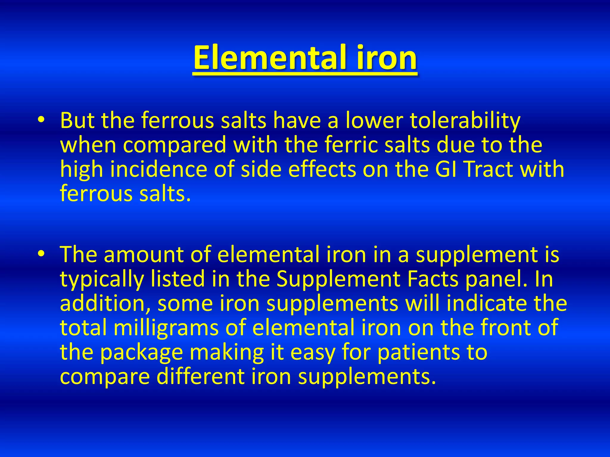


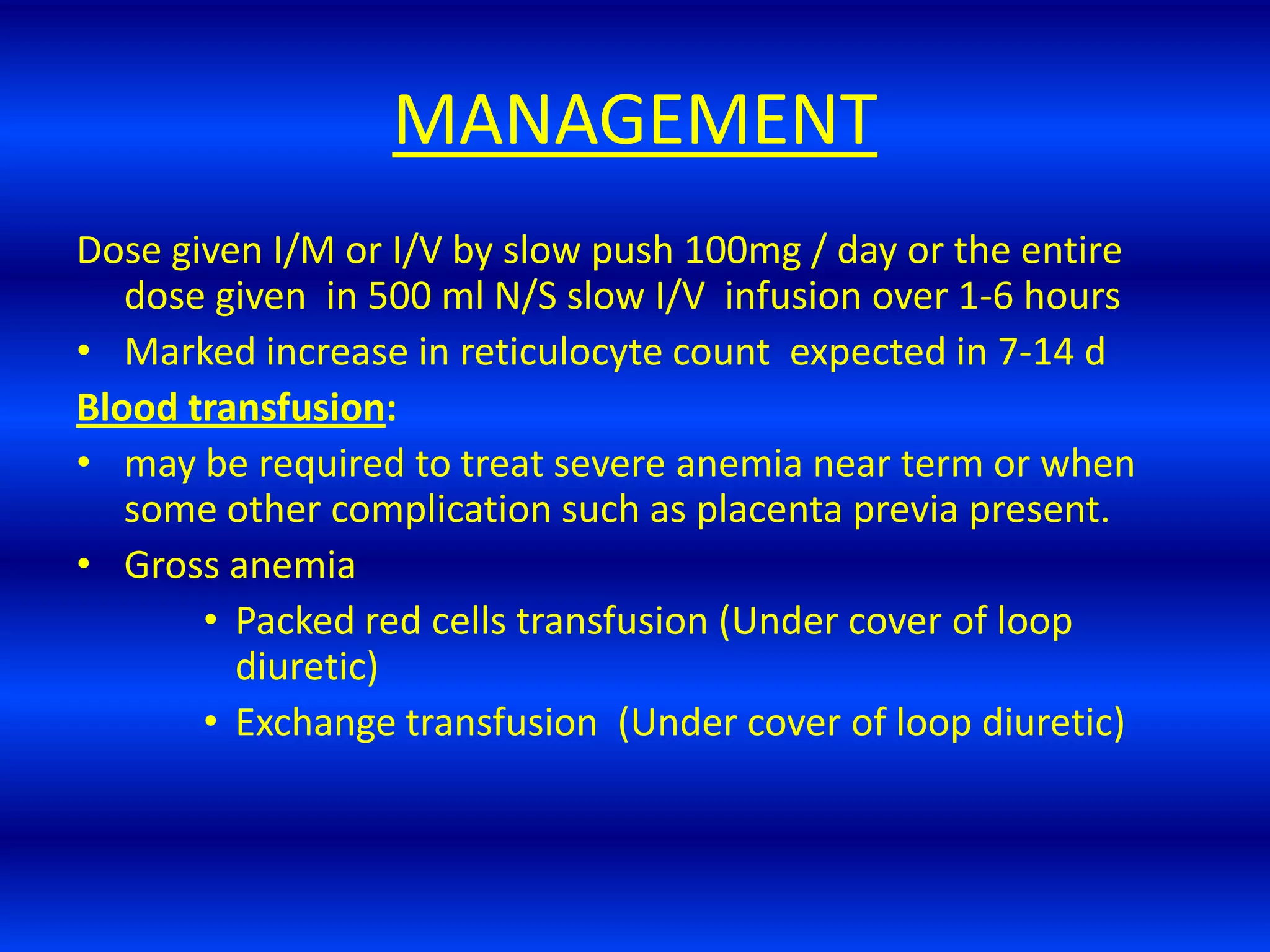


Anemia is a common medical disorder in pregnancy that increases risks for both mother and baby. The document defines anemia in pregnancy according to WHO standards and describes the main causes as decreased red blood cell production or increased destruction, with 90% of cases due to iron deficiency. Evaluation involves hematological indices and iron studies. Management focuses on iron supplementation orally or parenterally depending on severity, with blood transfusions for severe cases. Specific attention is given to nutritional deficiencies like iron, folate, vitamin B12 and hemoglobinopathies.


![Skin anatomy chc training 2012 [compatibility mode] [repaired]](https://cdn.slidesharecdn.com/ss_thumbnails/skinanatomychctraining2012compatibilitymoderepaired-131009014235-phpapp01-thumbnail.jpg?width=640&height=640&fit=bounds)


