Download to read offline


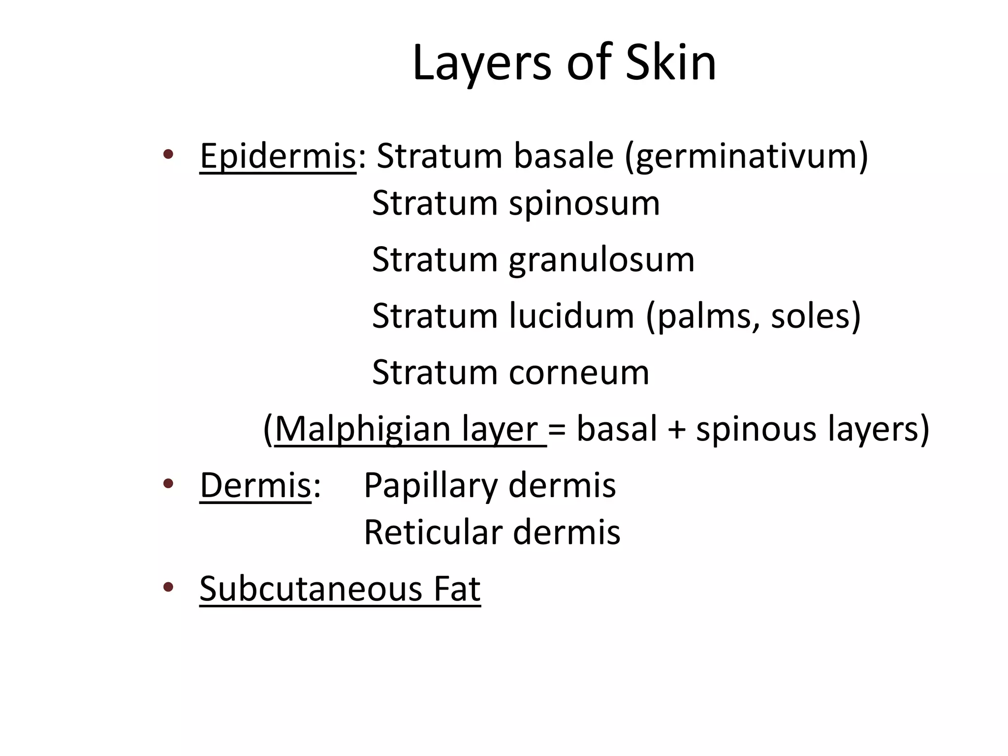


The document provides an overview of the structure and functions of the skin, highlighting the role of dermatology in diagnosing and treating various skin disorders, which can range from cosmetic issues to life-threatening conditions. It elaborates on the different layers of the skin, the types of cells within the epidermis, and the functions of skin components such as hair, nails, and sebaceous glands. Additionally, it discusses the skin's critical functions, including protection, thermoregulation, and sensory reception.