Downloaded 116 times
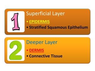
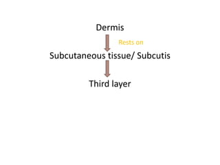
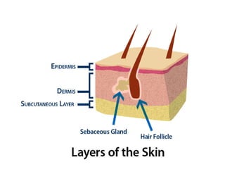

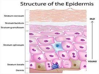
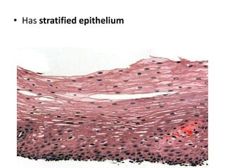
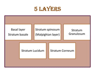
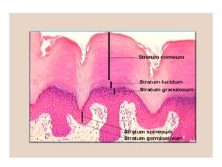
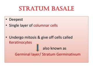
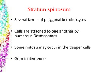
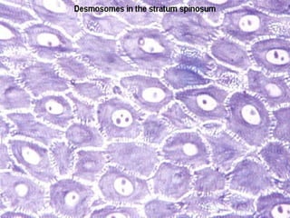
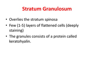
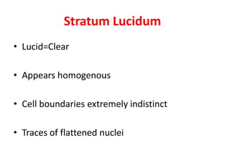
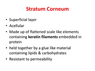
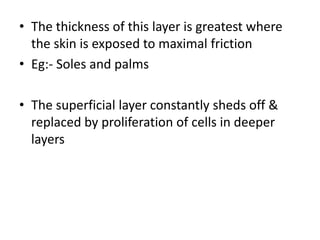
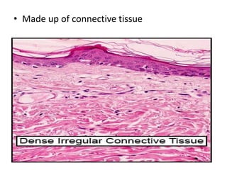
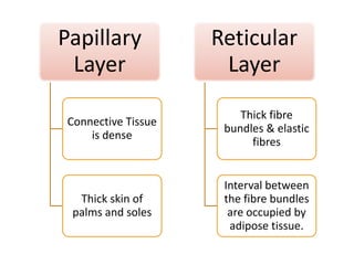
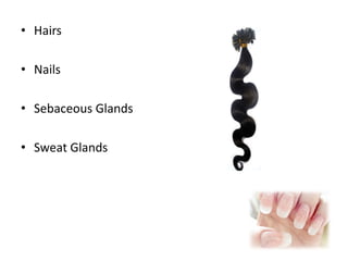
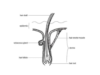
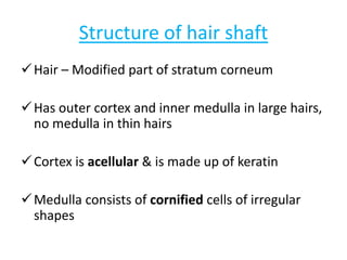
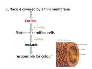
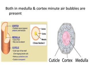
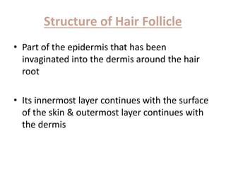
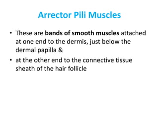
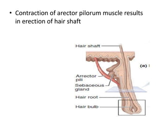
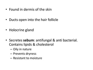
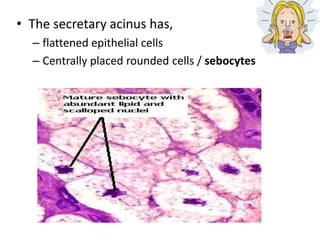
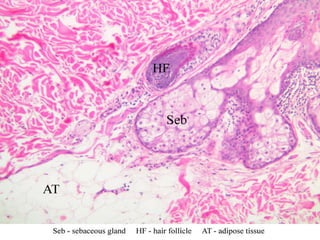
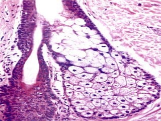

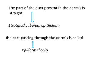
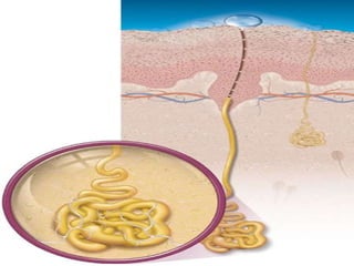
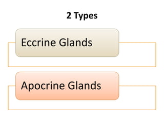
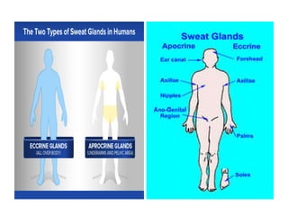
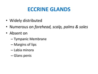
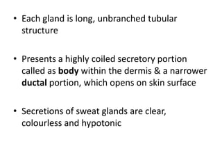
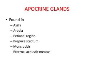
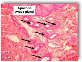
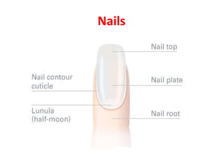


The document summarizes the structure and layers of skin. It describes the three main layers of skin - the epidermis, dermis, and subcutaneous layer. It provides details on the 5 layers of the epidermis including the stratum basale, stratum spinosum, stratum granulosum, stratum lucidum, and stratum corneum. It also discusses structures like hair follicles, sebaceous glands, sweat glands, and nails.


![ONFH[AVN HIP] -TRIPLE REGIME -A NOVAL SURGICAL CONCEPT .pptx](https://cdn.slidesharecdn.com/ss_thumbnails/onfhavnhip2026koaconcalicutdrgokuldevdrmashraf-260210064517-213ec005-thumbnail.jpg?width=640&height=640&fit=bounds)


