
Downloaded 209 times

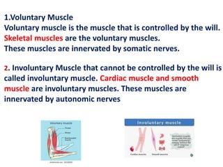
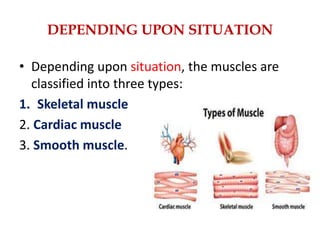
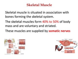
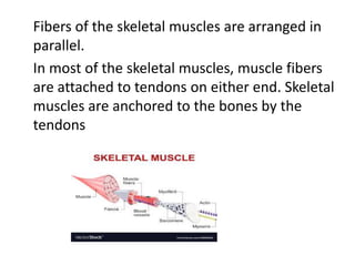
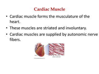
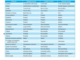
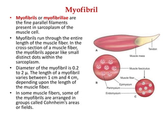

The muscular system consists of three types of muscle tissue - skeletal, smooth, and cardiac muscle. It permits movement, maintains posture, and circulates blood throughout the body. The muscular systems are controlled by the nervous system, though some muscles can contract autonomously. The skeletal muscle fibers are striated and arranged in bundles, with each fiber containing myofibrils made up of repeating sarcomere units consisting of actin and myosin filaments that slide past each other to cause contraction.


