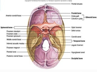
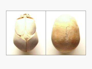
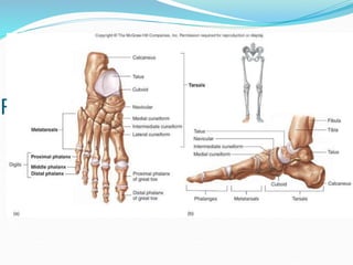
SKELETAL SYSTEM
BONES & JOINTS
Functions of the Bones
1. Support
2.Protection
3. Movement
4.Storage
5.Hematopoiesis
Table. 6.1
2 Types of Bone
Compact bone
Spongy bone
CLASSIFICATION of BONES
• According to structure
1. compact = solid mass; dense & hard
= forms the outer layer of bone structure
= functional unit --- Haversian system
2. cancellous or spongy = contain spaces
filled with bone marrow
= incomplete Haversian system
CLASSIFICATION of BONES
• According to shape
1. long bones = length is greater than breadth
= consists of shaft (diaphysis) &
two extremities (epiphysis)
diaphysis = filled with yellow marrow
= cylindrical, large space or canal at the center
= periosteum
epiphysis = made up of cancellous tissue
e.g.: femur, humerus, tibia, fibula, radius, ulna, phalanges
Membranes: 1. periosteum
2. endosteum
CLASSIFICATION of BONES cont’n.
• According to shape
2. short bones = cuboidal in shape
= spongy bone with thin coat of compact bone
= sesamoid bone -- short bone embedded in a
tendon e.g.: patella
e.g.: carpals (wrist), tarsal (ankle) bones
3. flat bones = broad or elongated flat plates
= for protection & muscle attachments
composition: 2 thin layers of compact tse. enclosing
a thin layer of spongy bone
e.g.: bones of the skull, sternum, ribs, scapula
CLASSIFICATION of BONES cont’n.
• According to shape
4. Irregular bones = all other bones not assigned to
the previous groups
e.g.: vertebrae
pelvic bones
bones of the base of the skull
CLASSIFICATION of BONES cont’n.
• According to location
A X I A L
skull 22
hyoid 1
ossicles 6
vertebrae 26
ribs & sternum 25_
80
CLASSIFICATION of BONES cont’n.
• According to location
APPENDICULAR
Upper Extremities Lower Extremities
clavicle 2 hip bone 2
scapulae 2 femur 2
humerus 2 patella 2
radius 2 tibia 2
ulna 2 fibula 2
carpals 16 tarsals 14
metacarpals 10 metatarsals 10
phalanges 28__ phalanges 28__
64 62
Fig. 6.10
AXIAL SKELETON
I. SKULL
= skeleton --- head & face
= united by joints (sutures)
cranium -- skull minus mandible
cavities: a. Cranial - contains the brain
b. Orbital - contains eyeball
& accessory organs
c. nasal
Fig. 6.14
Fontanelle
= membrane filled spaces found in the skull of
newborn infants
e.g.: 1. anterior = largest
2. posterior
3. anterolateral (sphenoidal)
4. posterolateral (mastoid)
Fig. 6.37
AXIAL SKELETON
I. VERTEBRAL COLUMN
= long, curved, slightly movable pillar
= united together by cartilage & ligaments
= formed by series of bones -- vertebrae
FUNCTION:
1. support of the trunk
2. contains & protects the spinal cord &
nerves
VERTEBRAL COLUMN
Classification of vertebra
cervical 7
thoracic 12
lumbar 5
sacral 5
coccygeal 4
33
Intervertebral discs = flattened plates of fibrocartilage that
are interposed between the adjacent surfaces of the
bodies of vertebrae
FUNCTION: 1. uniting medium between vertebrae
2. main shock absorber
3. give flexibility & movement to the whole
vertebral column
VERTEBRAL COLUMN
a. Cervical vertebrae (7) = forms the skeleton of
the neck
atypical cervical vertebrae:
1. atlas -- 1st
2. axis = 2nd
3. 7th cervical vertebrae = spinous process
b. Thoracic vertebrae (12) = costal pits - for rib
attachment
VERTEBRAL COLUMN
c. Lumbar vertebrae (5)
= triangular vertebral foramen
d. Sacrum = inverted triangular bone
situated between hip bones
e. Coccygeal vertebrae (1) = 4 small
incomplete vertebrae fused to form the
coccyx / tail bone; triangular
Fig. 6.17
Fig. 6.18
Fig. 6.20
AXIAL SKELETON
I. STERNUM (breast bone)
= flat bone, found -- anterior thoracic wall
PARTS: a. Manubrium
b. Body
c. Xiphoid process
2. RIBS (12 pairs)
= narrow arched flat bones with 2
ends
1. vertebral - posterior; attaches with thoracic
2. sternal - anterior; attaches with costal
cartilages
Fig. 6.21
Classification of ribs:
a. true ribs (1st to 7th)
- ribs whose costal cartilages are directly
attached to sternum
b. false ribs (8th to 12th)
- ribs whose costal cartilages are not attached
directly to the sternum but to 7th
APPENDICULAR SKELETON
BONES of the UPPER EXTREMITY (UE)
1. Clavicle (collar bone)
2. Scapula (shoulder blade)
3. Humerus (arm bone)
4. Radius - lateral bone of the forearm
5. Ulna - medial bone of the forearm
6. Carpals (wrist bone) - 8 bones arranged into 2 rows - proximal
& distal rows
7. Metacarpals (bones of the hand)
8. Phalanges (bones of the fingers) long bones of the fingers
Fig. 6.26
Fig. 6.22
Fig. 6.27
APPENDICULAR SKELETON
BONES of the LOWER EXTREMITY (LE)
1. Hip bone (innominate bone)
right & left hip bones + sacrum = pelvic girdle
3 bones: 1. ilium
2. ischium
3. pubis
2. Femur (thigh) = longest, strongest, largest bone in
the body
3. Tibia (shin bone) = long bone; anterior, medial,
4. Fibula (peroneal bone) = long slender bone placed
parallel with the tibia but located laterally
Fig. 6.29
Fig. 6.30
Fig. 6.33
Fig. 6.34
APPENDICULAR SKELETON
BONES of the LOWER EXTREMITY (LE) cont’n
5. Tarsals (ankle bone) = short bones;
2 rows: internal & external rows
6. Metatarsals (bones of foot) = 5 long bones
numbered from medial to lateral
7. Phalanges (bones of toes) = similar to bones of
the fingers
Fig. 6.35
END OF LECTURE
 It is impossible for a man to learn what he thinks
he already knows
 Epictetus.

SKELETAL SYSTEM-1.ppt

  • 1.
  • 3.
    Functions of theBones 1. Support 2.Protection 3. Movement 4.Storage 5.Hematopoiesis
  • 5.
  • 6.
    2 Types ofBone Compact bone Spongy bone
  • 7.
    CLASSIFICATION of BONES •According to structure 1. compact = solid mass; dense & hard = forms the outer layer of bone structure = functional unit --- Haversian system 2. cancellous or spongy = contain spaces filled with bone marrow = incomplete Haversian system
  • 9.
    CLASSIFICATION of BONES •According to shape 1. long bones = length is greater than breadth = consists of shaft (diaphysis) & two extremities (epiphysis) diaphysis = filled with yellow marrow = cylindrical, large space or canal at the center = periosteum epiphysis = made up of cancellous tissue e.g.: femur, humerus, tibia, fibula, radius, ulna, phalanges Membranes: 1. periosteum 2. endosteum
  • 10.
    CLASSIFICATION of BONEScont’n. • According to shape 2. short bones = cuboidal in shape = spongy bone with thin coat of compact bone = sesamoid bone -- short bone embedded in a tendon e.g.: patella e.g.: carpals (wrist), tarsal (ankle) bones 3. flat bones = broad or elongated flat plates = for protection & muscle attachments composition: 2 thin layers of compact tse. enclosing a thin layer of spongy bone e.g.: bones of the skull, sternum, ribs, scapula
  • 11.
    CLASSIFICATION of BONEScont’n. • According to shape 4. Irregular bones = all other bones not assigned to the previous groups e.g.: vertebrae pelvic bones bones of the base of the skull
  • 12.
    CLASSIFICATION of BONEScont’n. • According to location A X I A L skull 22 hyoid 1 ossicles 6 vertebrae 26 ribs & sternum 25_ 80
  • 13.
    CLASSIFICATION of BONEScont’n. • According to location APPENDICULAR Upper Extremities Lower Extremities clavicle 2 hip bone 2 scapulae 2 femur 2 humerus 2 patella 2 radius 2 tibia 2 ulna 2 fibula 2 carpals 16 tarsals 14 metacarpals 10 metatarsals 10 phalanges 28__ phalanges 28__ 64 62
  • 14.
  • 15.
    AXIAL SKELETON I. SKULL =skeleton --- head & face = united by joints (sutures) cranium -- skull minus mandible cavities: a. Cranial - contains the brain b. Orbital - contains eyeball & accessory organs c. nasal
  • 18.
  • 19.
    Fontanelle = membrane filledspaces found in the skull of newborn infants e.g.: 1. anterior = largest 2. posterior 3. anterolateral (sphenoidal) 4. posterolateral (mastoid)
  • 21.
  • 22.
    AXIAL SKELETON I. VERTEBRALCOLUMN = long, curved, slightly movable pillar = united together by cartilage & ligaments = formed by series of bones -- vertebrae FUNCTION: 1. support of the trunk 2. contains & protects the spinal cord & nerves
  • 23.
    VERTEBRAL COLUMN Classification ofvertebra cervical 7 thoracic 12 lumbar 5 sacral 5 coccygeal 4 33 Intervertebral discs = flattened plates of fibrocartilage that are interposed between the adjacent surfaces of the bodies of vertebrae FUNCTION: 1. uniting medium between vertebrae 2. main shock absorber 3. give flexibility & movement to the whole vertebral column
  • 24.
    VERTEBRAL COLUMN a. Cervicalvertebrae (7) = forms the skeleton of the neck atypical cervical vertebrae: 1. atlas -- 1st 2. axis = 2nd 3. 7th cervical vertebrae = spinous process b. Thoracic vertebrae (12) = costal pits - for rib attachment
  • 26.
    VERTEBRAL COLUMN c. Lumbarvertebrae (5) = triangular vertebral foramen d. Sacrum = inverted triangular bone situated between hip bones e. Coccygeal vertebrae (1) = 4 small incomplete vertebrae fused to form the coccyx / tail bone; triangular
  • 27.
  • 28.
  • 29.
  • 30.
    AXIAL SKELETON I. STERNUM(breast bone) = flat bone, found -- anterior thoracic wall PARTS: a. Manubrium b. Body c. Xiphoid process 2. RIBS (12 pairs) = narrow arched flat bones with 2 ends 1. vertebral - posterior; attaches with thoracic 2. sternal - anterior; attaches with costal cartilages
  • 31.
  • 32.
    Classification of ribs: a.true ribs (1st to 7th) - ribs whose costal cartilages are directly attached to sternum b. false ribs (8th to 12th) - ribs whose costal cartilages are not attached directly to the sternum but to 7th
  • 33.
    APPENDICULAR SKELETON BONES ofthe UPPER EXTREMITY (UE) 1. Clavicle (collar bone) 2. Scapula (shoulder blade) 3. Humerus (arm bone) 4. Radius - lateral bone of the forearm 5. Ulna - medial bone of the forearm 6. Carpals (wrist bone) - 8 bones arranged into 2 rows - proximal & distal rows 7. Metacarpals (bones of the hand) 8. Phalanges (bones of the fingers) long bones of the fingers
  • 36.
  • 37.
  • 38.
  • 39.
    APPENDICULAR SKELETON BONES ofthe LOWER EXTREMITY (LE) 1. Hip bone (innominate bone) right & left hip bones + sacrum = pelvic girdle 3 bones: 1. ilium 2. ischium 3. pubis 2. Femur (thigh) = longest, strongest, largest bone in the body 3. Tibia (shin bone) = long bone; anterior, medial, 4. Fibula (peroneal bone) = long slender bone placed parallel with the tibia but located laterally
  • 40.
  • 41.
  • 42.
  • 43.
  • 44.
    APPENDICULAR SKELETON BONES ofthe LOWER EXTREMITY (LE) cont’n 5. Tarsals (ankle bone) = short bones; 2 rows: internal & external rows 6. Metatarsals (bones of foot) = 5 long bones numbered from medial to lateral 7. Phalanges (bones of toes) = similar to bones of the fingers
  • 45.
  • 46.
  • 47.
     It isimpossible for a man to learn what he thinks he already knows  Epictetus.