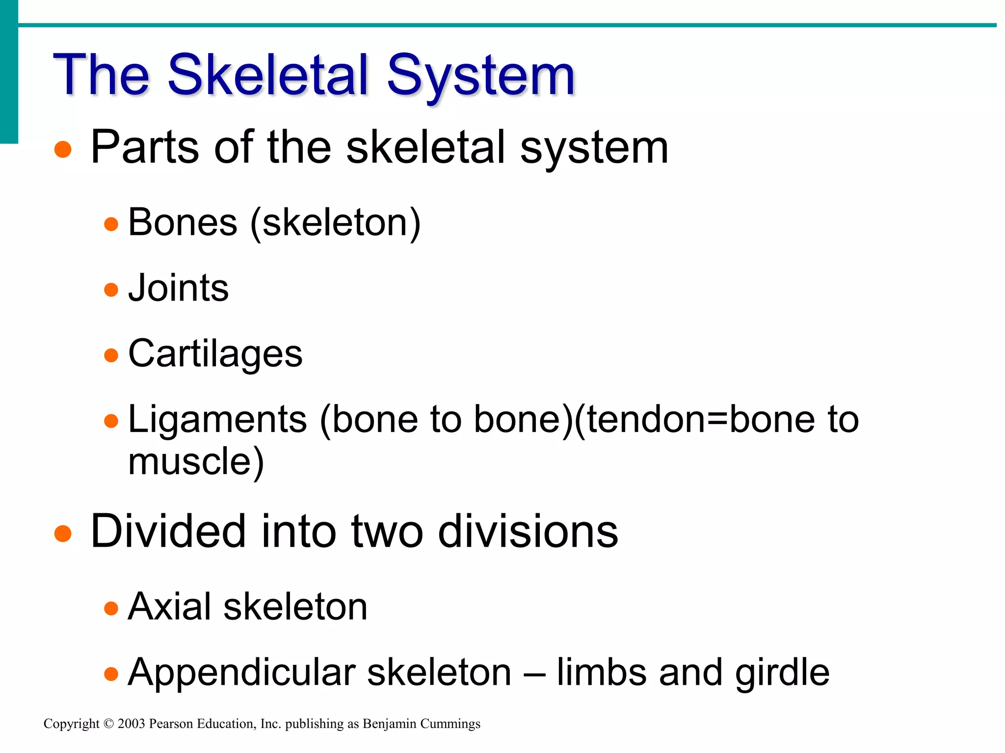
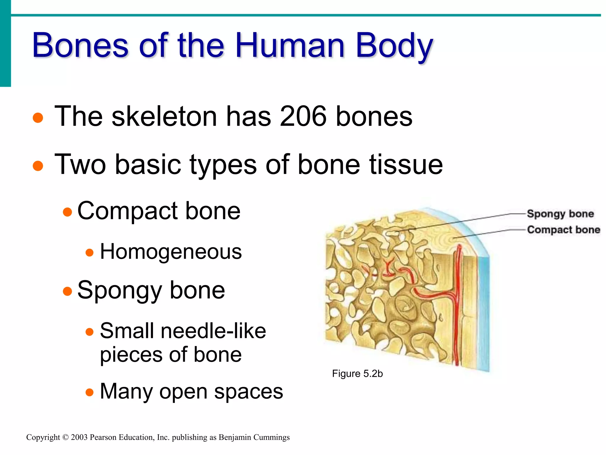
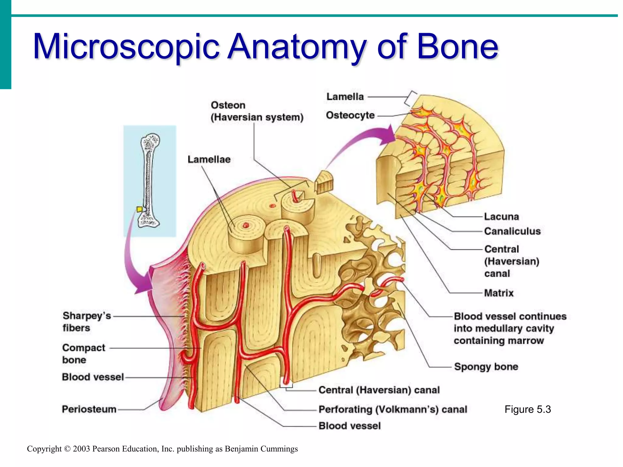
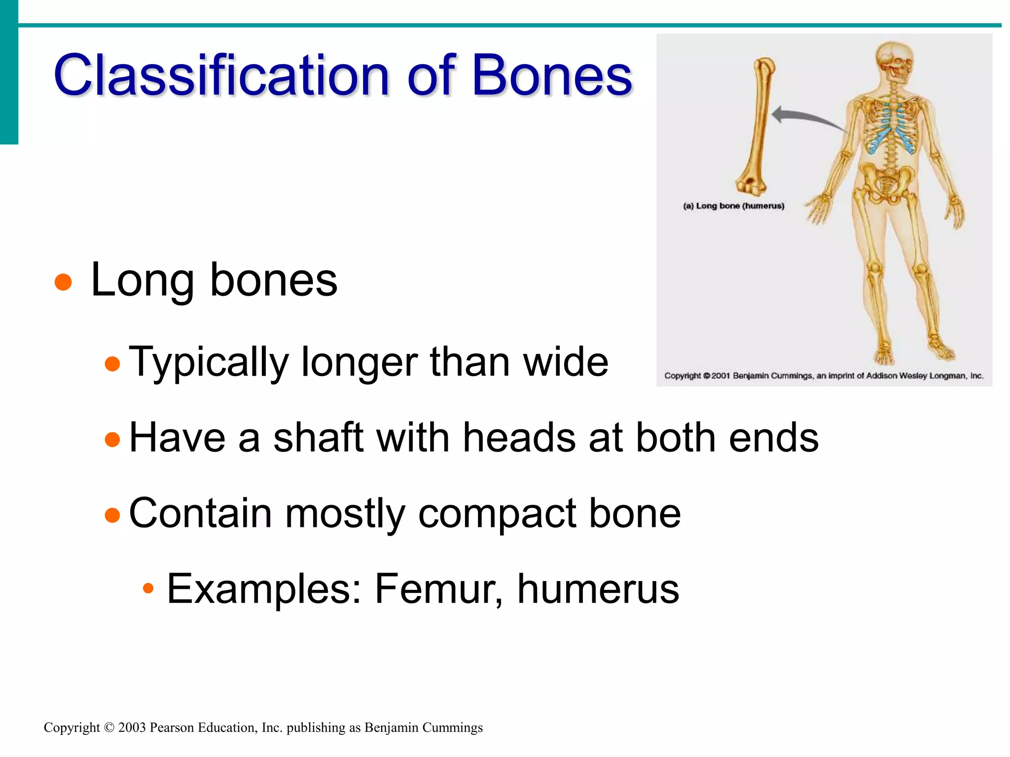
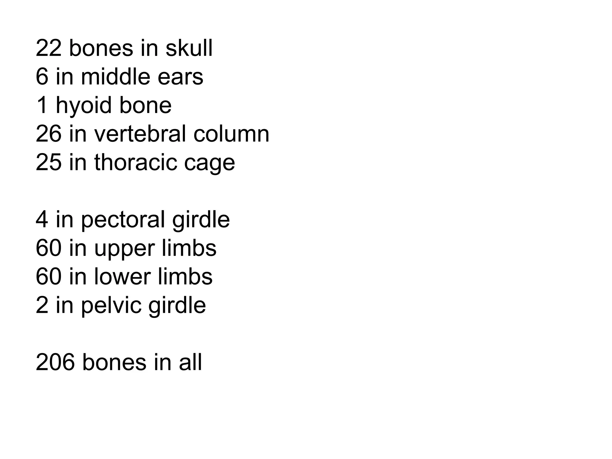
By: Dr Krishna Bhatt, Assistant Professor
ARIP, Charusat
Human Anatomy:
The Skeletal System:
Structure, Function, and
Diseases
of the bones and joints
Is this the correct anatomical position?
BONES AND BONE TISSUES
Introduction
• One of the most remarkable tissues of the
human body
• Far from inert and lifeless, bones are living,
dynamic structures
• Bones serve a wide variety of very diverse
functions within us
• Noted for their strength and resiliency
during life, bones will remain after we are
long gone
The Skeletal System
Copyright © 2003 Pearson Education, Inc. publishing as Benjamin Cummings
 Parts of the skeletal system
 Bones (skeleton)
 Joints
 Cartilages
 Ligaments (bone to bone)(tendon=bone to
muscle)
 Divided into two divisions
 Axial skeleton
 Appendicular skeleton – limbs and girdle
Bones of the Human Body
Copyright © 2003 Pearson Education, Inc. publishing as Benjamin Cummings
 The skeleton has 206 bones
 Two basic types of bone tissue
Compact bone
 Homogeneous
Spongy bone
 Small needle-like
pieces of bone
 Many open spaces
Figure 5.2b
Microscopic Anatomy of Bone
Copyright © 2003 Pearson Education, Inc. publishing as Benjamin Cummings
Figure 5.3
SKELETAL CARTILAGES
Skeletal Cartilages
• Initially our skeleton is made up of
cartilages and fibrous membranes
• Gradually our skeletal cartilages are
replaced by bone
• Upon reaching adulthood the skeleton
becomes almost fully ossified
• Only a few cartilages remain in the adult
skeleton
Basic structure, type & location
• A skeletal cartilage is made of some variety
of cartilage tissue
• Each type contains a high proportion of
water which makes them resilient
• Cartilage has no nerves or blood supply
• It is surrounded by a dense tissue
membrane called a perichondrium
Basic structure, type & location
• There are three types of cartilage tissue:
hyaline, elastic, and fibrocartilage
• Each contains a matrix of jellylike ground
substance and fibers
Cartilages
Functions of Bones
Copyright © 2003 Pearson Education, Inc. publishing as Benjamin Cummings
 Support of the body
 Protection of soft organs
 Movement due to attached skeletal
muscles
 Storage of minerals and fats
 Blood cell formation
Function of Bones
• Support • Bones provide a hard
framework that
supports the body
• Bones provide support
for internal organs
Function of Bone
• Protection • Fused bones provide a
brain case that protects
this vital tissue
• Spinal cord is
surrounded by
vertebrae
• Rib cage protects vital
organs
Function of Bone
• Movement • Skeletal muscle
attached to bones use
the bones as levers to
move the body
• Arrangement of bones
and joints determine
the movements
possible
Function of Bones
• Mineral Storage • Bone serves as a
mineral reservoir
• Phosphate and calcium
ions can be released
into the blood steam for
distribution
• Deposition and removal
are ongoing
Function of Bones
• Blood cell formation • Hematopoiesis occurs
within the marrow
cavities of the long
bones
• The majority of
hematopoiesis occurs in
bones
CLASSIFICATION OF BONE
Classification of Bone:
• Bones vary in shape and size
• The unique shape of each bone fulfills a
particular need
• Bones are classified by their shape as long,
short, flat, or irregular bone
• Bones differ in the distribution of compact
and spongy osseous tissues
Classification of Bones
Bones are classified by
their shape:
1.long
2.short
3.flat
4.irregular
Classification of Bones on the
Basis of Shape
Copyright © 2003 Pearson Education, Inc. publishing as Benjamin Cummings
Figure 5.1
Classification of Bones
Copyright © 2003 Pearson Education, Inc. publishing as Benjamin Cummings
 Long bones
Typically longer than wide
Have a shaft with heads at both ends
Contain mostly compact bone
• Examples: Femur, humerus
Types of Bone Cells
Copyright © 2003 Pearson Education, Inc. publishing as Benjamin Cummings
 Osteocytes
 Mature bone cells
 Osteoblasts
 Bone-forming cells
 Osteoclasts
 Bone-destroying cells
 Break down bone matrix for remodeling and
release of calcium
 Bone remodeling is a process by both
osteoblasts and osteoclasts
Bone Fractures
Copyright © 2003 Pearson Education, Inc. publishing as Benjamin Cummings
 A break in a bone
 Types of bone fractures
 Closed (simple) fracture – break that does not
penetrate the skin
 Open (compound) fracture – broken bone
penetrates through the skin
 Bone fractures are treated by reduction
and immobilization
 Realignment of the bone
Common Types of Fractures
Slide 5.17Copyright © 2003 Pearson Education, Inc. publishing as Benjamin Cummings
Table 5.2
Stages in the Healing of a Bone
Fracture
Slide 5.19Copyright © 2003 Pearson Education, Inc. publishing as Benjamin Cummings
Figure 5.5
Axial skeleton supports and protects organs of head,
neck and trunk
Axial skeleton:
skull (cranium and facial bones)
hyoid bone (anchors tongue and muscles
associated with swallowing)
vertebral column (vertebrae and disks)
bony thorax (ribs and sternum)
Appendicular skeleton includes bones of limbs and
bones that anchor them to the axial skeleton
Appendicular skeleton:
pectoral girdle (clavicles and scapulae)
upper limbs (arms)
pelvic girdle (sacrum, coccyx)
lower limbs (legs)
Articulation- where joints meet, connect, and are formed.
22 bones in skull
6 in middle ears
1 hyoid bone
26 in vertebral column
25 in thoracic cage
4 in pectoral girdle
60 in upper limbs
60 in lower limbs
2 in pelvic girdle
206 bones in all
The Axial Skeleton
SlideCopyright © 2003 Pearson Education, Inc. publishing as Benjamin Cummings
 Forms the longitudinal part of the body
 Divided into three parts
Skull
Vertebral column
Bony thorax
The Axial Skeleton
SlideCopyright © 2003 Pearson Education, Inc. publishing as Benjamin Cummings
Figure 5.6
The skull
8 sutured bones in cranium
Facial bones: 13 sutured bones, 1 mandible
Cranium
encases brain
attachments for muscles
sinuses
Bones of the Skull
Copyright © 2003 Pearson Education, Inc. publishing as Benjamin Cummings
Figure 5.11
Allows for
growth
Human Skull, Superior View
Slide 5.23Copyright © 2003 Pearson Education, Inc. publishing as Benjamin Cummings
Figure 5.8
Human Skull, Inferior View
Slide 5.24Copyright © 2003 Pearson Education, Inc. publishing as Benjamin Cummings
Figure 5.9
Paranasal Sinuses
SlideCopyright © 2003 Pearson Education, Inc. publishing as Benjamin Cummings
 Hollow portions of bones surrounding
the nasal cavity
Figure 5.10
The Vertebral Column
Slide 5.28Copyright © 2003 Pearson Education, Inc. publishing as Benjamin Cummings
 Vertebrae
separated by
intervertebral discs
 The spine has a
normal curvature
 Each vertebrae is
given a name
according to its
location Figure 5.14
Vertebral column
7 cervial vertebrae
12 thoracic
5 lumbar
1 sacrum (5 fused
1 coccyx (4 fused)
Vertebrae vary in size and morphology
Structure of a Typical Vertebrae
Slide 5.29Copyright © 2003 Pearson Education, Inc. publishing as Benjamin Cummings
Figure 5.16
Thoracic cage
ribs
thoracic vertebrae
sternum
costal cartilages
True ribs are directly attached to the sternum
(first seven pairs)
Three false ribs are joined to the 7th rib
Two pairs of floating ribs
Joints
• Fibrous-Fibrous joints connect bones without
allowing any movement. The bones of your
skull and pelvis are held together by fibrous
joints.
• Cartilaginous-Cartilaginous joints are joints
in which the bones are attached by cartilage.
These joints allow for only a little movement,
such as in the spine or ribs.
• Synovial-Synovial joints allow for much more
movement than cartilaginous joints. Cavities
between bones in synovial joints are filled
with synovial fluid. This fluid helps lubricate
and protect the bones. Bursa sacks contain
the synovial fluid. within fixed limits
• A joint, or articulation, is the place
where two bones come together.
• There are three types of joints
classified by the amount of
movement they allow:
 Immovable
 slightly movable
 freely movable
Types of Joints
Hinge- A hinge joint allows extension and
retraction of an appendage. (Elbow, Knee)
Ball and Socket- A ball and socket joint
allows for radial movement in almost
any direction. They are found in the hips
and shoulders. (Hip, Shoulder)
Gliding- In a gliding or plane joint bones
slide past each other. Mid-carpal and mid-
tarsal joints are gliding joints. (Hands,
Feet)
Saddle- This type of joint occurs when the
touching surfaces of two bones have both
concave and convex regions with the shapes
of the two bones complementing one other
and allowing a wide range of movement.
(Thumb)
Types of Synovial Joints Based on
Shape
SlideCopyright © 2003 Pearson Education, Inc. publishing as Benjamin Cummings
Figure 5.29a–c
Types of Synovial Joints Based on
Shape
SlideCopyright © 2003 Pearson Education, Inc. publishing as Benjamin Cummings
Figure 5.29d–f
Diseases and Conditions
of the Skeletal System
Arthritis
Anatomy of skeletal system

Anatomy of skeletal system

  • 1.
    By: Dr KrishnaBhatt, Assistant Professor ARIP, Charusat Human Anatomy: The Skeletal System:
  • 2.
  • 3.
    Is this thecorrect anatomical position?
  • 4.
  • 5.
    Introduction • One ofthe most remarkable tissues of the human body • Far from inert and lifeless, bones are living, dynamic structures • Bones serve a wide variety of very diverse functions within us • Noted for their strength and resiliency during life, bones will remain after we are long gone
  • 6.
    The Skeletal System Copyright© 2003 Pearson Education, Inc. publishing as Benjamin Cummings  Parts of the skeletal system  Bones (skeleton)  Joints  Cartilages  Ligaments (bone to bone)(tendon=bone to muscle)  Divided into two divisions  Axial skeleton  Appendicular skeleton – limbs and girdle
  • 7.
    Bones of theHuman Body Copyright © 2003 Pearson Education, Inc. publishing as Benjamin Cummings  The skeleton has 206 bones  Two basic types of bone tissue Compact bone  Homogeneous Spongy bone  Small needle-like pieces of bone  Many open spaces Figure 5.2b
  • 8.
    Microscopic Anatomy ofBone Copyright © 2003 Pearson Education, Inc. publishing as Benjamin Cummings Figure 5.3
  • 9.
  • 10.
    Skeletal Cartilages • Initiallyour skeleton is made up of cartilages and fibrous membranes • Gradually our skeletal cartilages are replaced by bone • Upon reaching adulthood the skeleton becomes almost fully ossified • Only a few cartilages remain in the adult skeleton
  • 11.
    Basic structure, type& location • A skeletal cartilage is made of some variety of cartilage tissue • Each type contains a high proportion of water which makes them resilient • Cartilage has no nerves or blood supply • It is surrounded by a dense tissue membrane called a perichondrium
  • 12.
    Basic structure, type& location • There are three types of cartilage tissue: hyaline, elastic, and fibrocartilage • Each contains a matrix of jellylike ground substance and fibers
  • 13.
  • 14.
    Functions of Bones Copyright© 2003 Pearson Education, Inc. publishing as Benjamin Cummings  Support of the body  Protection of soft organs  Movement due to attached skeletal muscles  Storage of minerals and fats  Blood cell formation
  • 15.
    Function of Bones •Support • Bones provide a hard framework that supports the body • Bones provide support for internal organs
  • 16.
    Function of Bone •Protection • Fused bones provide a brain case that protects this vital tissue • Spinal cord is surrounded by vertebrae • Rib cage protects vital organs
  • 17.
    Function of Bone •Movement • Skeletal muscle attached to bones use the bones as levers to move the body • Arrangement of bones and joints determine the movements possible
  • 18.
    Function of Bones •Mineral Storage • Bone serves as a mineral reservoir • Phosphate and calcium ions can be released into the blood steam for distribution • Deposition and removal are ongoing
  • 19.
    Function of Bones •Blood cell formation • Hematopoiesis occurs within the marrow cavities of the long bones • The majority of hematopoiesis occurs in bones
  • 20.
  • 21.
    Classification of Bone: •Bones vary in shape and size • The unique shape of each bone fulfills a particular need • Bones are classified by their shape as long, short, flat, or irregular bone • Bones differ in the distribution of compact and spongy osseous tissues
  • 22.
  • 23.
    Bones are classifiedby their shape: 1.long 2.short 3.flat 4.irregular
  • 24.
    Classification of Boneson the Basis of Shape Copyright © 2003 Pearson Education, Inc. publishing as Benjamin Cummings Figure 5.1
  • 25.
    Classification of Bones Copyright© 2003 Pearson Education, Inc. publishing as Benjamin Cummings  Long bones Typically longer than wide Have a shaft with heads at both ends Contain mostly compact bone • Examples: Femur, humerus
  • 26.
    Types of BoneCells Copyright © 2003 Pearson Education, Inc. publishing as Benjamin Cummings  Osteocytes  Mature bone cells  Osteoblasts  Bone-forming cells  Osteoclasts  Bone-destroying cells  Break down bone matrix for remodeling and release of calcium  Bone remodeling is a process by both osteoblasts and osteoclasts
  • 27.
    Bone Fractures Copyright ©2003 Pearson Education, Inc. publishing as Benjamin Cummings  A break in a bone  Types of bone fractures  Closed (simple) fracture – break that does not penetrate the skin  Open (compound) fracture – broken bone penetrates through the skin  Bone fractures are treated by reduction and immobilization  Realignment of the bone
  • 28.
    Common Types ofFractures Slide 5.17Copyright © 2003 Pearson Education, Inc. publishing as Benjamin Cummings Table 5.2
  • 29.
    Stages in theHealing of a Bone Fracture Slide 5.19Copyright © 2003 Pearson Education, Inc. publishing as Benjamin Cummings Figure 5.5
  • 30.
    Axial skeleton supportsand protects organs of head, neck and trunk Axial skeleton: skull (cranium and facial bones) hyoid bone (anchors tongue and muscles associated with swallowing) vertebral column (vertebrae and disks) bony thorax (ribs and sternum) Appendicular skeleton includes bones of limbs and bones that anchor them to the axial skeleton Appendicular skeleton: pectoral girdle (clavicles and scapulae) upper limbs (arms) pelvic girdle (sacrum, coccyx) lower limbs (legs) Articulation- where joints meet, connect, and are formed.
  • 31.
    22 bones inskull 6 in middle ears 1 hyoid bone 26 in vertebral column 25 in thoracic cage 4 in pectoral girdle 60 in upper limbs 60 in lower limbs 2 in pelvic girdle 206 bones in all
  • 33.
    The Axial Skeleton SlideCopyright© 2003 Pearson Education, Inc. publishing as Benjamin Cummings  Forms the longitudinal part of the body  Divided into three parts Skull Vertebral column Bony thorax
  • 34.
    The Axial Skeleton SlideCopyright© 2003 Pearson Education, Inc. publishing as Benjamin Cummings Figure 5.6
  • 35.
    The skull 8 suturedbones in cranium Facial bones: 13 sutured bones, 1 mandible Cranium encases brain attachments for muscles sinuses
  • 37.
    Bones of theSkull Copyright © 2003 Pearson Education, Inc. publishing as Benjamin Cummings Figure 5.11
  • 38.
  • 39.
    Human Skull, SuperiorView Slide 5.23Copyright © 2003 Pearson Education, Inc. publishing as Benjamin Cummings Figure 5.8
  • 40.
    Human Skull, InferiorView Slide 5.24Copyright © 2003 Pearson Education, Inc. publishing as Benjamin Cummings Figure 5.9
  • 41.
    Paranasal Sinuses SlideCopyright ©2003 Pearson Education, Inc. publishing as Benjamin Cummings  Hollow portions of bones surrounding the nasal cavity Figure 5.10
  • 42.
    The Vertebral Column Slide5.28Copyright © 2003 Pearson Education, Inc. publishing as Benjamin Cummings  Vertebrae separated by intervertebral discs  The spine has a normal curvature  Each vertebrae is given a name according to its location Figure 5.14
  • 43.
    Vertebral column 7 cervialvertebrae 12 thoracic 5 lumbar 1 sacrum (5 fused 1 coccyx (4 fused) Vertebrae vary in size and morphology
  • 45.
    Structure of aTypical Vertebrae Slide 5.29Copyright © 2003 Pearson Education, Inc. publishing as Benjamin Cummings Figure 5.16
  • 46.
    Thoracic cage ribs thoracic vertebrae sternum costalcartilages True ribs are directly attached to the sternum (first seven pairs) Three false ribs are joined to the 7th rib Two pairs of floating ribs
  • 48.
    Joints • Fibrous-Fibrous jointsconnect bones without allowing any movement. The bones of your skull and pelvis are held together by fibrous joints. • Cartilaginous-Cartilaginous joints are joints in which the bones are attached by cartilage. These joints allow for only a little movement, such as in the spine or ribs. • Synovial-Synovial joints allow for much more movement than cartilaginous joints. Cavities between bones in synovial joints are filled with synovial fluid. This fluid helps lubricate and protect the bones. Bursa sacks contain the synovial fluid. within fixed limits
  • 49.
    • A joint,or articulation, is the place where two bones come together. • There are three types of joints classified by the amount of movement they allow:  Immovable  slightly movable  freely movable
  • 50.
    Types of Joints Hinge-A hinge joint allows extension and retraction of an appendage. (Elbow, Knee)
  • 51.
    Ball and Socket-A ball and socket joint allows for radial movement in almost any direction. They are found in the hips and shoulders. (Hip, Shoulder)
  • 52.
    Gliding- In agliding or plane joint bones slide past each other. Mid-carpal and mid- tarsal joints are gliding joints. (Hands, Feet)
  • 53.
    Saddle- This typeof joint occurs when the touching surfaces of two bones have both concave and convex regions with the shapes of the two bones complementing one other and allowing a wide range of movement. (Thumb)
  • 54.
    Types of SynovialJoints Based on Shape SlideCopyright © 2003 Pearson Education, Inc. publishing as Benjamin Cummings Figure 5.29a–c
  • 55.
    Types of SynovialJoints Based on Shape SlideCopyright © 2003 Pearson Education, Inc. publishing as Benjamin Cummings Figure 5.29d–f
  • 56.
    Diseases and Conditions ofthe Skeletal System
  • 57.