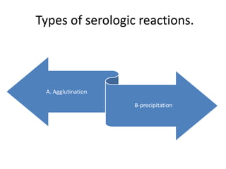
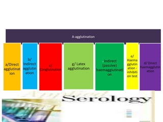
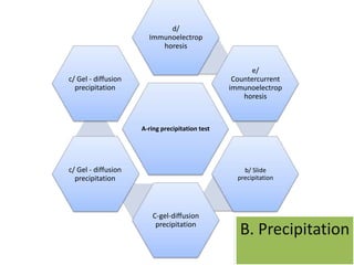
This document discusses various serological and immune assay techniques. It begins by defining serological assays and immune assays. It then describes different types of serological reactions including agglutination (direct, indirect, conglutination, latex agglutination) and precipitation (ring precipitation test, slide precipitation, gel diffusion precipitation, immunoelectrophoresis, countercurrent immunoelectrophoresis). The document also discusses immune assays including principles, qualitative vs quantitative assays, and different methods like immunoprecipitation, particle immunoassays, immunonephelometry, radioimmunoassay, enzyme immunoassay, fluorescent immunoassay, and chemiluminescent immunoassay. It provides examples of using these techniques to detect antigens and antibodies.
5. A. Agglutination
A corpuscular antigen - agglutinogen - is agglutinated when the specific antibody -
agglutinin - is added. Agglutination can be read either visually or in the microscope .
Presence or absence of clumping is noted.
a/Direct agglutination
tests the presence of antibodies in the serum. It is used most often as Widal reaction for
diagnosing typhus and paratyphus. Direct agglutination is also used for detecting
antibodies in tularemia, brucellosis (Wright reaction), listeriosis, rickettsial disease (Weil-
Felix reaction).
b/ Indirect agglutination
tests the presence of unknown microbial antigen structure with a diagnostic serum. Slide
agglutination is most often used method for the identification of the bacteria by means
of specific diagnostic sera, especially in enterobacteria. Specific antisera are prepared by
immunization of animals with bacterial strain.
.
6. c/ Conglutination
Specific immunoglobulin G (e.g. against Streptococcus spp., Hemophilic
influenza, Neisseria meningitidis) is bound with Fc fragment to protein A
which is present on the surface of bacterial cell wall of Staphylococcus
aurous, strain Cowan I. After addition of the antigen the clumps are formed
on the slide within 1 minute.
This reaction is used for identifying serological types of Streptococcus spp.,
Neisseria meningitidis and Hemophilic influenza
7. d/ Direct haemagglutination
is an agglutination of red blood cells. Haemagglutinaytion can be caused by
antierythrocytic antibodies, by several viruses (e.g.myxoviruses,
paramyxoviruses) and bacteria (Bordetella pertussis) which contain antigen
called haemagglutinin. Haemagglutination is caused after their binding on the
receptors present on the erythrocyte surface.
Direct haemagglutination is used for the diagnosing of infectious
mononucleosis (Paul-Bunnel reaction), for the detection of cold agglutinins in
atypical pneumonia caused by Mycoplasma pneumonia (agglutination of
human group 0 red blood cells at low temperatures).
e/ Haemagglutination - inhibition test
is used in virology. Many viruses agglutinate red blood cells because of
containing haemagglutinin . Since the process is specifically inhibited by
antibodies against the virus, haemagglutination inhibition can be used as an
test for identifying viruses and measuring antibodies.
.
8. f/ Indirect (passive) haemagglutination
Red blood cells which are first treated with tannic acid or formalin are the
carriers of soluble antigens. Antigen is either adsorbed or bound on the
erythrocyte surface.
The method is sensitive and is used to detect antibodies against
enterobacteria, Clostridium tetani, Treponema pallidum (TPHA test) and
against some tissue antigens (e.g. thyroid gland antigens).
g/ Latex agglutination
Inert latex particles are the carriers of antigens. There are many commercial
latex particle tests, e.g. for grouping streptococci, detecting bacterial antigens
from cerebrospinal fluid, for detecting rheumatoid factors, for detecting some
viruses from obtained samples (rotaviruses and adenoviruses from the
stools). Also antibodies against rubella can be detected by latex particle test
9. Agglutination reactions
a. Agglutination reactions serve to detect ant quantitate agglutinins and to
identify cellular antigens
such as bacterial cells, white blood cells, and red blood cells.
(1) When cells interact in vitro with the appropriate antibody, they clump
together and eventually form
masses that become large enough to be seen. (When antibody agglutinates
bacteria in the body,
opsonization occurs).
(a) Agglutination occurs because antibodies are at least bivalent (i.e., they have at
least two combining
sites).
(b) Two sites on the antibody and multiple sites on the antigen result in antigen
antibody lattice
formation that can build up into increasingly larger complexes
10. (2) The aggregates may be seen in the test tube or under the microscope.
(3) If red blood cells are employed as the agglutinogen, the process is hem
agglutination.
b. The classic application of the agglutination reaction is the Widal test for the
diagnosis of typhoid
fever.
(1) In this test, the antibody content of the patient's serum is measured by
adding a constant amount of
antigen (e.g., Salmonella typhi) to serially diluted serum, that is, to
progressively more dilute
serum samples.
(2) After appropriate incubation, the serum samples are examined for visible
agglutination. The highest
dilution of serum that shows agglutination is referred to as the titer.
12. B-precipitation
a/ Ring precipitation test
A solution of antigen is layered on the surface of the antibody in a
small tube or capillary tube. A narrow ring of precipitate occurs near
the junction of two fluids. The result can be read visually. The
concentration of immunoprecipitate is possible to be measured by
this method or by laser nephelometry.
This type of test is used for grouping streptococci (according to C
polysaccharide), for determining unknown proteins in forensic
medicine.
b/ Slide precipitation
is carried out on a slide and the occurrence of precipitate is detected
in the light microscope . This type of precipitation test is used for
diagnosing of lues (quick reagin reaction).
13. c/ Gel - diffusion precipitation
Antigen and antibody meet in an agar medium and a thin line of precipitate is
produced there (antigen - antibody complex).
1. Single diffusion
Antigen diffuses in the agar medium (antibody is homogenously spread in the
agar). It is carried out either in the tubes - single gel - diffusion by Oudin or on
the slide - single radial immunodiffusion by Mancini.
The principle of the reaction: The antigen is placed in a well cut in an agar gel
containing suitable diluted antibody. A ring of precipitate forms where the
reactants meet in optimal proportions. The higher is the concentration of the
examined antigen, the greater is the diameter of the ring. According to the
diameter of the ring it is possible to count the concentration of the examined
antigen.
This type of immunodiffusion is used for quantitative determination of
immunoglobulins (IgM, IgG, IgA and IgD), complement components and other
serum proteins.
.
14. 2. Double immunodiffusion by Ouchterlony
is used more often. Antigen and antibody are allowed to diffuse
towards each other in an agar medium, e.g. from separate wells
cut in an agar plate or in a Petri dish. When antigen and antibody
meet in optimal proportions they produce a thin line of
precipitate. Position of the precipitate line depends on
concentrations of both antigen and antibody and on their
diffusion coefficient.
This reaction is used for diagnosing various bacterial, viral,
fungal and autoimmune diseases, for recognizing toxin
production by Corynebacterium diphtheria
15. d/ Immunoelectrophoresis
is a combination of electrophoresis and gel - diffusion precipitation. Antigens (most
usually serum proteins) are first divided by electrophoresis according to their electric
charge (albumins are directed towards the anode and globulins towards the cathode)
on an agar coated slide. After electrophoresis is finished the longitudinal troughs are
cut in the agar parallel to the axis of electrophoresis and filled with antibody. Diffusion
then takes place. When antigen and antibody meet precipitate lines of single
immunoglobulin classes occur. The lines are read after staining by amid black dye.
Immunoelectrophoresis is a delicate technique for analyzing complicated mixtures of
antigens and antibodies, e.g. serum immunoglobulins.
e/ Countercurrent immunoelectrophoresis
is a rapid and more sensitive variant of double diffusion method in which an electric
current is used to drive the antigen towards the antibody in negatively charged gel.
This method was used to detect hepatitis B surface antigen. It is used for the rapid
detection of bacterial antigens in clinical specimens, alfa-1-fetoprotein, etc. This
method is being replaced by the ELISA methods.
16. Immune assay
Immunoassays are chemical tests used to detect or quantify a specific substance, the
analyze, in a blood or body fluid sample, using an immunological reaction.
Immunoassays are highly sensitive and specific. Their high specificity results from the
use of antibodies and purified antigens as reagents. An antibody is a protein
(immunoglobulin) produced by B-lymphocytes (immune cells) in response to
stimulation by an antigen. Immunoassays measure the formation of antibody-antigen
complexes and detect them via an indicator reaction. High sensitivity is achieved by
using an indicator system (e.g., enzyme label) that results in amplification of the
measured product.
Immunoassays may be qualitative (positive or negative) or quantitative (amount
measured). An example of a qualitative assay is an immunoassay test for pregnancy.
Pregnancy tests detect the presence of human chorionic gonadotropin (hCG) in urine
or serum. Highly purified antibodies can detect pregnancy within two days of
fertilization. Quantitative immunoassays are performed by measuring the signal
produced by the indicator reaction. This same test for pregnancy can be made into a
quantitative assay of hCG by measuring the concentration of product formed.
17. The purpose of an immunoassay is to measure (or, in a qualitative
assay, to detect) an analyte
for measuring analytes normally present at
very low concentrations that cannot be
determined accurately by other less
expensive tests.
18. Qualitative immunoassays are often used to detect antigens
on infectious agents and antibodies that the body produces
to fight them.
For example, immunoassays are used to detect antigens on Hemophilic,
Cryptococcus , and Streptococcus organisms in the cerebrospinal fluid (CSF) of
meningitis patients. They are also used to detect antigens associated with
organisms that are difficult to culture, such as hepatitis B virus and Chlamydia
trichromatic . Immunoassays for antibodies produced in viral hepatitis, HIV, and
Lyme disease are commonly used to identify patients with these diseases.
19. There are several different methods used in
immunoassay tests.
Immunoprecipitation. The simplest immunoassay method measures the quantity
of precipitate, which forms after the reagent antibody (precipitin) has incubated
with the sample and reacted with its respective antigen to form an insoluble
aggregate. Immunoprecipitation reactions may be qualitative or quantitative.
20. Particle immunoassays. By linking several
antibodies to the particle, the particle is able to
bind many antigen molecules simultaneously.
This greatly accelerates the speed of the visible
reaction. This allows rapid and sensitive
detection of antibodies that are markers of
such diseases, as infectious mononucleosis and
rheumatoid arthritis.
21. Immunonephelometry. The immediate union of
antibody and antigen forms immune complexes that
are too small to precipitate. However, these
complexes will scatter incident light and can be
measured using an instrument called a
nephelometer. The antigen concentration can be
determined within minutes of the reaction
22. Radioimmunoassay (RIA) is a method employing
radioactive isotopes to label either the antigen or
antibody. This isotope emits gamma rays are, which are
usually measured following removal of unbound (free)
radiolabel. The major advantages of RIA, compared with
other immunoassays, are higher sensitivity, easy signal
detection, and well-established, rapid assays. The major
disadvantages are the health and safety risks posed by
the use of radiation and the time and expense associated
with maintaining a licensed radiation safety and disposal
program. For this reason, RIA has been largely replaced
in routine clinical laboratory practice by enzyme
immunoassay
23. Enzyme (EIA) immunoassay was developed as an alternative to
radioimmunoassay (RIA). These methods use an enzyme to label
either the antibody or antigen. The sensitivity of EIA approaches
that for RIA, without the danger posed by radioactive isotopes.
One of the most widely used EIA methods for detection of
infectious diseases is the enzyme-linked immunosorbent assay
(ELISA).
24. Fluorescent immunoassay (FIA) refers to
immunoassays which utilize a fluorescent label or an
enzyme label which acts on the substrate to form a
fluorescent product. Fluorescent measurements are
inherently more sensitive than colorimetric
(spectrophotometric) measurements. Therefore, FIA
methods have greater analytical sensitivity than EIA
methods, which employ absorbance (optical density)
measurement.
25. Chemiluminescent immunoassays utilize a
chemiluminescent label.
Chemiluminescent molecules produce light
when they are excited by chemical energy.
These emissions are measured by a light
detector.
26. Enzyme Immunoassays (EIA)
There are two general approaches to diagnosing diseases by
immunoassays: testing for specific antigens or testing for antigen-
specific antibodies. Enzyme linked immunosorbent assays
(ELISA), also known as enzyme immunoassays (EIA), are tests
designed to detect antigens or antibodies by producing an
enzyme triggered color change.
All of the EIAs performed in the Diagnostic Serology Section are
known as solid-phase assays. This assay requires the
immobilization of antigens or antibodies on solid surfaces such as
plastic beads or the wells of microtiter plates.
27. EIAs for Antigen Detection
The only tests performed in the Diagnostic Serology Section for antigen detection are
Hepatitis B EIAs. They are direct EIAs which have four steps:
*Antigen-specific antibody is attached to a solid-phase surface (plastic beads)
*Test specimen is added, which may or may not contain the antigen
*An enzyme-labeled antibody specific to the antigen is added (conjugate)
*Chromogenic substrate is added, which in the presence of the enzyme, changes
color. The amount of color that develops is proportional to the amount of antigen in
the test specimen.
28.
29. Noncompetitive EIA
Specific antigen is attached to a solid-phase surface (plastic bead or microtiter
well)
Test specimen is added, which may or may not contain the antibody
An enzyme-labeled antibody specific to the test antibody is added (conjugate)
Chromogenic substrate is added, which in the presence of the enzyme,
changes color. The amount of color that develops is proportional to the
amount of antibody in the test specimen.
30.
31. Competitive EIA
Specific antigen is attached to a solid-phase surface (plastic bead or microtiter well)
Test specimen, which may or may not contain the antibody, and an enzyme-labeled antibody
specific to the test antibody (conjugate) are added together
Chromogenic substrate is added, which in the presence of the enzyme, changes color. The
amount of color that develops is inversely proportional to the amount of antibody in the test
specimen.
32.
33. Capture EIA
A capture EIA is designed to detect a specific type of antibody, such as IgG or
IgM.
Antibody specific for IgG or IgM is attached to a solid-phase surface (plastic
bead or microtiter well)
Test specimen containing IgG or IgM is added
Specific antigen is added
An enzyme-labeled antibody specific to the test antigen is added (conjugate)
Chromogenic substrate is added, which in the presence of the enzyme,
changes color. The amount of color that develops is proportional to the
amount of antigen-specific IgG or IgM in the test specimen