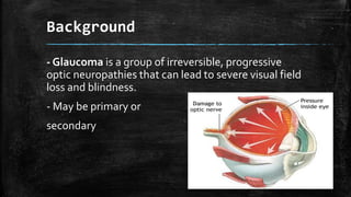
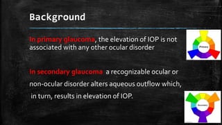
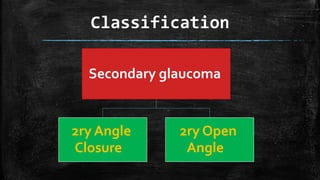
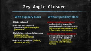
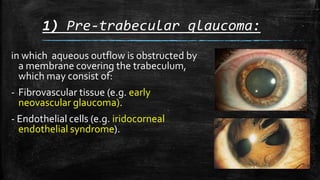
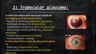
This presentation describes the secondary glaucoma and its different types .....you can find the illustrated video presentation in the following link:
https://www.youtube.com/watch?v=G1wkThV_za8
2. Background
- Glaucoma is a group of irreversible, progressive
optic neuropathies that can lead to severe visual field
loss and blindness.
- May be primary or
secondary
3. Background
In primary glaucoma, the elevation of IOP is not
associated with any other ocular disorder
In secondary glaucoma a recognizable ocular or
non-ocular disorder alters aqueous outflow which,
in turn, results in elevation of IOP.
6. 2ry Angle Closure
With pupillary block
- Miotic induced.
- Swollen lens induced
glaucoma (Phacomorphic
glaucoma).
- Mobile lens induced glaucoma
(ectopia lentis,
microspherophakia).
- Posterior synechiae (to lens,
vitreous, or IOL).
Without pupillary block
a) Anterior pulling mechanism:
pulling the iris forward by
contraction of a membrane e.g.
late neovascular glaucoma and
inflammatory membrane.
b) Posterior pushing mechanism:
pushing the iris forward by a
condition in the posterior
segment e.g. Malignant
glaucoma and intraocular
tumors.
7. 2ry Open Angle
Subdivided on the basis of the site of aqueous
outflow obstruction.
Pre-trabecular
glaucoma
Trabecular
Post-
trabecular
8. 1) Pre-trabecular glaucoma:
in which aqueous outflow is obstructed by
a membrane covering the trabeculum,
which may consist of:
- Fibrovascular tissue (e.g. early
neovascular glaucoma).
- Endothelial cells (e.g. iridocorneal
endothelial syndrome).
9. 2) Trabecular glaucoma:
in which the obstruction occurs as a result of:
a) Clogging up of the meshwork by:
- Pigment particles (e.g. pigmentary glaucoma).
- Red blood cells (e.g. red cell glaucoma).
- Degenerated red cells (e.g. ghost cell glaucoma).
- Macrophages and lens proteins (e.g. phacolytic
glaucoma).
- Proteins (e.g. hypertensive uveitis).
- Pseudoexfoliative material.
b) Alteration of the trabecular fibers themselves
by:
- Edema (e.g. herpes zoster iritis).
- Scarring (e.g. post-traumatic angle recession
glaucoma).
10. 3) Post-trabecular glaucoma
Aqueous outflow is impaired as a result
of elevated episcleral venous pressure
due to:
- Carotid-cavernous fistula.
- Obstruction of the superior vena cava.
12. Pseudoexofoliation Glaucoma
- Pseudoexofoliation (PEX) syndrome:
characterized by deposition of grey-white,
fibrillogranular material in the anterior lens
capsule, zonules, ciliary body,
iris, trabeculum,
anterior vitreous face and conjunctiva.
- Pseudoexofoliation glaucoma occurs when
trabecular block occur by clogging up of the
trabeculum by Pseudoexofoliation material
13. Pseudoexofoliation Glaucoma
Clinical picture:
1) Cornea: PEX materials on the posterior
surface.
2) Iris: Atrophy β PEX material.
3) Lens: PEX materials forming central disc and
peripheral band β cataract β subluxation.
4) Gonioscopy:Trabecular hyperpigmentation
and PEX materials.
Treatment: as POAG.
14. Neovascular Glaucoma
Retinal ischemia ο vasoproliferative growth
factor ο retinal neovascularization,
rubeosis iridis & neovascularization at the
angle of AC.
Causes:
1) Ischemic CRVO and CRAO.
2) PDR.
3) OLD RD.
4) IOTumors.
15. Neovascular Glaucoma: 3 stages
1) Rubeosis iridis: iris new vessles.
Treatment : Argon laser photocoagulation of the ischemic retina.
2) 2ry open angle glaucoma: due to neovascular membrane in
front of the trabeculum.
Treatment: 1) Medical: antiglaucoma β steroids β atropine.
2) Argon laser photocoagulation of the ischemic retina.
3) 2ry angle closure glaucoma: due to contraction of the
membrane.
Treatment:1) Argon laser photocoagulation of the ischemic retina.
2) Surgery: trabeculectomy with adjunctive Mitomycin C OR artificial
shunt.
3) Cyclodestruction: destruction of the ciliary processes by laser.
17. A) Phacolytic Glaucma:
Hypermature cataract ο lens proteins leak through
intact capsule ο blockage of the trabeculum with
proteins and macrophages filled with proteins.
Clinical picture:
- Corneal edema.
- Deep AC with floating white particles
(pseudohypopyon).
- Gonioscopy: open angle.
Treatment:
- 1st : Control IOP medically.
- Then: cataract surgery.