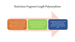
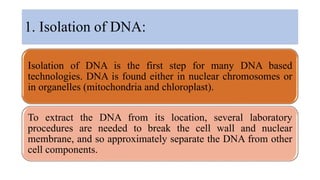
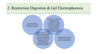
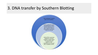
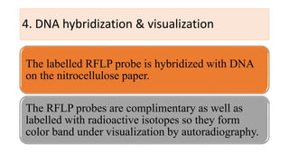
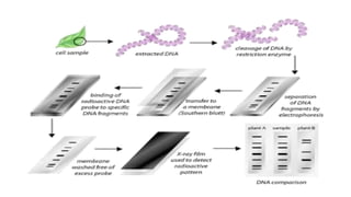
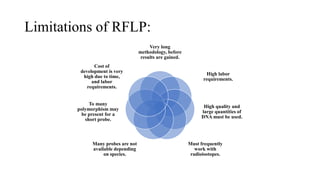
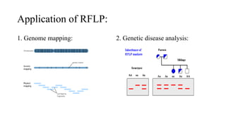
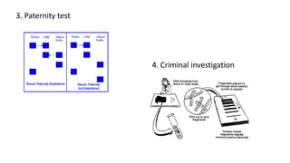
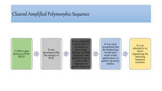
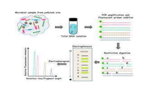
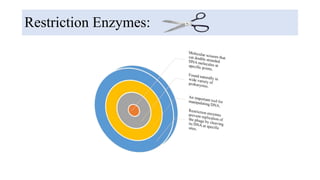
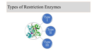
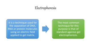
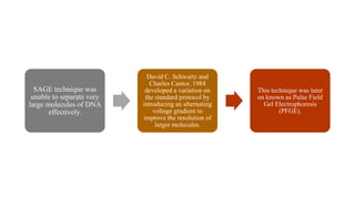
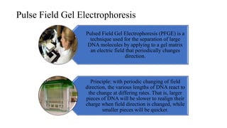
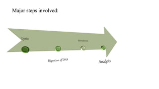
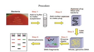
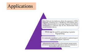
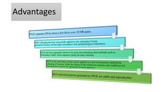
Molecular Techniques in Microbiology
Keezia Khurshid
Ph.D. Scholar (AAHM)
Unit V
Restriction Fragment Length Polymorphism
RFLP is a variation in the length
of a DNA fragment produced by
a specific restriction enzyme
acting on a DNA from different
individuals that usually results
from a genetic mutation.
The principle of RFLP markers is
that any genomic DNA can be
differentiated according to the
presence or absence of
restriction enzyme sites.
A restriction enzyme cuts the
DNA molecule at every
occurrence of a particular
sequence, called restriction
sites.
1. Isolation of DNA:
Isolation of DNA is the first step for many DNA based
technologies. DNA is found either in nuclear chromosomes or
in organelles (mitochondria and chloroplast).
To extract the DNA from its location, several laboratory
procedures are needed to break the cell wall and nuclear
membrane, and so approximately separate the DNA from other
cell components.
2. Restriction Digestion & Gel Electrophoresis
Extracted DNA is
digested with specific
restriction enzymes.
Each restriction enzyme
will recognize and cut
DNA in a predictable
way under appropriate
conditions resulting in a
reproducible set of
DNA fragments
(restriction fragments)
of different lengths.
The millions of restriction
fragments are then
separated by gel
electrophoresis, because
the fragments would be
seen as a continuous
smear if stained with
ethidium bromide,
staining alone cannot
detect the polymorphism.
Hybridization, therefore
must be used to detect
specific polymorphism.
3. DNA transfer by Southern Blotting
This technique is named
after E.M. Southern.
In this method the gel is
first denatured in a basic
solution and placed in a
tray. A porous nylon or
nitrocellulose membrane
is laid over the gel.
All the DNA restriction
fragments in the gel
transfer as single strand
will retain the same
pattern on the
membrane as on the gel
due to capillary action.
4. DNA hybridization & visualization
The labelled RFLP probe is hybridized with DNA
on the nitrocellulose paper.
The RFLP probes are complimentary as well as
labelled with radioactive isotopes so they form
color band under visualization by autoradiography.
Advantages of RFLP:
Produces semi-dominant markers, allowing determination of homozygosity or
heterozygosity.
Stable and reproducible, gives constant results over time and location
No prior information on DNA sequence is required.
Relatively simple technique.
Limitations of RFLP:
Very long
methodology, before
results are gained.
High labor
requirements.
High quality and
large quantities of
DNA must be used.
Must frequently
work with
radioisotopes.
Many probes are not
available depending
on species.
To many
polymorphism may
be present for a
short probe.
Cost of
development is very
high due to time,
and labor
requirements.
Application of RFLP:
1. Genome mapping: 2. Genetic disease analysis:
3. Paternity test
4. Criminal investigation
Cleaved Amplified Polymorphic Sequence
CAPS is also
known as PCR-
RFLP.
It was
developed after
the emerge of
PCR.
It was originally
conceived as a
technique to
detect base
changes in DNA
sequences and
therefore acts as
a tool in the
diagnosis of
genetic diseases.
It was soon
recognized that
the technology
would have
much wider
applications in
genetic diversity
studies.
It is an
alternative to
direct
sequencing for
detecting
sequence
variation.
Advantages:
Most CAPS markers are co-
dominant and locus-specific.
Most CAPS genotypes are
easily scored and interpreted.
CAPS markers are easily
shared between laboratories.
CAPS assay does not require
the use of radioactive
isotopes, and it is more
amenable, therefore, to
analyses in clinical settings.
Disadvantages:
Sequence the RFLP probe.
Design primers to amplify 800–2,000-bp DNA fragments. Targeting introns or 3' untranslated
regions should increase the chance of finding polymorphisms
The PCR product is cloned and sequenced.
PCR amplify DNA fragments from target genotypes, separately digest the amplicons with one
or more restriction enzymes.
Screen the digested amplicons for polymorphism on gels stained with ethidium bromide.
Restriction Enzymes:
Mechanism of Action
• Restriction Enzymes recognize a specific sequence of
nucleotides, and produce a double stranded cut in the DNA.
• These cuts are of two types:
• Blunt Ends
• Sticky Ends
Type
I
Type
II
Type
III
Types of Restriction Enzymes
Electrophoresis
It is a technique used for
the separation of DNA,
RNA or protein molecules
using an electric field
applied to gel matrix.
The most common
technique for this
purpose is that of
standard agarose gel
electrophoresis.
SAGE technique was
unable to separate very
large molecules of DNA
effectively.
David C. Schwartz and
Charles Cantor, 1984
developed a variation on
the standard protocol by
introducing an alternating
voltage gradient to
improve the resolution of
larger molecules.
This technique was later
on known as Pulse Field
Gel Electrophoresis
(PFGE).
Pulse Field Gel Electrophoresis
Pulsed Field Gel Electrophoresis (PFGE) is a
technique used for the separation of large
DNA molecules by applying to a gel matrix
an electric field that periodically changes
direction.
Principle: with periodic changing of field
direction, the various lengths of DNA react to
the change at differing rates. That is, larger
pieces of DNA will be slower to realign their
charge when field direction is changed, while
smaller pieces will be quicker.
Major steps involved:
Procedure
Applications
Since, field gel electrophoresis allows the separation of DNA
fragments containing up to 100,000 bp (100 kilobase pairs,
or kbp), characterization of such large fragments has allowed
construction of a physical map for the chromosomes from
several bacterial species.
PFGE may be used for genotyping or genetic
fingerprinting.
It is commonly considered a gold standard in epidemiological
studies of pathogenic organisms.
Subtyping has made it easier to discriminate among strains
of Listeria monocytogenes and thus to link environmental or
food isolates with clinical infections.
Advantages
Limitations

Molecular Techniques in Microbiology.pptx

  • 1.
    Molecular Techniques inMicrobiology Keezia Khurshid Ph.D. Scholar (AAHM) Unit V
  • 2.
    Restriction Fragment LengthPolymorphism RFLP is a variation in the length of a DNA fragment produced by a specific restriction enzyme acting on a DNA from different individuals that usually results from a genetic mutation. The principle of RFLP markers is that any genomic DNA can be differentiated according to the presence or absence of restriction enzyme sites. A restriction enzyme cuts the DNA molecule at every occurrence of a particular sequence, called restriction sites.
  • 4.
    1. Isolation ofDNA: Isolation of DNA is the first step for many DNA based technologies. DNA is found either in nuclear chromosomes or in organelles (mitochondria and chloroplast). To extract the DNA from its location, several laboratory procedures are needed to break the cell wall and nuclear membrane, and so approximately separate the DNA from other cell components.
  • 5.
    2. Restriction Digestion& Gel Electrophoresis Extracted DNA is digested with specific restriction enzymes. Each restriction enzyme will recognize and cut DNA in a predictable way under appropriate conditions resulting in a reproducible set of DNA fragments (restriction fragments) of different lengths. The millions of restriction fragments are then separated by gel electrophoresis, because the fragments would be seen as a continuous smear if stained with ethidium bromide, staining alone cannot detect the polymorphism. Hybridization, therefore must be used to detect specific polymorphism.
  • 6.
    3. DNA transferby Southern Blotting This technique is named after E.M. Southern. In this method the gel is first denatured in a basic solution and placed in a tray. A porous nylon or nitrocellulose membrane is laid over the gel. All the DNA restriction fragments in the gel transfer as single strand will retain the same pattern on the membrane as on the gel due to capillary action.
  • 7.
    4. DNA hybridization& visualization The labelled RFLP probe is hybridized with DNA on the nitrocellulose paper. The RFLP probes are complimentary as well as labelled with radioactive isotopes so they form color band under visualization by autoradiography.
  • 9.
    Advantages of RFLP: Producessemi-dominant markers, allowing determination of homozygosity or heterozygosity. Stable and reproducible, gives constant results over time and location No prior information on DNA sequence is required. Relatively simple technique.
  • 10.
    Limitations of RFLP: Verylong methodology, before results are gained. High labor requirements. High quality and large quantities of DNA must be used. Must frequently work with radioisotopes. Many probes are not available depending on species. To many polymorphism may be present for a short probe. Cost of development is very high due to time, and labor requirements.
  • 11.
    Application of RFLP: 1.Genome mapping: 2. Genetic disease analysis:
  • 12.
    3. Paternity test 4.Criminal investigation
  • 13.
    Cleaved Amplified PolymorphicSequence CAPS is also known as PCR- RFLP. It was developed after the emerge of PCR. It was originally conceived as a technique to detect base changes in DNA sequences and therefore acts as a tool in the diagnosis of genetic diseases. It was soon recognized that the technology would have much wider applications in genetic diversity studies. It is an alternative to direct sequencing for detecting sequence variation.
  • 15.
    Advantages: Most CAPS markersare co- dominant and locus-specific. Most CAPS genotypes are easily scored and interpreted. CAPS markers are easily shared between laboratories. CAPS assay does not require the use of radioactive isotopes, and it is more amenable, therefore, to analyses in clinical settings.
  • 16.
    Disadvantages: Sequence the RFLPprobe. Design primers to amplify 800–2,000-bp DNA fragments. Targeting introns or 3' untranslated regions should increase the chance of finding polymorphisms The PCR product is cloned and sequenced. PCR amplify DNA fragments from target genotypes, separately digest the amplicons with one or more restriction enzymes. Screen the digested amplicons for polymorphism on gels stained with ethidium bromide.
  • 17.
  • 18.
    Mechanism of Action •Restriction Enzymes recognize a specific sequence of nucleotides, and produce a double stranded cut in the DNA. • These cuts are of two types: • Blunt Ends • Sticky Ends
  • 19.
  • 20.
    Electrophoresis It is atechnique used for the separation of DNA, RNA or protein molecules using an electric field applied to gel matrix. The most common technique for this purpose is that of standard agarose gel electrophoresis.
  • 21.
    SAGE technique was unableto separate very large molecules of DNA effectively. David C. Schwartz and Charles Cantor, 1984 developed a variation on the standard protocol by introducing an alternating voltage gradient to improve the resolution of larger molecules. This technique was later on known as Pulse Field Gel Electrophoresis (PFGE).
  • 22.
    Pulse Field GelElectrophoresis Pulsed Field Gel Electrophoresis (PFGE) is a technique used for the separation of large DNA molecules by applying to a gel matrix an electric field that periodically changes direction. Principle: with periodic changing of field direction, the various lengths of DNA react to the change at differing rates. That is, larger pieces of DNA will be slower to realign their charge when field direction is changed, while smaller pieces will be quicker.
  • 23.
  • 24.
  • 25.
    Applications Since, field gelelectrophoresis allows the separation of DNA fragments containing up to 100,000 bp (100 kilobase pairs, or kbp), characterization of such large fragments has allowed construction of a physical map for the chromosomes from several bacterial species. PFGE may be used for genotyping or genetic fingerprinting. It is commonly considered a gold standard in epidemiological studies of pathogenic organisms. Subtyping has made it easier to discriminate among strains of Listeria monocytogenes and thus to link environmental or food isolates with clinical infections.
  • 26.
  • 27.