Downloaded 170 times
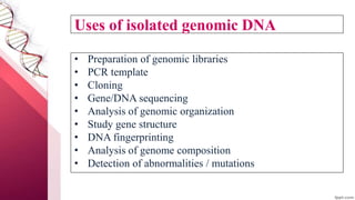
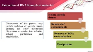
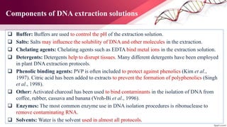
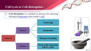
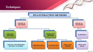


The document outlines various techniques for DNA extraction, purification, and quantification, emphasizing the importance of DNA isolation in biotechnology applications. It details several methods including chemical, physical, and enzymatic techniques, as well as approaches for purifying and quantifying DNA. Additionally, the paper discusses the use of different reagents and equipment necessary for successful DNA extraction and quantification.


![谷歌留痕技术 [ 𝙩𝙤𝙥 𝟮𝟯𝟯. 𝙘 𝙤𝙢 ]](https://cdn.slidesharecdn.com/ss_thumbnails/top233-260130174328-3833018c-thumbnail.jpg?width=640&height=640&fit=bounds)


