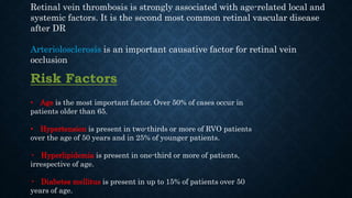
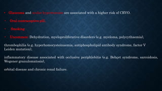
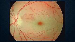
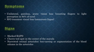
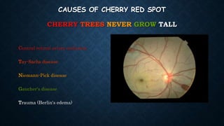
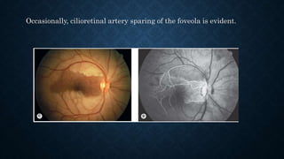
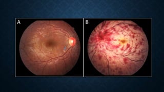
RETINAL VASCULAR DISEASES - I
By
Ahmed Alsherbeny
MD, EBO, CABOphth, MRCSEd Ophth, FRCS Ophth (Glasg), FICO, MSc
Retinal Vein Occlusion
Retinal vein thrombosis is strongly associated with age-related local and
systemic factors. It is the second most common retinal vascular disease
after DR
Arteriolosclerosis is an important causative factor for retinal vein
occlusion
Risk Factors
• Age is the most important factor. Over 50% of cases occur in
patients older than 65.
• Hypertension is present in two-thirds or more of RVO patients
over the age of 50 years and in 25% of younger patients.
• Hyperlipidemia is present in one-third or more of patients,
irrespective of age.
• Diabetes mellitus is present in up to 15% of patients over 50
years of age.
• Glaucoma and ocular hypertension are associated with a higher risk of CRVO.
• Oral contraceptive pill.
• Smoking
• Uncommon: Dehydration, myeloproliferative disorders (e.g. myeloma, polycythaemia),
thrombophilia (e.g. hyperhomocysteinaemia, antiphospholipid antibody syndrome, factor V
Leiden mutation),
inflammatory disease associated with occlusive periphlebitis (e.g. Behçet syndrome, sarcoidosis,
Wegener granulomatosis),
orbital disease and chronic renal failure.
Don’t Forget:
Systemic assessment (All patients)
1 Blood pressure.
2 Erythrocyte sedimentation rate (ESR) or plasma viscosity (PV).
3 Full blood count (FBC).
4 Random blood glucose. Further assessment for diabetes if indicated.
5 Lipid profile
6 Plasma protein electrophoresis. To detect dysproteinaemias such as multiple myeloma.
7 Urea, electrolytes and creatinine. Chronic renal failure is a rare cause of RVO, but renal disease may
occur in association with hypertension.
8 Thyroid function tests. Patients with RVO have a higher prevalence of thyroid disease than the general
population. Thyroid dysfunction is also associated with dyslipidaemia.
9 ECG.
Systemic assessment (Selected patients according to clinical indication)
Patients in whom these might be considered are those under the age of 50, those with bilateral RVO, a history of previous
thromboses or a family history of thrombosis, and possibly in other patients in whom investigation for the common
associations is negative
1 Chest X-ray. Sarcoidosis, tuberculosis, left ventricular hypertrophy in hypertension.
2 C-reactive protein (CRP). Sensitive indicator of inflammation.
3 'Thrombophilia screen’. By convention refers to heritable thrombophilias; might typically include thrombin time,
prothrombin time and activated partial thromboplastin time, antithrombin functional assay, protein C, protein S,
activated protein C resistance, factor V Leiden mutation, anticardiolipin antibody, lupus anticoagulant.
4 Autoantibodies. Rheumatoid factor, anti-nuclear antibody, anti-DNA antibody.
5 Serum angiotensin-converting enzyme (ACE). Sarcoidosis.
6 Fasting plasma homocysteine level. To exclude hyperhomocysteinaemia.
7 Carotid duplex imaging to exclude ocular ischaemic syndrome.
Retinal Vein
Occlusion
CRVO
Ischemic
Non-
ischemic
BRVO
Ischemic
Non-
ischemic
• BRVO are three times commoner than CRVO
• Division of non-ischaemic vs. ischaemic CRVO on FFA is a useful predictor
of visual outcome and risk of neovascularization
Branch Retinal Vein Occlusion
Classification:
1 Major branch retinal vein occlusion
(BRVO) at the disc (A) and away from the disc
(B).
2 Macular BRVO involving only a
macular branch (C).
3 Peripheral BRVO not involving the
macular circulation (D-F).
Symptoms
• Asymptomatic
• VF defect
• Loss of vision, usually unilateral
• Metamorphopsia
Signs
• Superficial hemorrhages in a sector of the retina along a retinal vein
• CWSs
• Dilated and tortuous retinal vein
• The superotemporal quadrant is most commonly affected.
• The site of occlusion is often identifiable as an arteriovenous crossing point.
• VH
• The acute features usually resolve within 6–12 months leaving venous
sheathing and sclerosis and variable persistent/recurrent haemorrhage
WORK-UP
 Ocular:
• Complete ocular examination: careful slit lamp examination, gonioscopy to rule out
NVI (Before dilation) and dilated fundus examination
 Systemic:
• History: Medical problems, medications (HTN, oral contraceptives, eye diseases
‘Glaucoma’)
• ABP
• FBS and Hb A1c, CBC with differential, (PT/PTT)
• Specialized investigations, which may include serum ACE, anticardiolipin, lupus
anticoagulant, autoantibodies (RF, ANA, anti- DNA, ANCA), fasting homocysteine,
CXR, and thrombophilia screen (e.g. proteins C and S, antithrombin, factor V).
Better carried out by the patient’s physician or a haematology specialist
 FFA:
• If uncertain diagnosis or to
assess ischaemia
• shows variable delayed
venous filling, blockage by
blood, staining of the vessel
wall, hypofluorescence due to
capillary non-perfusion in
the ischaemic areas
INVESTIGATIONS:
 OCT:
• demonstrates and allows quantification of the severity of
macular oedema and is a useful way of monitoring its course or
the response to treatment.
The acute features usually resolve within 6–12 months and may be replaced by the
following:
• Exudates, venous sheathing and sclerosis peripheral to the site of obstruction,
collaterals and variable residual haemorrhage
• Collateral are characterized by slightly tortuous veins that develop locally or
across the horizontal raphe between the inferior and superior vascular arcades and
are best detected on FA
• The severity of residual signs is highly variable.
COURSE
TREATMENT
• Underlying medical conditions Referral to a physician for
investigation and treatment
• IOP control if elevated
• Management of the 2 main complications:
1. Neovascularization
2. Macular edema
TREATMENT
1. Neovascularization:
• NVDs, NVEs or NVIs sectoral PRP to the ischemic area, which
corresponds to area of capillary nonperfusion on FFA
• Anti – VEGF may be used for temporarily halting of
neovascularization especially for NVIs till sectoral PRP takes action
TREATMENT
2. Macular edema:
• Focal retinal laser photocoagulation has historically been the gold-
standard treatment if edema is present for 3 to 6 months duration, and
visual acuity is below 20/40 with macular capillary perfusion.
• Anti-VEGF treatment is now largely favored
• Multiple pharmacologic trials (BRAVO and CRUISE) have validated
that early anti-VEGF treatment leads to better visual outcomes
FOLLOW-UP
• Every month initially, with gradual interval taper based on vision,
presence of macular edema, and response to treatment
• At each visit, the patient should be checked for neovascularization and
macular edema
Central Retinal Vein Occlusion
Symptoms
• Painless loss of vision
Signs
• Diffuse retinal hemorrhages in all four quadrants of the retina
• Dilated, tortuous retinal veins
• CWSs
• Disc edema and hemorrhages
• Macular edema
Conversion of Non-ischemic CRVO to Ischemic CRVO: 15% within 4 months and 34%
within 3 years
 Ocular & Systemic:
• The same as BRVO
 FFA:
 Normal arm to eye time, slow AV phase, acutely vein wall staining,
microaneurysms, dilated optic disc capillaries
 Ischaemic: Capillary closure (5 – 10 DD is borderline; >10 is significantly
ischaemic)
WORK-UP
WORK-UP
 OCT:
• Substantial retinal thickening, with inner and outer retinal cysts
and SRF at the fovea
• Allows rapid quantitative analysis of the macula and has become
important in the assessment and subsequent management of
retinal vein occlusion
WORK-UP
WORK-UP
WORK-UP
TREATMENT
• Underlying medical conditions Referral to a physician for
investigation and treatment
• IOP control if elevated
• Management of the 2 main complications:
1. Neovascularization
2. Macular edema
TREATMENT
1. Neovascularization:
• NVDs, NVEs or NVIs PRP
• Anti – VEGF may be used for temporarily halting of
neovascularization especially for NVIs till PRP takes action
• Prophylactic PRP not recommended
 No prevention of neovascularization
 Faster regression in non-treated eyes
TREATMENT
2. Macular edema:
• Intravitreal Anti-VEGF injection: ‘Monthly injection’
 Ranibizumab 0.5 mg Or Aflibercept 2 mg: FDA approved
 Bevacizumab 1.25 mg: Off-label
• Intravitreal steroid injection:
 Dexamethasone 0.7 mg ‘Ozurdex’: FDA approved
 Triamcinolone 1 mg: Off-label
• Macular grid laser No benefit
TREATMENT
2. Macular edema:
How to treat
 Anti-VEGF monthly injections are given until maximal V/A (stable
V/A on three consecutive visits)
 Discontinue injections if no improvement after six injections
 Dexamethasone implant (if anti- VEGF is not suitable),
retreatment may be required 4- to 6- monthly until V/A is stable
 Ischaemic CRVO with macular oedema, can be treated with anti-
VEGF/ dexamethasone, but guarded prognosis should be explained
FOLLOW-UP
• Every month initially, with gradual interval taper based on vision,
presence of macular edema, and response to treatment.
• At each follow-up visit, evaluate anterior segment for NVI followed by
careful dilated fundus examination looking for any retinal
neovascularization
• Evidence of early neovascularization should prompt immediate PRP
and/or anti-VEGF therapy and monthly follow-up until stabilized or
regressed
• Patients should be informed that there is an 8% to 10% risk for the
development of a BRVO or CRVO in the fellow eye
Retinal Artery Occlusion
AETIOLOGY
• Common:
 Atherosclerosis-related thrombosis: by
far the most common underlying
cause of central retinal artery
occlusion (CRAO), accounting for
about 80% of cases
 Carotid emboli: CCF
• Cholesterol (A)
• Fibrin-platelet (B&C)
• Calcific (D)
AETIOLOGY
• Less common:
 Giant cell (temporal) arteritis (GCA).
 Cardiac embolism
 Vasculitis (PAN – SLE)
 Thrombophilic disorders
 Sickling haemoglobinopathies.
 Retinal migraine
 Susac syndrome (retinocochleocerebral vasculopathy) is a microangiopathy characterized by the
triad of retinal artery occlusion, sensorineural deafness and encephalopathy.
Don’t Forget:
Systemic assessment (All patients)
1 Symptoms of GCA such as headache, jaw claudication, scalp tenderness, limb girdle pain, weight loss
and existing polymyalgia rheumatica
2 Pulse
3 Blood pressure.
4 Cardiac auscultation.
5 Carotid examination.
6 ECG
7 Erythrocyte sedimentation rate and C-reactive protein to detect the remote possibility of GCA.
8 Other blood tests include FBC, random glucose, lipids, urea and electrolytes.
9 Carotid duplex scanning
Systemic assessment (Selected patients according to clinical indication)
1 Further carotid imaging
2 Cranial MRI or CT may be indicated to rule out intracranial or orbital pathology; may be required prior
to fibrinolysis.
3 Echocardiography.
4 Chest X-ray.
5 24-hour ECG (Holter monitor) to exclude intermittent arrhythmia.
6 Additional blood tests:
a Plasma homocysteine level to exclude hyperhomocysteinaemia.
b 'Thrombophilia screen’.
c Plasma protein electrophoresis to detect dysproteinaemias such as multiple myeloma.
d Thyroid function tests, especially if atrial fibrillation is present; may be associated with dyslipidaemia.
e Autoantibodies. Rheumatoid factor, anti-nuclear antibody, anti-DNA antibody.
Symptoms
• Unilateral, painless, acute vision loss (counting fingers to light
perception in 94% of eyes)
• H/O transient visual loss (amaurosis fugax)
Signs
• Marked RAPD
• Cherry-red spot in the center of the macula
• Narrowed retinal arterioles; box-carring or segmentation of the blood
column in the arterioles
CAUSES OF CHERRY RED SPOT
CHERRY TREES NEVER GROW TALL
Central retinal artery occlusion
Tay-Sachs disease
Niemann-Pick disease
Gaucher's disease
Trauma (Berlin's edema)
Occasionally, cilioretinal artery sparing of the foveola is evident.
TREATMENT
None of these treatments have been proven effective in randomized, controlled
clinical trials, and should not be considered the standard of care.
• 1 Adoption of a supine posture might improve ocular perfusion.
• 2 Ocular massage using a three-mirror contact lens
• 3 Anterior chamber paracentesis should be carried out in most cases.
• 4 Intravenous acetazolamide 500 mg to achieve a more sustained lowering
of intraocular pressure.
TREATMENT
• 5 Sublingual isosorbide dinitrate to induce vasodilation.
• 6 ‘Rebreathing’ into a paper bag in order to elevate blood carbon dioxide and
respiratory acidosis has been advocated, as this may promote vasodilation.
• 7 Breathing a high oxygen (95%) and carbon dioxide (5%) mixture, ‘carbogen’, has
been advocated for a possible dual effect of retarding ischaemia and vasodilation.
• 8 Hyperosmotic agents. Mannitol or glycerol have been used for their possibly
more rapid IOP-lowering effect as well as increased intravascular volume.
TREATMENT
Management of systemic disease: 20% mortality over 5 years
WORK-UP
In the acute setting, the diagnosis is not usually in doubt, so the
urgent priority is to rule out an underlying disease (such as
GCA) that may threaten the contralateral eye.
• Immediate ESR, CRP, and platelets to rule out GCA if the patient is
>55 years and no embolus seen
• ABP
• Other blood tests: FBS and Hb A1c, CBC with differential, (PT/PTT).
• Carotid artery evaluation & Cardiac evaluation
FOLLOW-UP
• Follow as directed by managing internist and/or neurologist.
• Repeat eye examination checking for neovascularization
Thank You

Retinal Vascular Diseases - I

  • 1.
    RETINAL VASCULAR DISEASES- I By Ahmed Alsherbeny MD, EBO, CABOphth, MRCSEd Ophth, FRCS Ophth (Glasg), FICO, MSc
  • 2.
  • 3.
    Retinal vein thrombosisis strongly associated with age-related local and systemic factors. It is the second most common retinal vascular disease after DR Arteriolosclerosis is an important causative factor for retinal vein occlusion Risk Factors • Age is the most important factor. Over 50% of cases occur in patients older than 65. • Hypertension is present in two-thirds or more of RVO patients over the age of 50 years and in 25% of younger patients. • Hyperlipidemia is present in one-third or more of patients, irrespective of age. • Diabetes mellitus is present in up to 15% of patients over 50 years of age.
  • 4.
    • Glaucoma andocular hypertension are associated with a higher risk of CRVO. • Oral contraceptive pill. • Smoking • Uncommon: Dehydration, myeloproliferative disorders (e.g. myeloma, polycythaemia), thrombophilia (e.g. hyperhomocysteinaemia, antiphospholipid antibody syndrome, factor V Leiden mutation), inflammatory disease associated with occlusive periphlebitis (e.g. Behçet syndrome, sarcoidosis, Wegener granulomatosis), orbital disease and chronic renal failure.
  • 5.
  • 6.
    Systemic assessment (Allpatients) 1 Blood pressure. 2 Erythrocyte sedimentation rate (ESR) or plasma viscosity (PV). 3 Full blood count (FBC). 4 Random blood glucose. Further assessment for diabetes if indicated. 5 Lipid profile 6 Plasma protein electrophoresis. To detect dysproteinaemias such as multiple myeloma. 7 Urea, electrolytes and creatinine. Chronic renal failure is a rare cause of RVO, but renal disease may occur in association with hypertension. 8 Thyroid function tests. Patients with RVO have a higher prevalence of thyroid disease than the general population. Thyroid dysfunction is also associated with dyslipidaemia. 9 ECG.
  • 7.
    Systemic assessment (Selectedpatients according to clinical indication) Patients in whom these might be considered are those under the age of 50, those with bilateral RVO, a history of previous thromboses or a family history of thrombosis, and possibly in other patients in whom investigation for the common associations is negative 1 Chest X-ray. Sarcoidosis, tuberculosis, left ventricular hypertrophy in hypertension. 2 C-reactive protein (CRP). Sensitive indicator of inflammation. 3 'Thrombophilia screen’. By convention refers to heritable thrombophilias; might typically include thrombin time, prothrombin time and activated partial thromboplastin time, antithrombin functional assay, protein C, protein S, activated protein C resistance, factor V Leiden mutation, anticardiolipin antibody, lupus anticoagulant. 4 Autoantibodies. Rheumatoid factor, anti-nuclear antibody, anti-DNA antibody. 5 Serum angiotensin-converting enzyme (ACE). Sarcoidosis. 6 Fasting plasma homocysteine level. To exclude hyperhomocysteinaemia. 7 Carotid duplex imaging to exclude ocular ischaemic syndrome.
  • 8.
    Retinal Vein Occlusion CRVO Ischemic Non- ischemic BRVO Ischemic Non- ischemic • BRVOare three times commoner than CRVO • Division of non-ischaemic vs. ischaemic CRVO on FFA is a useful predictor of visual outcome and risk of neovascularization
  • 9.
  • 10.
    Classification: 1 Major branchretinal vein occlusion (BRVO) at the disc (A) and away from the disc (B). 2 Macular BRVO involving only a macular branch (C). 3 Peripheral BRVO not involving the macular circulation (D-F).
  • 11.
    Symptoms • Asymptomatic • VFdefect • Loss of vision, usually unilateral • Metamorphopsia Signs • Superficial hemorrhages in a sector of the retina along a retinal vein • CWSs • Dilated and tortuous retinal vein • The superotemporal quadrant is most commonly affected. • The site of occlusion is often identifiable as an arteriovenous crossing point. • VH • The acute features usually resolve within 6–12 months leaving venous sheathing and sclerosis and variable persistent/recurrent haemorrhage
  • 12.
    WORK-UP  Ocular: • Completeocular examination: careful slit lamp examination, gonioscopy to rule out NVI (Before dilation) and dilated fundus examination  Systemic: • History: Medical problems, medications (HTN, oral contraceptives, eye diseases ‘Glaucoma’) • ABP • FBS and Hb A1c, CBC with differential, (PT/PTT) • Specialized investigations, which may include serum ACE, anticardiolipin, lupus anticoagulant, autoantibodies (RF, ANA, anti- DNA, ANCA), fasting homocysteine, CXR, and thrombophilia screen (e.g. proteins C and S, antithrombin, factor V). Better carried out by the patient’s physician or a haematology specialist
  • 13.
     FFA: • Ifuncertain diagnosis or to assess ischaemia • shows variable delayed venous filling, blockage by blood, staining of the vessel wall, hypofluorescence due to capillary non-perfusion in the ischaemic areas INVESTIGATIONS:
  • 15.
     OCT: • demonstratesand allows quantification of the severity of macular oedema and is a useful way of monitoring its course or the response to treatment.
  • 16.
    The acute featuresusually resolve within 6–12 months and may be replaced by the following: • Exudates, venous sheathing and sclerosis peripheral to the site of obstruction, collaterals and variable residual haemorrhage • Collateral are characterized by slightly tortuous veins that develop locally or across the horizontal raphe between the inferior and superior vascular arcades and are best detected on FA • The severity of residual signs is highly variable. COURSE
  • 17.
    TREATMENT • Underlying medicalconditions Referral to a physician for investigation and treatment • IOP control if elevated • Management of the 2 main complications: 1. Neovascularization 2. Macular edema
  • 18.
    TREATMENT 1. Neovascularization: • NVDs,NVEs or NVIs sectoral PRP to the ischemic area, which corresponds to area of capillary nonperfusion on FFA • Anti – VEGF may be used for temporarily halting of neovascularization especially for NVIs till sectoral PRP takes action
  • 19.
    TREATMENT 2. Macular edema: •Focal retinal laser photocoagulation has historically been the gold- standard treatment if edema is present for 3 to 6 months duration, and visual acuity is below 20/40 with macular capillary perfusion. • Anti-VEGF treatment is now largely favored • Multiple pharmacologic trials (BRAVO and CRUISE) have validated that early anti-VEGF treatment leads to better visual outcomes
  • 20.
    FOLLOW-UP • Every monthinitially, with gradual interval taper based on vision, presence of macular edema, and response to treatment • At each visit, the patient should be checked for neovascularization and macular edema
  • 21.
  • 22.
    Symptoms • Painless lossof vision Signs • Diffuse retinal hemorrhages in all four quadrants of the retina • Dilated, tortuous retinal veins • CWSs • Disc edema and hemorrhages • Macular edema
  • 24.
    Conversion of Non-ischemicCRVO to Ischemic CRVO: 15% within 4 months and 34% within 3 years
  • 26.
     Ocular &Systemic: • The same as BRVO  FFA:  Normal arm to eye time, slow AV phase, acutely vein wall staining, microaneurysms, dilated optic disc capillaries  Ischaemic: Capillary closure (5 – 10 DD is borderline; >10 is significantly ischaemic) WORK-UP
  • 27.
    WORK-UP  OCT: • Substantialretinal thickening, with inner and outer retinal cysts and SRF at the fovea • Allows rapid quantitative analysis of the macula and has become important in the assessment and subsequent management of retinal vein occlusion
  • 28.
  • 29.
  • 30.
  • 31.
    TREATMENT • Underlying medicalconditions Referral to a physician for investigation and treatment • IOP control if elevated • Management of the 2 main complications: 1. Neovascularization 2. Macular edema
  • 32.
    TREATMENT 1. Neovascularization: • NVDs,NVEs or NVIs PRP • Anti – VEGF may be used for temporarily halting of neovascularization especially for NVIs till PRP takes action • Prophylactic PRP not recommended  No prevention of neovascularization  Faster regression in non-treated eyes
  • 33.
    TREATMENT 2. Macular edema: •Intravitreal Anti-VEGF injection: ‘Monthly injection’  Ranibizumab 0.5 mg Or Aflibercept 2 mg: FDA approved  Bevacizumab 1.25 mg: Off-label • Intravitreal steroid injection:  Dexamethasone 0.7 mg ‘Ozurdex’: FDA approved  Triamcinolone 1 mg: Off-label • Macular grid laser No benefit
  • 34.
    TREATMENT 2. Macular edema: Howto treat  Anti-VEGF monthly injections are given until maximal V/A (stable V/A on three consecutive visits)  Discontinue injections if no improvement after six injections  Dexamethasone implant (if anti- VEGF is not suitable), retreatment may be required 4- to 6- monthly until V/A is stable  Ischaemic CRVO with macular oedema, can be treated with anti- VEGF/ dexamethasone, but guarded prognosis should be explained
  • 35.
    FOLLOW-UP • Every monthinitially, with gradual interval taper based on vision, presence of macular edema, and response to treatment. • At each follow-up visit, evaluate anterior segment for NVI followed by careful dilated fundus examination looking for any retinal neovascularization • Evidence of early neovascularization should prompt immediate PRP and/or anti-VEGF therapy and monthly follow-up until stabilized or regressed • Patients should be informed that there is an 8% to 10% risk for the development of a BRVO or CRVO in the fellow eye
  • 38.
  • 39.
    AETIOLOGY • Common:  Atherosclerosis-relatedthrombosis: by far the most common underlying cause of central retinal artery occlusion (CRAO), accounting for about 80% of cases  Carotid emboli: CCF • Cholesterol (A) • Fibrin-platelet (B&C) • Calcific (D)
  • 40.
    AETIOLOGY • Less common: Giant cell (temporal) arteritis (GCA).  Cardiac embolism  Vasculitis (PAN – SLE)  Thrombophilic disorders  Sickling haemoglobinopathies.  Retinal migraine  Susac syndrome (retinocochleocerebral vasculopathy) is a microangiopathy characterized by the triad of retinal artery occlusion, sensorineural deafness and encephalopathy.
  • 41.
  • 42.
    Systemic assessment (Allpatients) 1 Symptoms of GCA such as headache, jaw claudication, scalp tenderness, limb girdle pain, weight loss and existing polymyalgia rheumatica 2 Pulse 3 Blood pressure. 4 Cardiac auscultation. 5 Carotid examination. 6 ECG 7 Erythrocyte sedimentation rate and C-reactive protein to detect the remote possibility of GCA. 8 Other blood tests include FBC, random glucose, lipids, urea and electrolytes. 9 Carotid duplex scanning
  • 43.
    Systemic assessment (Selectedpatients according to clinical indication) 1 Further carotid imaging 2 Cranial MRI or CT may be indicated to rule out intracranial or orbital pathology; may be required prior to fibrinolysis. 3 Echocardiography. 4 Chest X-ray. 5 24-hour ECG (Holter monitor) to exclude intermittent arrhythmia. 6 Additional blood tests: a Plasma homocysteine level to exclude hyperhomocysteinaemia. b 'Thrombophilia screen’. c Plasma protein electrophoresis to detect dysproteinaemias such as multiple myeloma. d Thyroid function tests, especially if atrial fibrillation is present; may be associated with dyslipidaemia. e Autoantibodies. Rheumatoid factor, anti-nuclear antibody, anti-DNA antibody.
  • 45.
    Symptoms • Unilateral, painless,acute vision loss (counting fingers to light perception in 94% of eyes) • H/O transient visual loss (amaurosis fugax) Signs • Marked RAPD • Cherry-red spot in the center of the macula • Narrowed retinal arterioles; box-carring or segmentation of the blood column in the arterioles
  • 46.
    CAUSES OF CHERRYRED SPOT CHERRY TREES NEVER GROW TALL Central retinal artery occlusion Tay-Sachs disease Niemann-Pick disease Gaucher's disease Trauma (Berlin's edema)
  • 47.
    Occasionally, cilioretinal arterysparing of the foveola is evident.
  • 50.
    TREATMENT None of thesetreatments have been proven effective in randomized, controlled clinical trials, and should not be considered the standard of care. • 1 Adoption of a supine posture might improve ocular perfusion. • 2 Ocular massage using a three-mirror contact lens • 3 Anterior chamber paracentesis should be carried out in most cases. • 4 Intravenous acetazolamide 500 mg to achieve a more sustained lowering of intraocular pressure.
  • 51.
    TREATMENT • 5 Sublingualisosorbide dinitrate to induce vasodilation. • 6 ‘Rebreathing’ into a paper bag in order to elevate blood carbon dioxide and respiratory acidosis has been advocated, as this may promote vasodilation. • 7 Breathing a high oxygen (95%) and carbon dioxide (5%) mixture, ‘carbogen’, has been advocated for a possible dual effect of retarding ischaemia and vasodilation. • 8 Hyperosmotic agents. Mannitol or glycerol have been used for their possibly more rapid IOP-lowering effect as well as increased intravascular volume.
  • 52.
    TREATMENT Management of systemicdisease: 20% mortality over 5 years
  • 53.
    WORK-UP In the acutesetting, the diagnosis is not usually in doubt, so the urgent priority is to rule out an underlying disease (such as GCA) that may threaten the contralateral eye. • Immediate ESR, CRP, and platelets to rule out GCA if the patient is >55 years and no embolus seen • ABP • Other blood tests: FBS and Hb A1c, CBC with differential, (PT/PTT). • Carotid artery evaluation & Cardiac evaluation
  • 54.
    FOLLOW-UP • Follow asdirected by managing internist and/or neurologist. • Repeat eye examination checking for neovascularization
  • 57.