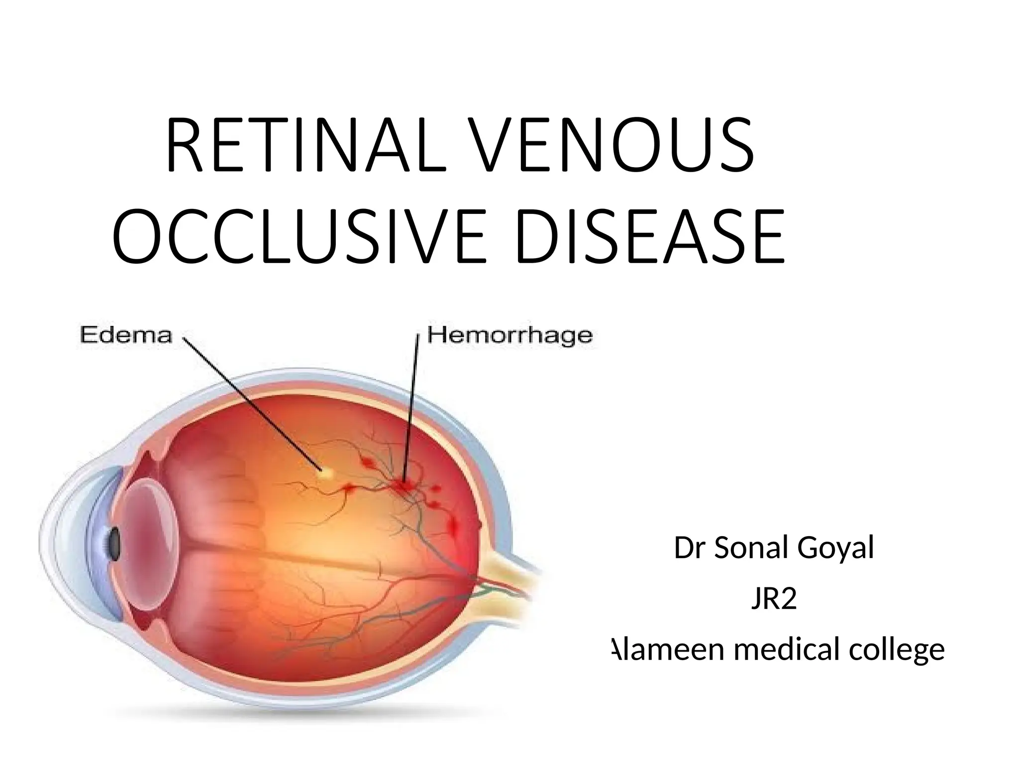
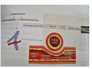
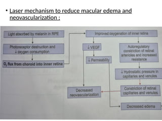
RETINAL VENOUS
OCCLUSIVE DISEASE
Dr Sonal Goyal
JR2
Alameen medical college
Introduction :
• It is the second most common vascular disease after
DR.
• It is caused by a thrombus located at any point along
the venous circulation and is named according to the
site of the occlusion.
• A branch retinal vein occlusion (BRVO) produces a
sectorial (typically quadrantic) area of damage due to
thrombosis in 1 of the branches of the intraretinal
portion of the venous tree.
• A central retinal vein occlusion (CRVO) arises from
thrombosis in the retrolaminar portion and, thus
affects the entire retina.
• Hemicentral retinal vein occlusion (HRVO) involves
one trunk of a dual- trunked CRV that has persisted in
the anterior part of the optic nerve head as a
congenital variant.
Pathogenesis:
• The pathogenesis of RVO mirrors Virchow’s triad for
thrombosis: endothelial injury, venous stasis, and
hypercoagulability.
• Normally, retinal arteries and veins share a common
adventitial sheath.
• Progressive arteriolosclerosis (as occurs in normal aging
and hypertension) leads to thickening of the arterial
wall, which in turn leads to compression and inelasticity
of the venous wall at the point of crossing.
• These changes to the venous wall (endothelial damage)
may produce turbulent blood flow and venous stasis,
which predispose to venous thrombosis.
• At the cellular level, retinal ischemia caused by
impairment of normal retinal circulation leads to retinal
hypoxia ->damage to the capillary endothelial cells -
>extravasation of blood constituents and the release of
hypoxia related factors into the retina and into the eye.
• The most salient of these are vascular endothelial
growth factor (VEGF) and various mediators of
inflammation.
• VEGF has 2 profound effects on the retinal vasculature:
(1) increased vessel wall permeability, leading to macular
edema; and
(2) growth of new blood vessels(neovascularization).
Risk factors:
The Eye Disease Case- Control Study and other studies have
enumerated risk factors associated with
BRVO and CRVO:
• increasing age (90% of patients are >50 years old)
• systemic arterial hypertension
• cigarette smoking
• open- angle glaucoma and ocular hypertension
• diabetes mellitus
• hyperlipidemia
• hypercoagulability
• hypothyroidism
• Chronic renal failure
• Although rare, predisposing hypercoagulable and
inflammatory conditions may be present like
a. Dehydration, thrombophilia, polycythemia,myeloma
,anemia
b. Behcet syndrome,sarcoidosis, wegener
granulomatosis, HIV ,SLE, syphilis
• Every patient with RVO should undergo medical
evaluation in addition to a comprehensive ocular
examination.
• In the absence of known cardiovascular disease, a
search for other causative or predisposing systemic
conditions should be considered, especially in patients
younger than 50 years or with bilateral CRVO.
Systemic assessment:
• The detection and management of associated
systemic Disease is aimed principally at reducing the
risk of future vascular occlusive events, both ocular
and systemic.
• Blood pressure
• erythrocyte Sedimentation rate (ESR)
• Random blood glucose- further assessment if diabetes
indicated.
• HDL ,cholesterol
• Plasma protein electrophoresis- to detect
dysproteinaemias such as multiple myeloma.
• Thyroid function test
• ECG
• Renal function test (RFT)
• Few investigations to be done in selected Patients:I.e.
age under 50, bilateral RVO, h/o thrombosis or family
history .
• Chest X-Ray
• CRP- inflammation indicator
• Thrombophilia screening
• Autoantibodies: RF, ANA, anti-DNA antibody
• Etc...
Clinical presentation
• Patients typically present usually with sudden, painless
loss of vision or visual field defect correlating to the area
of perfusion of the obstructed vessels.
• The classic ophthalmoscopic findings are dilated,
tortuous retinal veins and intraretinal hemorrhages in
the areas affected by the occlusion.
• Further clinical characteristics will be further discussed
under the headings of BRVO and CRVO.
Management:
Treatment goals
1.Treat systemic conditions :
Hypertension
Diabetes mellitus
Hyperlipidemia
Myeloproliferative disorders, thrombophilia and
anemia.
2.Treatment of macular edema
A. Anti-VEGF treatment- Primary therapy of choice
because of their excellent efficacy and safety profiles.
• Improvement and maintenance of visual acuity are
optimized by administering anti- VEGF treatment
immediately upon diagnosis of RVO- related macular
edema and continuing treatment long enough to prevent
recurrence of macular edema.
Intravitreal anti-VEGF injections:
• Ranibizumab(accentrix/ Lucentis):0.5mg/0.05ml
• Aflibercept (Eylea): 2mg/0.05ml
• Bevacizumab(Avastin): 1.25 mg/0.05ml( off label use)
Mechanism of action:
• Inhibit VEGF
• Enhanced retinal perfusion
• Results in lowered intravenous pressure and
• normalization of venous calibre and tortuosity.
B. Intravitreal steroid treatment:
-Triamcinolone acetonide(tricort/ aurocort)
• 4mg/0.1 ml
• 2mg /0.05ml
-Dexamethasone implant(ozurdex)
• 0.7mg
• Sustained release
• biodegradable implant
Mechanism of action:
• Anti-inflammatory effects by modulation of production of
cytokines and VEGF.
• Stabilizes blood retinal batrier with reduction of vascular
permeability .
• The Treatment can be repeated after 4-6 months.
Adverse effects include glaucoma and cataract Risk.
• Epidemiology: worldwide prevalence of BRVO is 13.9
million
• In BRVO, venous thrombosis and obstruction occur most
commonly at an arteriovenous crossing in the
superotemporal quadrant.
• When the occlusion does not occur at an arteriovenous
crossing, other inflammatory and infectious causes
should be considered.
• Visual acuity depends on the anatomical location of the
occlusion.
• The occlusion may also be restricted to the macula,
termed as macular BRVO.
Branch retinal vein occlusion
(BRVO)
• Peripheral occlusion may be asymptomatic.
• Iris neovascularization and neovascular glaucoma
are much less common in BRVO than in CRVO.
Fundus:
• Acute signs:
a. Dilated and tortuosity of the affected venous
segments.
b. Wedge shaped distribution of intraretinal
hemorrhages(flame shaped and dot blot), cotton
wool spots.
These changes are in the retinal area drained by the
thrombosed vein.
• The acute features usually resolve within 6-12 months
leaving resolved intraretinal hemorrhages, venous
sheathing ,sclerosis of the vessels and collateral vessels
formation.
Complications:
• Macular oedema
• Macular ischemia
• Severe sequelae:
a. Iris neovascularization
b. Retinal neovascularization
c. Vitreous haemorrhage
d. Tractional retinal detachment
e. neovascular Glaucoma
Management of BRVO:
A. Treat systemic conditions using medical therapy
B. Treat macular edema :Pharmacologic management is
currently the mainstay treatment of BRVO:
• Anti-VEGF Treatment
• Intravitreal steroid treatment
BRVO trials are:
1. BRAVO study: efficacy and safety of Ranibizumab
2. VIBRANT study: Intravitreal Aflibercept vs macular grid
laser.
3. Geneva study: Global Evaluation of Implantable
Dexamethasone in retinal Vein Occlusion With
Macular Edema
4. SCORE study:The Standard Care vs Corticosteroid for Retinal
Vein Occlusion,.
5. COMRADE study: compared the dexamethasone implant
against monthly ranibizumab
1.BRAVO study- efficacy and safety of Ranibizumab for Macular
Edema Following Branch Retinal Vein occlusion (2008)
• Mean change in BCVA at 6 months
• 1. Monthly intravitreal ranibizumab, 0.3 mg
• 2. Monthly intravitreal ranibizumab, 0.5 mg
• 3. Sham injection
• Monthly treatment with ranibizumab is superior to sham
treatment.
• Mean gains of 16 and18 letters for the 0.3-mg and 0.5-mg
ranibizumab groups respectively, compared with 7 letters for sham
group;
• 61% of eyes treated with 0.5-mg ranibizumab gained at least 15
letters.
2.VIBRANT study: Intravitreal Aflibercept vs macular grid
laser for Macular Edema Following BRVO(2012)
• Gain in >15 letters of BCVA at 6 months
• 1. Monthly intravitreal aflibercept, 2 mg
• 2. Macular laser photocoagulation
• Monthly treatment with aflibercept is superior to laser.
• 53% of eyes treated with aflibercept gained ≥15 letters.
3.Geneva study: Global Evaluation of Implantable
Dexamethasone in retinal Vein Occlusion With Macular
Edema (2008)
• 1. Single intravitreal dexamethasone implant, 0.7 mg
• 2. Single intravitreal dexamethasone implant, 0.35 mg
• 3. Sham injection
• Improvement in BCVA was greatest with dexamethasone
implant at 60 d, but effectiveness waned by 6 mo.
• Risk of ocular hypertension was 16% after 1
dexamethasone injection.
4. SCORE study:The Standard Care vs Corticosteroid for
Retinal Vein Occlusion, for BRVO (2008)
• Gain in >15 letters of BCVA at 12 months
• 1. Intravitreal triamcinolone,1 mg q4mo if re- treatment
criteria were met
• 2. Intravitreal triamcinolone, 4 mg q4mo if re- treatment
criteria were met
• 3. Grid macular laser photocoagulation
• Early visual acuity gains with steroid treatment were not
sustained.
• Cataract progression and need for IOP- lowering
medications were greater following steroid treatment and
were dose dependent
5. COMRADE study: compared the dexamethasone
implant against monthly ranibizumab treatment.
• The efficacy of the 2 treatments was similar early,
• But at months 4–6, the eyes treated with ranibizumab
had significantly better visual acuity.
. Laser treatment: Macular laser photocoagulation and
scatter panretinal photocoagulation may address macular
edema and retinal neovascularization, respectively.
• By local destruction of ischemic retina and reduction in
local pro- inflammatory and proangiogenic factors.
• But with the advent of intravitreal treatment ,macular
laser has become less popular and should be regarded as
an adjunctive or combination modality.
Grid laser in macular oedema(BRVO):
• If VA remains 6/12 or worse after 3-6 months because of
macular oedema that is associated with good central
macular perfusion on FA, laser may be considered:
• Argon blue-green laser
• 20-100 mild burns of diameter 50-100 um
• duration 0.01-0.05 s
• concentrating on areas of leakage on FA.
• Treatment should not encroach within 0.5 disc diameters of the
foveal centre.
• Retinal haemorrhages and blood vessels -especially collaterals-
should not be treated.
• Mechanism of laser to reduce macular edema :
• Activates retinal pigment epithelium (RPE) pump to reduce
edema
• Thinning of retina: Effective perfusion by choroidal vasculature
increases oxygenation.
• Autoregulatory constriction of retinal vasculature in the leaking
area.
C. Treatment of Neovascularization
• BVOS study recommendations :scatter photocoagulation to
the area of retinal capillary nonperfusion is effective in
causing regression of the new vessels in eyes with retinal,
ONH, or iris neovascularization.
• If neovascularization already exists, then laser can reduce
subsequent vitreous hemorrhage(60% to 30%).
• Parameters for scatter laser photocoagulation (Argon blue-
green laser)
Spot size: 200-500 um
Intensity: Medium white color
One burn width apart
Covering capillary nonperfusion areas
No closer than 2-disc diameter (DD) from foveal center
• Laser mechanism to reduce macular edema and
neovascularization :
Systemic anticoagulation :
• Systemic anticoagulation is not recommended for the
treatment of RVO.
• Case series suggest that patients may experience worse
outcomes as a result of increased bleeding in the retina.
Review :
• If early interventions not required, review should be done
after 3 months and then at 3-monthly intervals for upto 2
years mainly to detect neovascularization.
Prognosis of BRVO:
• In acute disease, the presence or absence of macular or
foveal involvement determines the visual prognosis.
• Over the long term, vision loss may be related to macular
ischemia, lipid residues (hard exudates) in the fovea, CME,
vitreous hemorrhage,tractional retinal detachment, and
rhegmatogenous retinal detachment.
• BRVO generally has a good visual prognosis,with 50to 60%
of patients maintaining a final VA of >6/15, even without
treatment.
• About 25% patients are left with a VA of 6/60 or worse.
• Developing BRVO in one eye slightly increases the risk of
a similar event occurring in the fellow eye.
Central retinal vein occlusion (CRVO):
Epidemiology:
• Greater prevalence: 65+ years of age
• Usually unilateral- may present in other eye in 1%
patients in 1 year and 7% patients in 5 years.
• CRVO is 30% of all retinal vein occlusion.
Clinical features:
Symptoms
• Sudden unilateral, acute,painless loss of vision ranging
from 20/20 to hand motions and metamorphopsia.
Acute signs:
• Intraretinal hemorrhages
Flame shaped,dot,blot hemorrhages
In all 4 quadrants
Radiating from the optic nerve head
Blood and thunder appearance.
• Dilated tortious retinal veins
• significant Macular edema
Chronic signs:
• Partial or complete resolution of retinal hemorrhages.
• Persistent macular edema
• Disc collaterals , appearing as a small vascular loop on the
optic nerve head(optociliary shunt vessels).
Complications:
• Macular oedema
• Sequelae of retinal ischemia and neovascularization
Iris neovascularization
Neovascularization of angle
Retinal neovascularization
Vitreous hemorrhage
Tractional retinal detachment
Neovascular glaucoma
• 75% CRVOs are Non ischemic, but 34% convert to
ischemic type in 3 years.
Ischemic vs. Non-ischemic CRVO
Ocular investigations:
• Optical coherence tomography (OCT) is useful in the
assessment of CMO.
• Fluorescein angiography:
Nonischemic CRVO: capillary nonperfusion areas of <10 disc
diameter
Ischemic CRVO: Capillary nonperfusion areas >10disc
diameter.
Hemi-central RVO:
• Prognosis depends on the severity of retinal ischaemia.
• Managed in the same way as ischaemic CRVO
• Symptoms:
• A sudden onset visual field defect .
• VA reduction varies depending of the site of the
occlusion.
• NVI is more common than in BRVO but less than in CRVO.
• Fundus shows the features of BRVO, involving the
superior or inferior retinal hemisphere .
• NVD may be more common than in either CRVO or
BRVO.
• FA shows hyperfluorescence due leakage and variable
capillary non- perfusion in the retrolaminar portion of
the central retinal vein, affecting either the superior or
the inferior half of the retina.
Differential diagnosis of CRVO:
• Hyperviscosity retinopathy can mimic the appearance of
CRVO. However, the retinal findings are generally
bilateral.
• Ocular ischemic syndrome can also resemble CRVO, but
hemorrhages in this syndrome are limited to the deeper
retinal layers, and vascular tortuosity is absent.
Management of CRVO:
• The management of non-ischaemic CRVO is
generally much less aggressive than ischaemic
CRVO.
Treatment goals :
1. Treat systemic conditions using medical
therapy
2. Treatment of macular oedema:Treatment is
generally indicated for VA worse than 6/9
and/or with significant central macular
thickening(e.g >250 um) on OCT, but is unlikely
to be of benefit if 6/120 or worse.
• Intravitreal anti-VEGF agents or dexamethasone implant
are the current standard of care for macular edema.
A. CRUISE study: Ranibizumab for Macular Edema
Following Central Retinal Vein Occlusion (2008)
• Mean change in BCVA at 6 mo
• 1. Monthly intravitreal ranibizumab, 0.3 mg
• 2. Monthly intravitreal ranibizumab, 0.5 mg
• 3. Sham injection
• Monthly treatment with ranibizumab for 6 months is
superior to sham treatment.
• Mean gains of 13 and 15 letters for the 0.3-mg and 0.5-
mg ranibizumab groups, respectively, compared with 0.8
letters for sham group; 48% of eyes treated with 0.5-mg
ranibizumab gained at least 15 letters.
B. GALILEO/COPERNICUS study: Evaluation of efficacy of
intravitreal aflibercept for Macular Edema secondary to
Central Retinal Vein Occlusion (2010)
• Gain in >15 letters of BCVA at 6 months
1. Monthly intravitreal aflibercept, 2 mg
2. Sham injection
• Monthly treatment with aflibercept is superior to sham
treatment. 56% of eyes treated with aflibercept gained
≥15 letters vs 12% in the sham group.
C. The GENEVA trial of a sustained-release biodegradable
dexamethasone intravitreal implant (Ozurdex) shows
Improvement in BCVA following single implantation and
was greatest at 60 d, but effectiveness waned by 6 months.
D. COMRADE study: concludes that eyes treated with
ranibizumab have significantly better VA than those
receiving intravitreal dexamethasone (0.7 mg) at 6 months.
E. Score study: improvement of 3 or 4 lines of vision at 1
year in over 25% of patients treated with an average of 2
injections of 1mg triamcinolone.
MACULAR LASER SURGERY: The CVOS showed that no
prophylactic laser photocoagulation in ischemic CRVO .
3. Treatment of ocular neovascularization:
• PRP should be performed without delay in eyes with NVI
or angle neovascularization.
• 1500- 2000 burns
• 0.5 -0.1 sec duration
• Spot size: 500 microns
• spaced one burn width apart
• avoiding areas of haemorrhage .
• Some specialists offer prophylactic PRP in patients with
ischaemic CRVO to reduce the risk of iris new vessel
formation, but the published evidence does not support
this practice.
• Adjunctive intravitreal anti- VEGF injections every 6
weeks until the eye stabilizes - more rapid resolution of
NV than PRP alone. This also has the advantage of
reducing macular oedema.
• Topical steroids to reduce inflammation.
• Topical/systemic antiglaucoma medication to reduce
IOP.
• Cycloplegics for pain management and prevention of
posterior synechiae.
• Vitreous haemorrhage may respond to intravitreal anti-
VEGF, but definitive treatment is with vitrectomy and
endolaser.
Review:
• Ischaemic CRVO: Where possible, patients
should be seen monthly for 6 months.
Subsequent monitoring should usually be for 2-
3years to detect significant ischaemia, macular
oedema and NVI. Once a disc collateral has
developed, the risk of neovascularization
reduces.
• Non-ischaemic CRVO: Initial follow-up should take
place after 3 months.The patient should be
instructed to make contact if the vision
deteriorates as this may indicate the
development of significant ischaemia. Pain or
redness, which may indicate neovascular
glaucoma and occasionally inflammation without
rubeosis, should also be reported.
• Subsequent review is dependent on the clinical
picture and response to treatment.
Thank You

Retinal venous occlusive disease....pptx

  • 1.
    RETINAL VENOUS OCCLUSIVE DISEASE DrSonal Goyal JR2 Alameen medical college
  • 2.
    Introduction : • Itis the second most common vascular disease after DR. • It is caused by a thrombus located at any point along the venous circulation and is named according to the site of the occlusion. • A branch retinal vein occlusion (BRVO) produces a sectorial (typically quadrantic) area of damage due to thrombosis in 1 of the branches of the intraretinal portion of the venous tree. • A central retinal vein occlusion (CRVO) arises from thrombosis in the retrolaminar portion and, thus affects the entire retina.
  • 3.
    • Hemicentral retinalvein occlusion (HRVO) involves one trunk of a dual- trunked CRV that has persisted in the anterior part of the optic nerve head as a congenital variant.
  • 4.
    Pathogenesis: • The pathogenesisof RVO mirrors Virchow’s triad for thrombosis: endothelial injury, venous stasis, and hypercoagulability. • Normally, retinal arteries and veins share a common adventitial sheath. • Progressive arteriolosclerosis (as occurs in normal aging and hypertension) leads to thickening of the arterial wall, which in turn leads to compression and inelasticity of the venous wall at the point of crossing. • These changes to the venous wall (endothelial damage) may produce turbulent blood flow and venous stasis, which predispose to venous thrombosis.
  • 6.
    • At thecellular level, retinal ischemia caused by impairment of normal retinal circulation leads to retinal hypoxia ->damage to the capillary endothelial cells - >extravasation of blood constituents and the release of hypoxia related factors into the retina and into the eye. • The most salient of these are vascular endothelial growth factor (VEGF) and various mediators of inflammation. • VEGF has 2 profound effects on the retinal vasculature: (1) increased vessel wall permeability, leading to macular edema; and (2) growth of new blood vessels(neovascularization).
  • 7.
    Risk factors: The EyeDisease Case- Control Study and other studies have enumerated risk factors associated with BRVO and CRVO: • increasing age (90% of patients are >50 years old) • systemic arterial hypertension • cigarette smoking • open- angle glaucoma and ocular hypertension • diabetes mellitus • hyperlipidemia • hypercoagulability • hypothyroidism • Chronic renal failure
  • 8.
    • Although rare,predisposing hypercoagulable and inflammatory conditions may be present like a. Dehydration, thrombophilia, polycythemia,myeloma ,anemia b. Behcet syndrome,sarcoidosis, wegener granulomatosis, HIV ,SLE, syphilis • Every patient with RVO should undergo medical evaluation in addition to a comprehensive ocular examination. • In the absence of known cardiovascular disease, a search for other causative or predisposing systemic conditions should be considered, especially in patients younger than 50 years or with bilateral CRVO.
  • 9.
    Systemic assessment: • Thedetection and management of associated systemic Disease is aimed principally at reducing the risk of future vascular occlusive events, both ocular and systemic. • Blood pressure • erythrocyte Sedimentation rate (ESR) • Random blood glucose- further assessment if diabetes indicated. • HDL ,cholesterol • Plasma protein electrophoresis- to detect dysproteinaemias such as multiple myeloma. • Thyroid function test
  • 10.
    • ECG • Renalfunction test (RFT) • Few investigations to be done in selected Patients:I.e. age under 50, bilateral RVO, h/o thrombosis or family history . • Chest X-Ray • CRP- inflammation indicator • Thrombophilia screening • Autoantibodies: RF, ANA, anti-DNA antibody • Etc...
  • 11.
    Clinical presentation • Patientstypically present usually with sudden, painless loss of vision or visual field defect correlating to the area of perfusion of the obstructed vessels. • The classic ophthalmoscopic findings are dilated, tortuous retinal veins and intraretinal hemorrhages in the areas affected by the occlusion. • Further clinical characteristics will be further discussed under the headings of BRVO and CRVO.
  • 12.
    Management: Treatment goals 1.Treat systemicconditions : Hypertension Diabetes mellitus Hyperlipidemia Myeloproliferative disorders, thrombophilia and anemia. 2.Treatment of macular edema A. Anti-VEGF treatment- Primary therapy of choice because of their excellent efficacy and safety profiles.
  • 13.
    • Improvement andmaintenance of visual acuity are optimized by administering anti- VEGF treatment immediately upon diagnosis of RVO- related macular edema and continuing treatment long enough to prevent recurrence of macular edema. Intravitreal anti-VEGF injections: • Ranibizumab(accentrix/ Lucentis):0.5mg/0.05ml • Aflibercept (Eylea): 2mg/0.05ml • Bevacizumab(Avastin): 1.25 mg/0.05ml( off label use) Mechanism of action: • Inhibit VEGF • Enhanced retinal perfusion • Results in lowered intravenous pressure and • normalization of venous calibre and tortuosity.
  • 14.
    B. Intravitreal steroidtreatment: -Triamcinolone acetonide(tricort/ aurocort) • 4mg/0.1 ml • 2mg /0.05ml -Dexamethasone implant(ozurdex) • 0.7mg • Sustained release • biodegradable implant Mechanism of action: • Anti-inflammatory effects by modulation of production of cytokines and VEGF. • Stabilizes blood retinal batrier with reduction of vascular permeability . • The Treatment can be repeated after 4-6 months. Adverse effects include glaucoma and cataract Risk.
  • 15.
    • Epidemiology: worldwideprevalence of BRVO is 13.9 million • In BRVO, venous thrombosis and obstruction occur most commonly at an arteriovenous crossing in the superotemporal quadrant. • When the occlusion does not occur at an arteriovenous crossing, other inflammatory and infectious causes should be considered. • Visual acuity depends on the anatomical location of the occlusion. • The occlusion may also be restricted to the macula, termed as macular BRVO. Branch retinal vein occlusion (BRVO)
  • 16.
    • Peripheral occlusionmay be asymptomatic. • Iris neovascularization and neovascular glaucoma are much less common in BRVO than in CRVO. Fundus: • Acute signs: a. Dilated and tortuosity of the affected venous segments. b. Wedge shaped distribution of intraretinal hemorrhages(flame shaped and dot blot), cotton wool spots. These changes are in the retinal area drained by the thrombosed vein.
  • 18.
    • The acutefeatures usually resolve within 6-12 months leaving resolved intraretinal hemorrhages, venous sheathing ,sclerosis of the vessels and collateral vessels formation.
  • 19.
    Complications: • Macular oedema •Macular ischemia • Severe sequelae: a. Iris neovascularization b. Retinal neovascularization c. Vitreous haemorrhage d. Tractional retinal detachment e. neovascular Glaucoma
  • 21.
    Management of BRVO: A.Treat systemic conditions using medical therapy B. Treat macular edema :Pharmacologic management is currently the mainstay treatment of BRVO: • Anti-VEGF Treatment • Intravitreal steroid treatment BRVO trials are: 1. BRAVO study: efficacy and safety of Ranibizumab 2. VIBRANT study: Intravitreal Aflibercept vs macular grid laser. 3. Geneva study: Global Evaluation of Implantable Dexamethasone in retinal Vein Occlusion With Macular Edema
  • 22.
    4. SCORE study:TheStandard Care vs Corticosteroid for Retinal Vein Occlusion,. 5. COMRADE study: compared the dexamethasone implant against monthly ranibizumab
  • 23.
    1.BRAVO study- efficacyand safety of Ranibizumab for Macular Edema Following Branch Retinal Vein occlusion (2008) • Mean change in BCVA at 6 months • 1. Monthly intravitreal ranibizumab, 0.3 mg • 2. Monthly intravitreal ranibizumab, 0.5 mg • 3. Sham injection • Monthly treatment with ranibizumab is superior to sham treatment. • Mean gains of 16 and18 letters for the 0.3-mg and 0.5-mg ranibizumab groups respectively, compared with 7 letters for sham group; • 61% of eyes treated with 0.5-mg ranibizumab gained at least 15 letters.
  • 24.
    2.VIBRANT study: IntravitrealAflibercept vs macular grid laser for Macular Edema Following BRVO(2012) • Gain in >15 letters of BCVA at 6 months • 1. Monthly intravitreal aflibercept, 2 mg • 2. Macular laser photocoagulation • Monthly treatment with aflibercept is superior to laser. • 53% of eyes treated with aflibercept gained ≥15 letters.
  • 26.
    3.Geneva study: GlobalEvaluation of Implantable Dexamethasone in retinal Vein Occlusion With Macular Edema (2008) • 1. Single intravitreal dexamethasone implant, 0.7 mg • 2. Single intravitreal dexamethasone implant, 0.35 mg • 3. Sham injection • Improvement in BCVA was greatest with dexamethasone implant at 60 d, but effectiveness waned by 6 mo. • Risk of ocular hypertension was 16% after 1 dexamethasone injection.
  • 27.
    4. SCORE study:TheStandard Care vs Corticosteroid for Retinal Vein Occlusion, for BRVO (2008) • Gain in >15 letters of BCVA at 12 months • 1. Intravitreal triamcinolone,1 mg q4mo if re- treatment criteria were met • 2. Intravitreal triamcinolone, 4 mg q4mo if re- treatment criteria were met • 3. Grid macular laser photocoagulation • Early visual acuity gains with steroid treatment were not sustained. • Cataract progression and need for IOP- lowering medications were greater following steroid treatment and were dose dependent
  • 28.
    5. COMRADE study:compared the dexamethasone implant against monthly ranibizumab treatment. • The efficacy of the 2 treatments was similar early, • But at months 4–6, the eyes treated with ranibizumab had significantly better visual acuity.
  • 29.
    . Laser treatment:Macular laser photocoagulation and scatter panretinal photocoagulation may address macular edema and retinal neovascularization, respectively. • By local destruction of ischemic retina and reduction in local pro- inflammatory and proangiogenic factors. • But with the advent of intravitreal treatment ,macular laser has become less popular and should be regarded as an adjunctive or combination modality. Grid laser in macular oedema(BRVO): • If VA remains 6/12 or worse after 3-6 months because of macular oedema that is associated with good central macular perfusion on FA, laser may be considered:
  • 30.
    • Argon blue-greenlaser • 20-100 mild burns of diameter 50-100 um • duration 0.01-0.05 s • concentrating on areas of leakage on FA. • Treatment should not encroach within 0.5 disc diameters of the foveal centre. • Retinal haemorrhages and blood vessels -especially collaterals- should not be treated. • Mechanism of laser to reduce macular edema : • Activates retinal pigment epithelium (RPE) pump to reduce edema • Thinning of retina: Effective perfusion by choroidal vasculature increases oxygenation. • Autoregulatory constriction of retinal vasculature in the leaking area.
  • 32.
    C. Treatment ofNeovascularization • BVOS study recommendations :scatter photocoagulation to the area of retinal capillary nonperfusion is effective in causing regression of the new vessels in eyes with retinal, ONH, or iris neovascularization. • If neovascularization already exists, then laser can reduce subsequent vitreous hemorrhage(60% to 30%). • Parameters for scatter laser photocoagulation (Argon blue- green laser) Spot size: 200-500 um Intensity: Medium white color One burn width apart Covering capillary nonperfusion areas No closer than 2-disc diameter (DD) from foveal center
  • 33.
    • Laser mechanismto reduce macular edema and neovascularization :
  • 34.
    Systemic anticoagulation : •Systemic anticoagulation is not recommended for the treatment of RVO. • Case series suggest that patients may experience worse outcomes as a result of increased bleeding in the retina. Review : • If early interventions not required, review should be done after 3 months and then at 3-monthly intervals for upto 2 years mainly to detect neovascularization.
  • 35.
    Prognosis of BRVO: •In acute disease, the presence or absence of macular or foveal involvement determines the visual prognosis. • Over the long term, vision loss may be related to macular ischemia, lipid residues (hard exudates) in the fovea, CME, vitreous hemorrhage,tractional retinal detachment, and rhegmatogenous retinal detachment. • BRVO generally has a good visual prognosis,with 50to 60% of patients maintaining a final VA of >6/15, even without treatment. • About 25% patients are left with a VA of 6/60 or worse.
  • 36.
    • Developing BRVOin one eye slightly increases the risk of a similar event occurring in the fellow eye.
  • 37.
    Central retinal veinocclusion (CRVO): Epidemiology: • Greater prevalence: 65+ years of age • Usually unilateral- may present in other eye in 1% patients in 1 year and 7% patients in 5 years. • CRVO is 30% of all retinal vein occlusion. Clinical features: Symptoms • Sudden unilateral, acute,painless loss of vision ranging from 20/20 to hand motions and metamorphopsia.
  • 38.
    Acute signs: • Intraretinalhemorrhages Flame shaped,dot,blot hemorrhages In all 4 quadrants Radiating from the optic nerve head Blood and thunder appearance. • Dilated tortious retinal veins • significant Macular edema Chronic signs: • Partial or complete resolution of retinal hemorrhages. • Persistent macular edema • Disc collaterals , appearing as a small vascular loop on the optic nerve head(optociliary shunt vessels).
  • 42.
    Complications: • Macular oedema •Sequelae of retinal ischemia and neovascularization Iris neovascularization Neovascularization of angle Retinal neovascularization Vitreous hemorrhage Tractional retinal detachment Neovascular glaucoma • 75% CRVOs are Non ischemic, but 34% convert to ischemic type in 3 years.
  • 44.
  • 45.
    Ocular investigations: • Opticalcoherence tomography (OCT) is useful in the assessment of CMO. • Fluorescein angiography: Nonischemic CRVO: capillary nonperfusion areas of <10 disc diameter Ischemic CRVO: Capillary nonperfusion areas >10disc diameter.
  • 46.
    Hemi-central RVO: • Prognosisdepends on the severity of retinal ischaemia. • Managed in the same way as ischaemic CRVO • Symptoms: • A sudden onset visual field defect . • VA reduction varies depending of the site of the occlusion. • NVI is more common than in BRVO but less than in CRVO. • Fundus shows the features of BRVO, involving the superior or inferior retinal hemisphere .
  • 47.
    • NVD maybe more common than in either CRVO or BRVO. • FA shows hyperfluorescence due leakage and variable capillary non- perfusion in the retrolaminar portion of the central retinal vein, affecting either the superior or the inferior half of the retina.
  • 48.
    Differential diagnosis ofCRVO: • Hyperviscosity retinopathy can mimic the appearance of CRVO. However, the retinal findings are generally bilateral. • Ocular ischemic syndrome can also resemble CRVO, but hemorrhages in this syndrome are limited to the deeper retinal layers, and vascular tortuosity is absent.
  • 49.
    Management of CRVO: •The management of non-ischaemic CRVO is generally much less aggressive than ischaemic CRVO. Treatment goals : 1. Treat systemic conditions using medical therapy 2. Treatment of macular oedema:Treatment is generally indicated for VA worse than 6/9 and/or with significant central macular thickening(e.g >250 um) on OCT, but is unlikely to be of benefit if 6/120 or worse.
  • 50.
    • Intravitreal anti-VEGFagents or dexamethasone implant are the current standard of care for macular edema.
  • 51.
    A. CRUISE study:Ranibizumab for Macular Edema Following Central Retinal Vein Occlusion (2008) • Mean change in BCVA at 6 mo • 1. Monthly intravitreal ranibizumab, 0.3 mg • 2. Monthly intravitreal ranibizumab, 0.5 mg • 3. Sham injection • Monthly treatment with ranibizumab for 6 months is superior to sham treatment. • Mean gains of 13 and 15 letters for the 0.3-mg and 0.5- mg ranibizumab groups, respectively, compared with 0.8 letters for sham group; 48% of eyes treated with 0.5-mg ranibizumab gained at least 15 letters.
  • 52.
    B. GALILEO/COPERNICUS study:Evaluation of efficacy of intravitreal aflibercept for Macular Edema secondary to Central Retinal Vein Occlusion (2010) • Gain in >15 letters of BCVA at 6 months 1. Monthly intravitreal aflibercept, 2 mg 2. Sham injection • Monthly treatment with aflibercept is superior to sham treatment. 56% of eyes treated with aflibercept gained ≥15 letters vs 12% in the sham group. C. The GENEVA trial of a sustained-release biodegradable dexamethasone intravitreal implant (Ozurdex) shows Improvement in BCVA following single implantation and was greatest at 60 d, but effectiveness waned by 6 months.
  • 53.
    D. COMRADE study:concludes that eyes treated with ranibizumab have significantly better VA than those receiving intravitreal dexamethasone (0.7 mg) at 6 months. E. Score study: improvement of 3 or 4 lines of vision at 1 year in over 25% of patients treated with an average of 2 injections of 1mg triamcinolone. MACULAR LASER SURGERY: The CVOS showed that no prophylactic laser photocoagulation in ischemic CRVO .
  • 55.
    3. Treatment ofocular neovascularization: • PRP should be performed without delay in eyes with NVI or angle neovascularization. • 1500- 2000 burns • 0.5 -0.1 sec duration • Spot size: 500 microns • spaced one burn width apart • avoiding areas of haemorrhage . • Some specialists offer prophylactic PRP in patients with ischaemic CRVO to reduce the risk of iris new vessel formation, but the published evidence does not support this practice.
  • 56.
    • Adjunctive intravitrealanti- VEGF injections every 6 weeks until the eye stabilizes - more rapid resolution of NV than PRP alone. This also has the advantage of reducing macular oedema. • Topical steroids to reduce inflammation. • Topical/systemic antiglaucoma medication to reduce IOP. • Cycloplegics for pain management and prevention of posterior synechiae. • Vitreous haemorrhage may respond to intravitreal anti- VEGF, but definitive treatment is with vitrectomy and endolaser.
  • 57.
    Review: • Ischaemic CRVO:Where possible, patients should be seen monthly for 6 months. Subsequent monitoring should usually be for 2- 3years to detect significant ischaemia, macular oedema and NVI. Once a disc collateral has developed, the risk of neovascularization reduces.
  • 58.
    • Non-ischaemic CRVO:Initial follow-up should take place after 3 months.The patient should be instructed to make contact if the vision deteriorates as this may indicate the development of significant ischaemia. Pain or redness, which may indicate neovascular glaucoma and occasionally inflammation without rubeosis, should also be reported. • Subsequent review is dependent on the clinical picture and response to treatment.
  • 59.