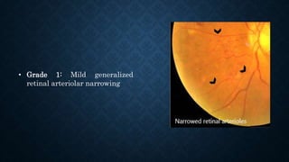
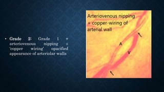
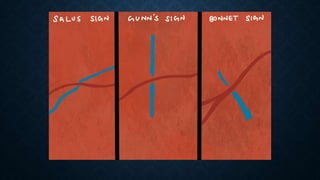
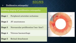
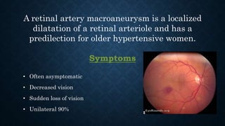
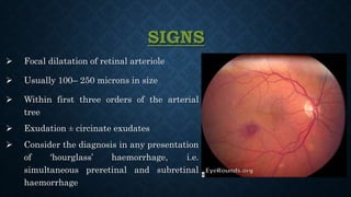
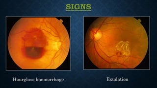
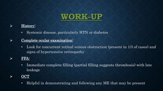
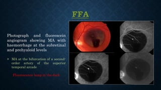
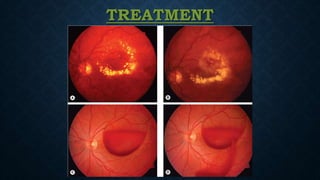
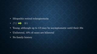
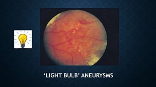
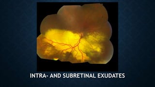
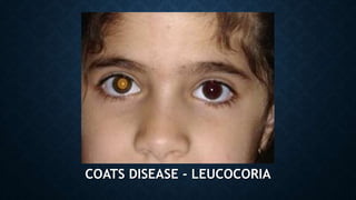
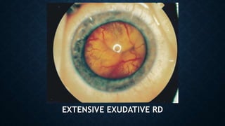
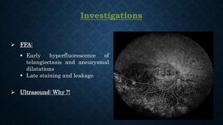
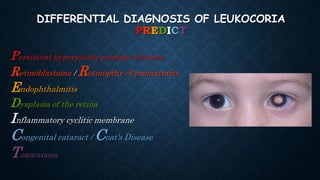
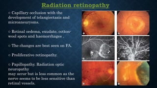
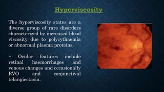
RETINAL VASCULAR DISEASES - II
By
Ahmed Alsherbeny
MD, EBO, CABOphth, MRCSEd Ophth, FRCS Ophth (Glasg), FICO, MSc
All the following may be associated findings after central retinal
vein occlusion, except:
A. Optic disc edema.
B. Optic disc venous–venous collateral (optociliary shunt) vessels.
C. Diffuse macular edema.
D. Neovascular glaucoma.
E. “Box-car” bloodstream in arterioles
Most common cause of neovascular glaucoma is :
A. Ischemic central retinal vein occlusion (CRVO).
B. Central retinal artery occlusion (CRAO).
C. Long-standing retinal detachment.
D. Uveitis.
E. Diabetes mellitus
Ophthalmic artery occlusion, rather than central retinal artery
occlusion is suggested by all the following findings, except:
A. Severe visual loss (bare to no light perception).
B. Marked choroidal perfusion defects on fluorescein angiography.
C. Intense ischemic retinal whitening that extends beyond the
macular area.
D. Few or no cherry-red spots.
E. Mildly decreased amplitude of the electroretinogram.
Hypertensive Retinopathy
Ocular associations and complications of hypertension:
RVO
RAO
RAM
AION
NP
Progression of DR
Posterior segment effects can be observed in the retina, choroid and
optic nerve
RETINOPATHY
• Grade 1: Mild generalized
retinal arteriolar narrowing
• Grade 2: Grade 1 +
arteriovenous nipping ±
‘copper wiring’ opacified
appearance of arteriolar walls
• Grade 3: Grade 2 + retinal
haemorrhages, exudates
‘macular star’ and cotton-wool
spots.
• Grade 4 ‘Malignant Hypertension’:
Grade 3 + optic disc swelling, which is a marker of ‘malignant’
hypertension Hypertensive emergency
CHOROIDOPATHY
• Siegrist streaks are flecks
arranged linearly along
choroidal vessels and are
indicative of fibrinoid necrosis
associated with malignant
hypertension
• Elschnig spots are focal choroidal
infarcts seen as small black spots
surrounded by yellow haloes
• Exudative retinal detachment,
sometimes bilateral, may occur in
acute severe hypertension such as
that associated with toxaemia of
pregnancy.
 When UNILATERAL, suspect carotid artery
obstruction on the side of the normal appearing
eye, sparing the retina from the effects of the
HTN
WORK-UP
 History:
• HTN, diabetes, or adnexal radiation?
 Complete ocular examination
 Check blood pressure
 Refer patient Medical internist
Emergency department
• Diastolic blood pressure of
≥110 mm hg
• Presence of chest pain
• Difficulty breathing
• Headache
• Change in mental status
• Blurred vision with optic disc
swelling
Sickle-cell Retinopathy
Sickling haemoglobinopathies are caused by one or more abnormal
haemoglobins which induce the red blood cell to adopt an anomalous
shape
1 SS (sickle-cell disease, sickle-cell anaemia) affects 0.4% of black Americans. The
disease is characterized by severe chronic haemolytic anaemia. Despite the severity of
systemic manifestations ocular complications are usually mild and asymptomatic.
2 AS (sickle-cell trait) is present in about 10% of black Americans. It is the mildest
form and usually requires severe hypoxia or other abnormal conditions to produce
sickling.
3 SC (sickle-cell C disease) is present in 0.2% of black Americans. It is
characterized by haemolytic anaemia and infarctive crises that are less severe than in
SS disease but may be associated with severe retinopathy.
4 SThal (sickle-cell thalassaemia) is characterized by mild anaemia but may be
associated with severe retinopathy.
SIGNS
 Anterior Segment:
• Conjunctiva: Dark red corkscrew- or comma-shaped vessels
• Iris: Patches of ischaemic atrophy
• Hyphaema may be spontaneous or follow minor trauma. Careful IOP
control (avoiding CAI) is critical
SIGNS
 Non- proliferative retinopathy:
• Venous changes. Tortuosity is very common and is thought to be due to peripheral arteriovenous shunting.
• Arteriolar changes. Occlusions can involve branch, central or macular vessels. ‘Silver wiring’ of arterioles in
the peripheral retina signifies previously occluded vessels. Corkscrewing of peripheral vessels may be seen.
• Optic disc ‘sign of sickling’. Dark red blots on the disc surface due to small vessel occlusion.
• ‘Salmon patches’. Orange–red mid-peripheral superficial intraretinal haemorrhages that may break through
to become preretinal or subretinal. The initiating event is thought to be a vascular occlusion.
• Black sunbursts. Patches of peripheral RPE hyperplasia and chorioretinal atrophy that evolve from some
salmon patches. The extent and morphology of pigmentation is variable, but an outer pale band is generally
present.
• Macular (retinal) depression sign. An oval depression in the temporal macular retina due to retinal thinning
following arteriolar occlusion, with irregularity of the light reflex.
• Peripheral areas of whitening or darkening.
• Angioid streaks occur in up to 6%.
SIGNS
 Proliferative retinopathy:
Goldberg staging of proliferative retinopathy
Stage 1 Peripheral arteriolar occlusions
Stage 2 AV anastomoses
Stage 3 Neovascular proliferation (‘sea- fans’)
Stage 4 Vitreous haemorrhage
Stage 5 Retinal detachment
Stage 1
Stage 2
Stage 3
Stage 4
Stage 5
TREATMENT
 Management of hyphema ‘High risk of optic nerve damage compared to
normal person’ IOP >24 mmHg, >24 hours AC wash
 There are no well-established indications or guidelines for retinopathy
treatment
 No treatment required for small peripheral lesions, as high probability of
spontaneous regression following autoinfarction
TREATMENT
 Scatter laser photocoagulation:
• Severe visual loss from the disease in the fellow eye
• Rapid growth of large elevated sea-fans with spontaneous haemorrhage
 Vitrectomy:
• Vitreous hemorrhage
• Retinal detatchment
Results are generally disappointing
Scleral buckling is not preferred to prevent anterior segment ischemia
TREATMENT
 Anti- VEGF therapy:
• May have a role, although evidence to date is limited
• Caution should be used in cases with significant traction
FOLLOW-UP
 No retinopathy: Annual dilated fundus examinations.
 Retinopathy present: Repeat dilated fundus examination every 3 to 6
months, depending on severity.
Retinal Artery Macroaneurysm
A retinal artery macroaneurysm is a localized
dilatation of a retinal arteriole and has a
predilection for older hypertensive women.
Symptoms
• Often asymptomatic
• Decreased vision
• Sudden loss of vision
• Unilateral 90%
 Focal dilatation of retinal arteriole
 Usually 100– 250 microns in size
 Within first three orders of the arterial
tree
 Exudation ± circinate exudates
 Consider the diagnosis in any presentation
of ‘hourglass’ haemorrhage, i.e.
simultaneous preretinal and subretinal
haemorrhage
SIGNS
SIGNS
Hourglass haemorrhage Exudation
WORK-UP
 History:
• Systemic disease, particularly HTN or diabetes
 Complete ocular examination:
• Look for concurrent retinal venous obstruction (present in 1/3 of cases) and
signs of hypertensive retinopathy
 FFA:
• Immediate complete filling (partial filling suggests thrombosis) with late
leakage
 OCT
• Helpful in demonstrating and following any ME that may be present
FFA
Photograph and fluorescein
angiogram showing MA with
haemorrhage at the subretinal
and prehyaloid levels
˃ MA at the bifurcation of a second-
order artery of the superior
temporal arcade
Fluorescence lamp in the dark
OCT
Hemorrhagic retinal arterial macroaneurysm
(RAM) visible on the inferior temporal branch,
surrounded by a dense subretinal hemorrhage,
and responsible for retrohyaloidal hematoma
and serohematic fluid
Horizontal OCT passing through the
macula. Retrohyaloidal
hyperreflectivity, dense in the lower
area, with visible serohematic fluid
CAUSES OF MULTILEVEL HAEMORRHAGES
(SUBRETINAL, RETINAL, SUBHYALOID, VITREOUS)
Retinal Artery Macroaneurysm
Hemorrhagic CNV
Trauma
Choroidal Melanoma
Valsalva retinopathy
Idiopathic polypoidal choroidal vasculopathy
Terson syndrome
TREATMENT
 Observation is indicated in eyes with good VA in which the macula is not threatened and in
those with mild retinal haemorrhage without significant oedema. In many cases
macroaneurysms will spontaneously involute
 Laser treatment may be considered if oedema or exudates threaten or involve the fovea,
particularly if there is documented visual deterioration.
 Intravitreal bevacizumab closed 95% of macroaneurysms in a case series, with resolution of
macular oedema.
 Nd:YAG laser hyaloidotomy may be considered for a persistent premacular haemorrhage in
order to disperse the blood into the vitreous cavity (Fig. 13.55C and D), from where it may be
absorbed more quickly.
TREATMENT
TREATMENT
 Intravitreal gas injection with face-down positioning may shift subretinal haemorrhage
away from the macula. Adjunctive intravitreal recombinant tissue plasminogen activator
(rtPA) may be used.
 Vitrectomy may be necessary for persistent vitreous haemorrhage.
FOLLOW-UP
 Based on the amount and location of exudate and hemorrhage
Coats Disease
• Idiopathic retinal telangiectasia
• ♂:♀ 3:1
• Young, although up to 1/3 may be asymptomatic until their 30s
• Unilateral, 10% of cases are bilateral
• No family history
Symptoms
• Asymptomatic
• V/A
• Strabismus
• Leucocoria
Signs
 Telangiectasia and fusiform focal aneurysmal arteriolar
dilatations:
• ‘Light bulb’ aneurysms
• Often in the inferior and temporal quadrant
 Intra- and subretinal exudates:
• Remote from the vascular abnormalities
• Progression to extensive exudative retinal detachment
‘LIGHT BULB’ ANEURYSMS
INTRA- AND SUBRETINAL EXUDATES
COATS DISEASE - LEUCOCORIA
EXTENSIVE EXUDATIVE RD
Investigations
 FFA:
 Early hyperfluorescence of
telangiectasis and aneurysmal
dilatations
 Late staining and leakage
 Ultrasound: Why ?!
DIFFERENTIAL DIAGNOSIS OF LEUKOCORIA
PREDICT
Persistent hyperplastic primary vitreous
Retinoblastoma / Retinopthy of prematurity
Endophthalmitis
Dysplasia of the retina
Inflammatory cyclitic membrane
Congenital cataract / Coat’s Disease
Toxocaiasis
TREATMENT
 Observation in patients with mild, non-vision threatening disease and in those with a
comfortable eye with total retinal detachment for whom there is no potential for
restoration of useful vision.
 Laser ablation of points of leakage should be considered if progressive exudation is
documented
 Anti-VEGF therapy. Limited studies of anti-VEGF therapy have been carried out, but
initial results are promising.
 Cryotherapy
 Vitrectomy may be considered in eyes with significant tractional preretinal fibrosis or
total exudative detachment.
 Enucleation may be required in painful eyes with neovascular glaucoma.
Other Retinopathies
Radiation retinopathy
○ Capillary occlusion with the
development of telangiectasis and
microaneurysms.
○ Retinal oedema, exudate, cotton-
wool spots and haemorrhages .
○ The changes are best seen on FA.
○ Proliferative retinopathy.
○ Papillopathy. Radiation optic
neuropathy
may occur but is less common as the
nerve seems to be less sensitive than
retinal vessels.
Purtscher retinopathy
Purtscher retinopathy is
caused by microvascular
damage with occlusion and
ischaemia associated with
severe trauma, especially to
the head and in chest
compressive injury
Valsalva retinopathy
The Valsalva manoeuvre consists of
forcible exhalation against a closed
glottis, thereby creating a sudden
increase in intrathoracic and intra-
abdominal pressure (e.g. weight-lifting,
blowing up balloons).
The associated sudden rise in venous
pressure may rupture perifoveal
capillaries leading to premacular
haemorrhage of varying severity
Retinopathy in Blood Disorders
Anaemia
• Retinopathy:
○ Retinal venous tortuosity is related to the severity of anaemia but may occur in
isolation.
○ Dot, blot and flame haemorrhages, cotton-wool spots and Roth spots are more
common with co-existing thrombocytopenia.
• Optic neuropathy may occur in pernicious anaemia.
Leukaemia
○ Retinal haemorrhages and cotton-wool spots are
common.
○ Roth spots are retinal haemorrhages with white
centres
○ Peripheral retinal neovascularization is an
occasional feature of chronic myeloid leukaemia
(A).
○ Retinal and choroidal infiltrates may occur, most
commonly posterior to the equator (B) and may
masquerade as posterior uveitis. In chronic
leukaemia they may give rise to a ‘leopard skin’
appearance (C).
○ Scattered haemorrhages may occur in patients
with thrombocytopenia (D).
○ Optic nerve infiltration may cause swelling and
visual loss. (DD of CNS Infiltration Vs Local)
Hyperviscosity
The hyperviscosity states are a
diverse group of rare disorders
characterized by increased blood
viscosity due to polycythaemia
or abnormal plasma proteins.
• Ocular features include
retinal haemorrhages and
venous changes and occasionally
RVO and conjunctival
telangiectasia.
Thank You

Retinal Vascular Diseases - II

  • 1.
    RETINAL VASCULAR DISEASES- II By Ahmed Alsherbeny MD, EBO, CABOphth, MRCSEd Ophth, FRCS Ophth (Glasg), FICO, MSc
  • 2.
    All the followingmay be associated findings after central retinal vein occlusion, except: A. Optic disc edema. B. Optic disc venous–venous collateral (optociliary shunt) vessels. C. Diffuse macular edema. D. Neovascular glaucoma. E. “Box-car” bloodstream in arterioles
  • 3.
    Most common causeof neovascular glaucoma is : A. Ischemic central retinal vein occlusion (CRVO). B. Central retinal artery occlusion (CRAO). C. Long-standing retinal detachment. D. Uveitis. E. Diabetes mellitus
  • 4.
    Ophthalmic artery occlusion,rather than central retinal artery occlusion is suggested by all the following findings, except: A. Severe visual loss (bare to no light perception). B. Marked choroidal perfusion defects on fluorescein angiography. C. Intense ischemic retinal whitening that extends beyond the macular area. D. Few or no cherry-red spots. E. Mildly decreased amplitude of the electroretinogram.
  • 5.
  • 6.
    Ocular associations andcomplications of hypertension: RVO RAO RAM AION NP Progression of DR Posterior segment effects can be observed in the retina, choroid and optic nerve
  • 7.
  • 8.
    • Grade 1:Mild generalized retinal arteriolar narrowing
  • 9.
    • Grade 2:Grade 1 + arteriovenous nipping ± ‘copper wiring’ opacified appearance of arteriolar walls
  • 11.
    • Grade 3:Grade 2 + retinal haemorrhages, exudates ‘macular star’ and cotton-wool spots.
  • 12.
    • Grade 4‘Malignant Hypertension’: Grade 3 + optic disc swelling, which is a marker of ‘malignant’ hypertension Hypertensive emergency
  • 13.
  • 14.
    • Siegrist streaksare flecks arranged linearly along choroidal vessels and are indicative of fibrinoid necrosis associated with malignant hypertension
  • 15.
    • Elschnig spotsare focal choroidal infarcts seen as small black spots surrounded by yellow haloes • Exudative retinal detachment, sometimes bilateral, may occur in acute severe hypertension such as that associated with toxaemia of pregnancy.
  • 16.
     When UNILATERAL,suspect carotid artery obstruction on the side of the normal appearing eye, sparing the retina from the effects of the HTN
  • 17.
    WORK-UP  History: • HTN,diabetes, or adnexal radiation?  Complete ocular examination  Check blood pressure  Refer patient Medical internist Emergency department • Diastolic blood pressure of ≥110 mm hg • Presence of chest pain • Difficulty breathing • Headache • Change in mental status • Blurred vision with optic disc swelling
  • 18.
  • 19.
    Sickling haemoglobinopathies arecaused by one or more abnormal haemoglobins which induce the red blood cell to adopt an anomalous shape 1 SS (sickle-cell disease, sickle-cell anaemia) affects 0.4% of black Americans. The disease is characterized by severe chronic haemolytic anaemia. Despite the severity of systemic manifestations ocular complications are usually mild and asymptomatic. 2 AS (sickle-cell trait) is present in about 10% of black Americans. It is the mildest form and usually requires severe hypoxia or other abnormal conditions to produce sickling. 3 SC (sickle-cell C disease) is present in 0.2% of black Americans. It is characterized by haemolytic anaemia and infarctive crises that are less severe than in SS disease but may be associated with severe retinopathy. 4 SThal (sickle-cell thalassaemia) is characterized by mild anaemia but may be associated with severe retinopathy.
  • 20.
    SIGNS  Anterior Segment: •Conjunctiva: Dark red corkscrew- or comma-shaped vessels • Iris: Patches of ischaemic atrophy • Hyphaema may be spontaneous or follow minor trauma. Careful IOP control (avoiding CAI) is critical
  • 21.
    SIGNS  Non- proliferativeretinopathy: • Venous changes. Tortuosity is very common and is thought to be due to peripheral arteriovenous shunting. • Arteriolar changes. Occlusions can involve branch, central or macular vessels. ‘Silver wiring’ of arterioles in the peripheral retina signifies previously occluded vessels. Corkscrewing of peripheral vessels may be seen. • Optic disc ‘sign of sickling’. Dark red blots on the disc surface due to small vessel occlusion. • ‘Salmon patches’. Orange–red mid-peripheral superficial intraretinal haemorrhages that may break through to become preretinal or subretinal. The initiating event is thought to be a vascular occlusion. • Black sunbursts. Patches of peripheral RPE hyperplasia and chorioretinal atrophy that evolve from some salmon patches. The extent and morphology of pigmentation is variable, but an outer pale band is generally present. • Macular (retinal) depression sign. An oval depression in the temporal macular retina due to retinal thinning following arteriolar occlusion, with irregularity of the light reflex. • Peripheral areas of whitening or darkening. • Angioid streaks occur in up to 6%.
  • 24.
    SIGNS  Proliferative retinopathy: Goldbergstaging of proliferative retinopathy Stage 1 Peripheral arteriolar occlusions Stage 2 AV anastomoses Stage 3 Neovascular proliferation (‘sea- fans’) Stage 4 Vitreous haemorrhage Stage 5 Retinal detachment
  • 25.
  • 26.
  • 27.
  • 28.
  • 29.
  • 30.
    TREATMENT  Management ofhyphema ‘High risk of optic nerve damage compared to normal person’ IOP >24 mmHg, >24 hours AC wash  There are no well-established indications or guidelines for retinopathy treatment  No treatment required for small peripheral lesions, as high probability of spontaneous regression following autoinfarction
  • 31.
    TREATMENT  Scatter laserphotocoagulation: • Severe visual loss from the disease in the fellow eye • Rapid growth of large elevated sea-fans with spontaneous haemorrhage  Vitrectomy: • Vitreous hemorrhage • Retinal detatchment Results are generally disappointing Scleral buckling is not preferred to prevent anterior segment ischemia
  • 32.
    TREATMENT  Anti- VEGFtherapy: • May have a role, although evidence to date is limited • Caution should be used in cases with significant traction
  • 33.
    FOLLOW-UP  No retinopathy:Annual dilated fundus examinations.  Retinopathy present: Repeat dilated fundus examination every 3 to 6 months, depending on severity.
  • 34.
  • 35.
    A retinal arterymacroaneurysm is a localized dilatation of a retinal arteriole and has a predilection for older hypertensive women. Symptoms • Often asymptomatic • Decreased vision • Sudden loss of vision • Unilateral 90%
  • 36.
     Focal dilatationof retinal arteriole  Usually 100– 250 microns in size  Within first three orders of the arterial tree  Exudation ± circinate exudates  Consider the diagnosis in any presentation of ‘hourglass’ haemorrhage, i.e. simultaneous preretinal and subretinal haemorrhage SIGNS
  • 37.
  • 38.
    WORK-UP  History: • Systemicdisease, particularly HTN or diabetes  Complete ocular examination: • Look for concurrent retinal venous obstruction (present in 1/3 of cases) and signs of hypertensive retinopathy  FFA: • Immediate complete filling (partial filling suggests thrombosis) with late leakage  OCT • Helpful in demonstrating and following any ME that may be present
  • 39.
    FFA Photograph and fluorescein angiogramshowing MA with haemorrhage at the subretinal and prehyaloid levels ˃ MA at the bifurcation of a second- order artery of the superior temporal arcade Fluorescence lamp in the dark
  • 40.
    OCT Hemorrhagic retinal arterialmacroaneurysm (RAM) visible on the inferior temporal branch, surrounded by a dense subretinal hemorrhage, and responsible for retrohyaloidal hematoma and serohematic fluid Horizontal OCT passing through the macula. Retrohyaloidal hyperreflectivity, dense in the lower area, with visible serohematic fluid
  • 41.
    CAUSES OF MULTILEVELHAEMORRHAGES (SUBRETINAL, RETINAL, SUBHYALOID, VITREOUS) Retinal Artery Macroaneurysm Hemorrhagic CNV Trauma Choroidal Melanoma Valsalva retinopathy Idiopathic polypoidal choroidal vasculopathy Terson syndrome
  • 42.
    TREATMENT  Observation isindicated in eyes with good VA in which the macula is not threatened and in those with mild retinal haemorrhage without significant oedema. In many cases macroaneurysms will spontaneously involute  Laser treatment may be considered if oedema or exudates threaten or involve the fovea, particularly if there is documented visual deterioration.  Intravitreal bevacizumab closed 95% of macroaneurysms in a case series, with resolution of macular oedema.  Nd:YAG laser hyaloidotomy may be considered for a persistent premacular haemorrhage in order to disperse the blood into the vitreous cavity (Fig. 13.55C and D), from where it may be absorbed more quickly.
  • 43.
  • 44.
    TREATMENT  Intravitreal gasinjection with face-down positioning may shift subretinal haemorrhage away from the macula. Adjunctive intravitreal recombinant tissue plasminogen activator (rtPA) may be used.  Vitrectomy may be necessary for persistent vitreous haemorrhage.
  • 45.
    FOLLOW-UP  Based onthe amount and location of exudate and hemorrhage
  • 46.
  • 47.
    • Idiopathic retinaltelangiectasia • ♂:♀ 3:1 • Young, although up to 1/3 may be asymptomatic until their 30s • Unilateral, 10% of cases are bilateral • No family history
  • 48.
    Symptoms • Asymptomatic • V/A •Strabismus • Leucocoria
  • 49.
    Signs  Telangiectasia andfusiform focal aneurysmal arteriolar dilatations: • ‘Light bulb’ aneurysms • Often in the inferior and temporal quadrant  Intra- and subretinal exudates: • Remote from the vascular abnormalities • Progression to extensive exudative retinal detachment
  • 50.
  • 51.
  • 52.
    COATS DISEASE -LEUCOCORIA
  • 53.
  • 54.
    Investigations  FFA:  Earlyhyperfluorescence of telangiectasis and aneurysmal dilatations  Late staining and leakage  Ultrasound: Why ?!
  • 55.
    DIFFERENTIAL DIAGNOSIS OFLEUKOCORIA PREDICT Persistent hyperplastic primary vitreous Retinoblastoma / Retinopthy of prematurity Endophthalmitis Dysplasia of the retina Inflammatory cyclitic membrane Congenital cataract / Coat’s Disease Toxocaiasis
  • 56.
    TREATMENT  Observation inpatients with mild, non-vision threatening disease and in those with a comfortable eye with total retinal detachment for whom there is no potential for restoration of useful vision.  Laser ablation of points of leakage should be considered if progressive exudation is documented  Anti-VEGF therapy. Limited studies of anti-VEGF therapy have been carried out, but initial results are promising.  Cryotherapy  Vitrectomy may be considered in eyes with significant tractional preretinal fibrosis or total exudative detachment.  Enucleation may be required in painful eyes with neovascular glaucoma.
  • 57.
  • 58.
    Radiation retinopathy ○ Capillaryocclusion with the development of telangiectasis and microaneurysms. ○ Retinal oedema, exudate, cotton- wool spots and haemorrhages . ○ The changes are best seen on FA. ○ Proliferative retinopathy. ○ Papillopathy. Radiation optic neuropathy may occur but is less common as the nerve seems to be less sensitive than retinal vessels.
  • 59.
    Purtscher retinopathy Purtscher retinopathyis caused by microvascular damage with occlusion and ischaemia associated with severe trauma, especially to the head and in chest compressive injury
  • 60.
    Valsalva retinopathy The Valsalvamanoeuvre consists of forcible exhalation against a closed glottis, thereby creating a sudden increase in intrathoracic and intra- abdominal pressure (e.g. weight-lifting, blowing up balloons). The associated sudden rise in venous pressure may rupture perifoveal capillaries leading to premacular haemorrhage of varying severity
  • 61.
  • 62.
    Anaemia • Retinopathy: ○ Retinalvenous tortuosity is related to the severity of anaemia but may occur in isolation. ○ Dot, blot and flame haemorrhages, cotton-wool spots and Roth spots are more common with co-existing thrombocytopenia. • Optic neuropathy may occur in pernicious anaemia.
  • 63.
    Leukaemia ○ Retinal haemorrhagesand cotton-wool spots are common. ○ Roth spots are retinal haemorrhages with white centres ○ Peripheral retinal neovascularization is an occasional feature of chronic myeloid leukaemia (A). ○ Retinal and choroidal infiltrates may occur, most commonly posterior to the equator (B) and may masquerade as posterior uveitis. In chronic leukaemia they may give rise to a ‘leopard skin’ appearance (C). ○ Scattered haemorrhages may occur in patients with thrombocytopenia (D). ○ Optic nerve infiltration may cause swelling and visual loss. (DD of CNS Infiltration Vs Local)
  • 64.
    Hyperviscosity The hyperviscosity statesare a diverse group of rare disorders characterized by increased blood viscosity due to polycythaemia or abnormal plasma proteins. • Ocular features include retinal haemorrhages and venous changes and occasionally RVO and conjunctival telangiectasia.
  • 65.