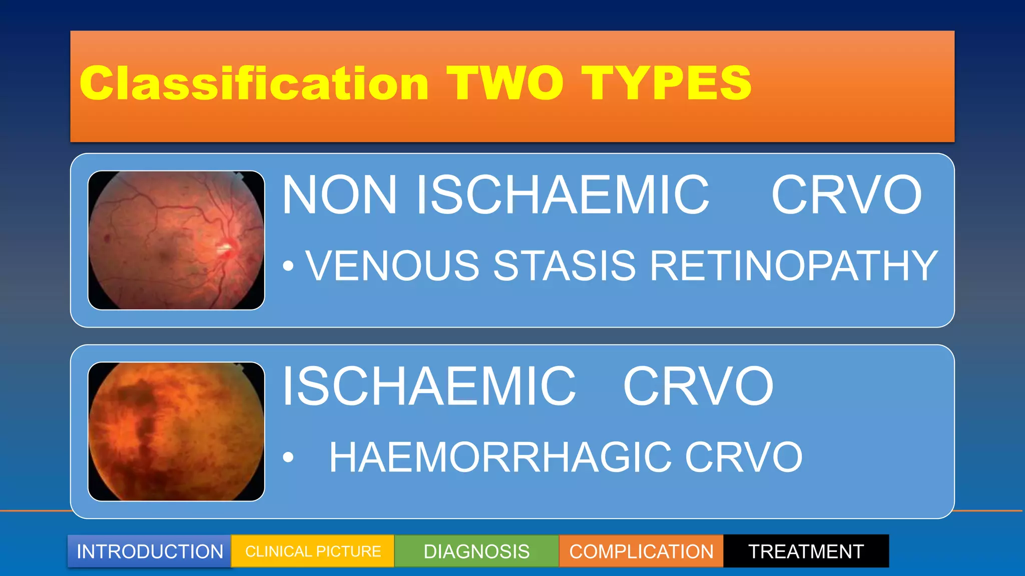
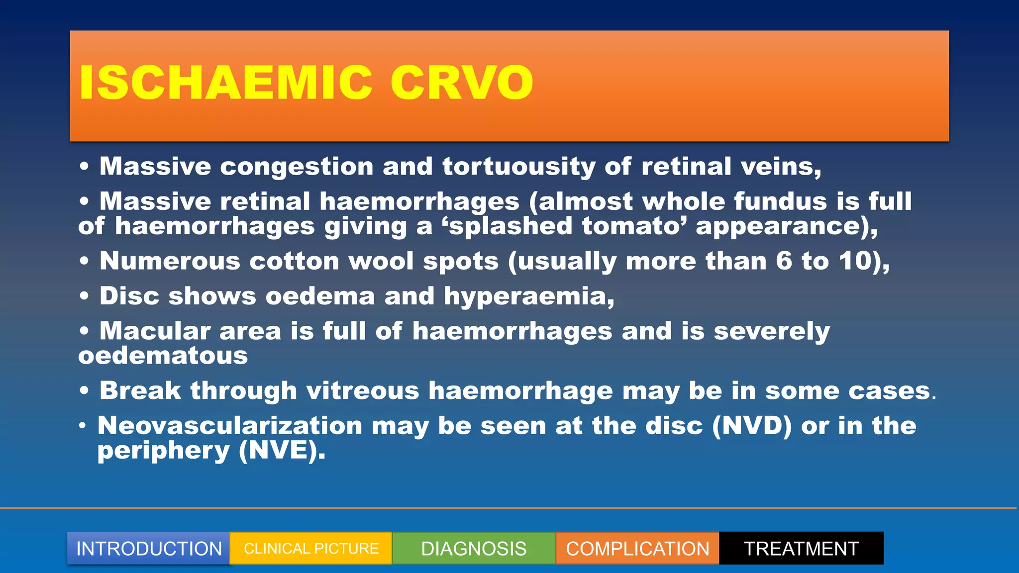
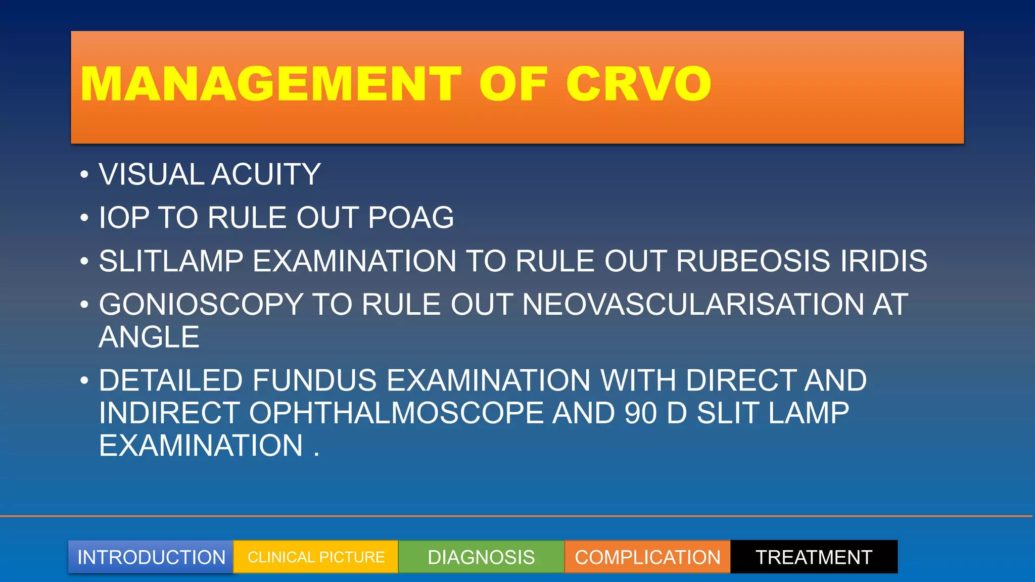
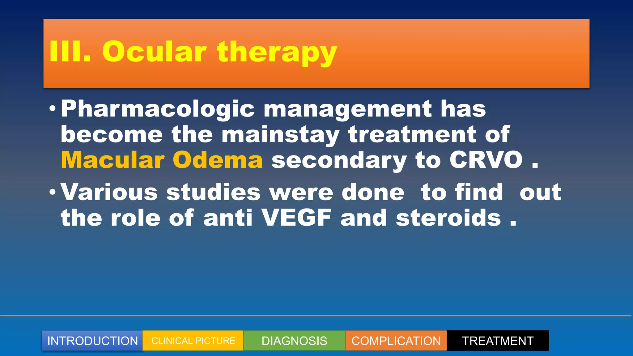
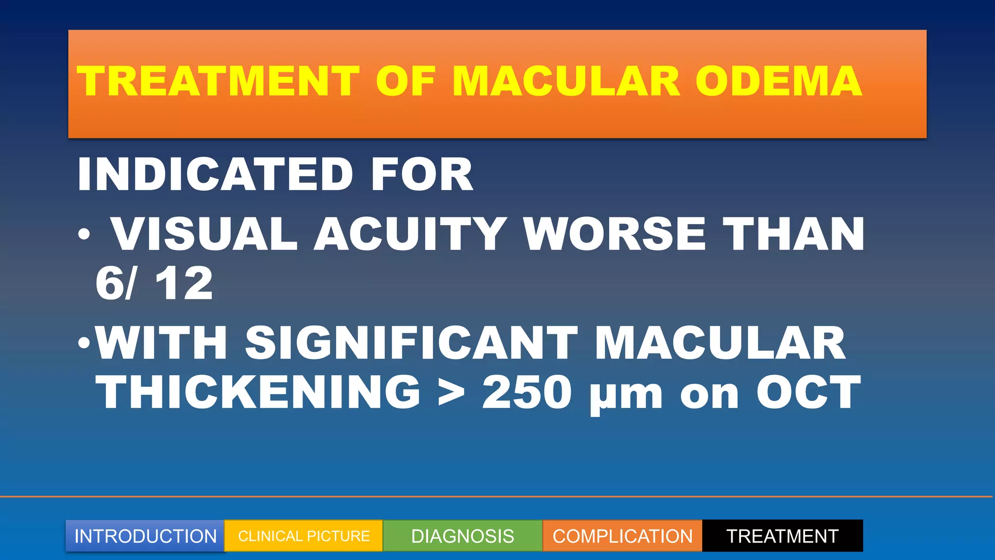
updating in diabetic macular edema including old and new approach era, including DRCR protocol
how to approach, how to treat, when to surgery
plus knownledge about anti-VEGF therapy up to date
Retinal vein occlusion (RVO) is an obstruction of the retinal venous system by thrombus formation and may involve the central, hemi-central or branch retinal vein.
The most common aetiological factor is compression by adjacent atherosclerotic retinal arteries.
Other possible causes are external compression or disease of the vein wall e.g. vasculitis.
Branched Retinal Vein Occlusion (BRVO) for undergraduate MBBS Students.
Covers the basics of Aetiology, pathophysiology, clinical features, types, associated conditions and management of BRVO.
Also encompasses salient points for PGMEE
Ischemic condition affecting the eye.
The ischemia can occur secondary to systemically problem [or] particulary the eye.
Many retinal vascular disorders {like CRAO,CRVO,Diabetic retinopathy,Hypertensive Retinopathy} shows ischemic signs.
Central Retinal Vein Occlusion (CRVO) for undergraduate MBBS Students.
Covers the basics of Aetiology, pathophysiology, clinical features, types, associated conditions and management of CRVO.
Also encompasses salient points for PGMEE
Central Retinal Artery Occlusion (CRAO) for undergraduate MBBS Students.
Covers the basics of Aetiology, pathophysiology, clinical features, types, associated conditions and management of CRAO.
Also encompasses salient points for PGMEE
Coats' disease, (also known as exudative retinitis or retinal telangiectasis, sometimes spelled Coates' disease), is a rare congenital, nonhereditary eye disorder, causing full or partial blindness, characterized by abnormal development of blood vessels behind the retina.
Duane syndrome, also called Duane retraction syndrome (DRS), is a congenital and non-progressive type of strabismus due to abnormal development of the 6th cranial nerve.
It is characterized by difficulty rotating one or both eyes outward (abduction) or inward (adduction).
On the other hands Duane Retraction Syndrome is a congenital strabismus syndrome occurring in isolated or syndromic forms. It presents with a variety of clinical features including diplopia, anisometropia, and amblyopia.
65 million (17%) of 387 million persons with diabetes mellitus (DM) globally reside in India.The prevalence of DR in urban areas is between 13–18% and in rural areas is 9–10%.In India, in 1970–1975 DR was the 20th cause of blindness, and today, it is the 6th cause of blindness.
TEST BANK for Operations Management, 14th Edition by William J. Stevenson, Ve...kevinkariuki227
TEST BANK for Operations Management, 14th Edition by William J. Stevenson, Verified Chapters 1 - 19, Complete Newest Version.pdf
- Video recording of this lecture in English language: https://youtu.be/lK81BzxMqdo
- Video recording of this lecture in Arabic language: https://youtu.be/Ve4P0COk9OI
- Link to download the book free: https://nephrotube.blogspot.com/p/nephrotube-nephrology-books.html
- Link to NephroTube website: www.NephroTube.com
- Link to NephroTube social media accounts: https://nephrotube.blogspot.com/p/join-nephrotube-on-social-media.html
Flu Vaccine Alert in Bangalore Karnatakaaddon Scans
As flu season approaches, health officials in Bangalore, Karnataka, are urging residents to get their flu vaccinations. The seasonal flu, while common, can lead to severe health complications, particularly for vulnerable populations such as young children, the elderly, and those with underlying health conditions.
Dr. Vidisha Kumari, a leading epidemiologist in Bangalore, emphasizes the importance of getting vaccinated. "The flu vaccine is our best defense against the influenza virus. It not only protects individuals but also helps prevent the spread of the virus in our communities," he says.
This year, the flu season is expected to coincide with a potential increase in other respiratory illnesses. The Karnataka Health Department has launched an awareness campaign highlighting the significance of flu vaccinations. They have set up multiple vaccination centers across Bangalore, making it convenient for residents to receive their shots.
To encourage widespread vaccination, the government is also collaborating with local schools, workplaces, and community centers to facilitate vaccination drives. Special attention is being given to ensuring that the vaccine is accessible to all, including marginalized communities who may have limited access to healthcare.
Residents are reminded that the flu vaccine is safe and effective. Common side effects are mild and may include soreness at the injection site, mild fever, or muscle aches. These side effects are generally short-lived and far less severe than the flu itself.
Healthcare providers are also stressing the importance of continuing COVID-19 precautions. Wearing masks, practicing good hand hygiene, and maintaining social distancing are still crucial, especially in crowded places.
Protect yourself and your loved ones by getting vaccinated. Together, we can help keep Bangalore healthy and safe this flu season. For more information on vaccination centers and schedules, residents can visit the Karnataka Health Department’s official website or follow their social media pages.
Stay informed, stay safe, and get your flu shot today!
Title: Sense of Taste
Presenter: Dr. Faiza, Assistant Professor of Physiology
Qualifications:
MBBS (Best Graduate, AIMC Lahore)
FCPS Physiology
ICMT, CHPE, DHPE (STMU)
MPH (GC University, Faisalabad)
MBA (Virtual University of Pakistan)
Learning Objectives:
Describe the structure and function of taste buds.
Describe the relationship between the taste threshold and taste index of common substances.
Explain the chemical basis and signal transduction of taste perception for each type of primary taste sensation.
Recognize different abnormalities of taste perception and their causes.
Key Topics:
Significance of Taste Sensation:
Differentiation between pleasant and harmful food
Influence on behavior
Selection of food based on metabolic needs
Receptors of Taste:
Taste buds on the tongue
Influence of sense of smell, texture of food, and pain stimulation (e.g., by pepper)
Primary and Secondary Taste Sensations:
Primary taste sensations: Sweet, Sour, Salty, Bitter, Umami
Chemical basis and signal transduction mechanisms for each taste
Taste Threshold and Index:
Taste threshold values for Sweet (sucrose), Salty (NaCl), Sour (HCl), and Bitter (Quinine)
Taste index relationship: Inversely proportional to taste threshold
Taste Blindness:
Inability to taste certain substances, particularly thiourea compounds
Example: Phenylthiocarbamide
Structure and Function of Taste Buds:
Composition: Epithelial cells, Sustentacular/Supporting cells, Taste cells, Basal cells
Features: Taste pores, Taste hairs/microvilli, and Taste nerve fibers
Location of Taste Buds:
Found in papillae of the tongue (Fungiform, Circumvallate, Foliate)
Also present on the palate, tonsillar pillars, epiglottis, and proximal esophagus
Mechanism of Taste Stimulation:
Interaction of taste substances with receptors on microvilli
Signal transduction pathways for Umami, Sweet, Bitter, Sour, and Salty tastes
Taste Sensitivity and Adaptation:
Decrease in sensitivity with age
Rapid adaptation of taste sensation
Role of Saliva in Taste:
Dissolution of tastants to reach receptors
Washing away the stimulus
Taste Preferences and Aversions:
Mechanisms behind taste preference and aversion
Influence of receptors and neural pathways
Impact of Sensory Nerve Damage:
Degeneration of taste buds if the sensory nerve fiber is cut
Abnormalities of Taste Detection:
Conditions: Ageusia, Hypogeusia, Dysgeusia (parageusia)
Causes: Nerve damage, neurological disorders, infections, poor oral hygiene, adverse drug effects, deficiencies, aging, tobacco use, altered neurotransmitter levels
Neurotransmitters and Taste Threshold:
Effects of serotonin (5-HT) and norepinephrine (NE) on taste sensitivity
Supertasters:
25% of the population with heightened sensitivity to taste, especially bitterness
Increased number of fungiform papillae
Ethanol (CH3CH2OH), or beverage alcohol, is a two-carbon alcohol
that is rapidly distributed in the body and brain. Ethanol alters many
neurochemical systems and has rewarding and addictive properties. It
is the oldest recreational drug and likely contributes to more morbidity,
mortality, and public health costs than all illicit drugs combined. The
5th edition of the Diagnostic and Statistical Manual of Mental Disorders
(DSM-5) integrates alcohol abuse and alcohol dependence into a single
disorder called alcohol use disorder (AUD), with mild, moderate,
and severe subclassifications (American Psychiatric Association, 2013).
In the DSM-5, all types of substance abuse and dependence have been
combined into a single substance use disorder (SUD) on a continuum
from mild to severe. A diagnosis of AUD requires that at least two of
the 11 DSM-5 behaviors be present within a 12-month period (mild
AUD: 2–3 criteria; moderate AUD: 4–5 criteria; severe AUD: 6–11 criteria).
The four main behavioral effects of AUD are impaired control over
drinking, negative social consequences, risky use, and altered physiological
effects (tolerance, withdrawal). This chapter presents an overview
of the prevalence and harmful consequences of AUD in the U.S.,
the systemic nature of the disease, neurocircuitry and stages of AUD,
comorbidities, fetal alcohol spectrum disorders, genetic risk factors, and
pharmacotherapies for AUD.
micro teaching on communication m.sc nursing.pdfAnurag Sharma
Microteaching is a unique model of practice teaching. It is a viable instrument for the. desired change in the teaching behavior or the behavior potential which, in specified types of real. classroom situations, tends to facilitate the achievement of specified types of objectives.
The prostate is an exocrine gland of the male mammalian reproductive system
It is a walnut-sized gland that forms part of the male reproductive system and is located in front of the rectum and just below the urinary bladder
Function is to store and secrete a clear, slightly alkaline fluid that constitutes 10-30% of the volume of the seminal fluid that along with the spermatozoa, constitutes semen
A healthy human prostate measures (4cm-vertical, by 3cm-horizontal, 2cm ant-post ).
It surrounds the urethra just below the urinary bladder. It has anterior, median, posterior and two lateral lobes
It’s work is regulated by androgens which are responsible for male sex characteristics
Generalised disease of the prostate due to hormonal derangement which leads to non malignant enlargement of the gland (increase in the number of epithelial cells and stromal tissue)to cause compression of the urethra leading to symptoms (LUTS
These lecture slides, by Dr Sidra Arshad, offer a quick overview of physiological basis of a normal electrocardiogram.
Learning objectives:
1. Define an electrocardiogram (ECG) and electrocardiography
2. Describe how dipoles generated by the heart produce the waveforms of the ECG
3. Describe the components of a normal electrocardiogram of a typical bipolar leads (limb II)
4. Differentiate between intervals and segments
5. Enlist some common indications for obtaining an ECG
Study Resources:
1. Chapter 11, Guyton and Hall Textbook of Medical Physiology, 14th edition
2. Chapter 9, Human Physiology - From Cells to Systems, Lauralee Sherwood, 9th edition
3. Chapter 29, Ganong’s Review of Medical Physiology, 26th edition
4. Electrocardiogram, StatPearls - https://www.ncbi.nlm.nih.gov/books/NBK549803/
5. ECG in Medical Practice by ABM Abdullah, 4th edition
6. ECG Basics, http://www.nataliescasebook.com/tag/e-c-g-basics
Pulmonary Thromboembolism - etilogy, types, medical- Surgical and nursing man...VarunMahajani
Disruption of blood supply to lung alveoli due to blockage of one or more pulmonary blood vessels is called as Pulmonary thromboembolism. In this presentation we will discuss its causes, types and its management in depth.
MANAGEMENT OF ATRIOVENTRICULAR CONDUCTION BLOCK.pdfJim Jacob Roy
Cardiac conduction defects can occur due to various causes.
Atrioventricular conduction blocks ( AV blocks ) are classified into 3 types.
This document describes the acute management of AV block.
2. INTRODUCTION CLINICAL PICTURE DIAGNOSIS COMPLICATION TREATMENT
CRVO
•CRVO is a common retinal vascular
disease typically affecting the
patients over the age of 60 years .
•More common than arterial
occlussion
3. INTRODUCTION CLINICAL PICTURE DIAGNOSIS COMPLICATION TREATMENT
Pathophysiology
• Occlusion of central retinal vein by
arteriosclerotic central retinal artery
when it crosses the vein at or behind
the lamina cribrosa, where the two
share a common adventitia .
• Occlusion by primary venous wall
degeneration or inflammation .
4. INTRODUCTION CLINICAL PICTURE DIAGNOSIS COMPLICATION TREATMENT
HAEMODYNAMIC DISTURBANCES
Retinal hypoxia
Damage of capillary
endothelial cells
Extravasation of blood
constituents
Liberation of
mediators such
as VEGF
venous occlusion
elevation of
venous and
capillary pressure
stagnation of
blood flow
5. INTRODUCTION CLINICAL PICTURE DIAGNOSIS COMPLICATION TREATMENT
RISK FACTORS
• 50 % ABOVE THE AGE OF 60 YRS
• HYPERTENSION +NT IN 2/3 OF RVO PTS ABOVE 50 YRS
AGE
• HYPERLIPIDAEMIA 1/3 OR GREATER IRRESPECTIVE OF
AGE
• DIABETES 15% ABOVE 50 YRS AGE
• GLAUCOMA AND OCULAR HYPERTENSION
• ORAL CONTRACEPTIVES YOUNGER FEMALES
• MYELOPROLIFERATIVE DISORDERS , SARCOIDOSIS , SLE
6. INTRODUCTION CLINICAL PICTURE DIAGNOSIS COMPLICATION TREATMENT
SYSTEMIC ASSESSMENT
EVERY PT OF CRVO SHOULD UNDERGO
• BLOOD PRESSURE
• ESR / PLASMA VISCOSITY
• TOTAL BLOOD COUNT
• FBS / RBS
• RANDOM TOTAL AND HIGH DENSITY LIPOPROTEINS ( HDL )
• PLASMA PROTEIN ELECTROPHORESIS
• CR PROTEIN
• THYROID FUNCTION TESTS
8. INTRODUCTION CLINICAL PICTURE DIAGNOSIS COMPLICATION TREATMENT
NON ISCHAEMIC CRVO
• MORE COMMON
• AFFECTING 75 TO 80 %
• MILD TO MODERATE VISUAL LOSS
• MILD OR NO RAPD (RELATIVE AFFERENT PUPILLARY DEFECT )
• MILD TORTUOSITY AND DILATATION OF ALL THE BRANCHES
OF CRV .
• DOT BLOT AND FLAME HAEMORRHAGES
• COTTON WOOL SPOTS
• MILD OR NO MACULAR ODEMA
10. INTRODUCTION CLINICAL PICTURE DIAGNOSIS COMPLICATION TREATMENT
NON ISCHAEMIC CRVO
About 15% cases of non-Ischaemic
CRVO are converted to
•Ischaemic CRVO in 4 months
•and about 30% in 3 years.
11. INTRODUCTION CLINICAL PICTURE DIAGNOSIS COMPLICATION TREATMENT
ISCHAEMIC CRVO
•Ischaemic CRVO refers to
acute ( sudden ) complete occlusion
of Central retinal vein .
•SUDDEN MONOOCULAR Painless
loss of vision VA REDUCED TO CF
OR HM .
•WITH RAPD .
12. INTRODUCTION CLINICAL PICTURE DIAGNOSIS COMPLICATION TREATMENT
ISCHAEMIC CRVO
• Ischaemic CRVO
refers to acute (
sudden ) complete
occlusion of Central
retinal vein
characterized by
marked sudden visual
loss and RAPD .
14. INTRODUCTION CLINICAL PICTURE DIAGNOSIS COMPLICATION TREATMENT
ISCHAEMIC CRVO
• Massive congestion and tortuousity of retinal veins,
• Massive retinal haemorrhages (almost whole fundus is full
of haemorrhages giving a ‘splashed tomato’ appearance),
• Numerous cotton wool spots (usually more than 6 to 10),
• Disc shows oedema and hyperaemia,
• Macular area is full of haemorrhages and is severely
oedematous
• Break through vitreous haemorrhage may be in some cases.
• Neovascularization may be seen at the disc (NVD) or in the
periphery (NVE).
16. INTRODUCTION CLINICAL PICTURE DIAGNOSIS COMPLICATION TREATMENT
ISCHAEMIC CRVO
Pathognomonic features for ischaemic CRVO
differentiating it from non-ischaemic CRVO are
• Presence of relative afferent pupillary defect
(RAPD),
• Visual field defects, and
• Reduced amplitude of b-wave of electroretinogram.
Complications.
RUBEOSIS IRIDIS AND NEOVASCULAR GLAUCOMA
OCCURS IN 25 % OF CASES .
17. INTRODUCTION CLINICAL PICTURE DIAGNOSIS COMPLICATION TREATMENT
MANAGEMENT OF CRVO
• VISUAL ACUITY
• IOP TO RULE OUT POAG
• SLITLAMP EXAMINATION TO RULE OUT RUBEOSIS IRIDIS
• GONIOSCOPY TO RULE OUT NEOVASCULARISATION AT
ANGLE
• DETAILED FUNDUS EXAMINATION WITH DIRECT AND
INDIRECT OPHTHALMOSCOPE AND 90 D SLIT LAMP
EXAMINATION .
18. INTRODUCTION CLINICAL PICTURE DIAGNOSIS COMPLICATION TREATMENT
MANAGEMENT OF CRVO
• ERG Reduced amplitude of b-wave of
electroretinogram IN PHOTOPIC SCOPTIC
CONDITIONS & prolonged a & b wave
implicit time
• On FA AREA OF CAPILLARY NON PERFUSION
GREATER THAN 10 DD AREA IN ISCHAEMIC
CRVO
• MACULAR CHANGES PETALOID LEAKAGE &
INCREASED FAZ IN MACULAR ISCHAEMIA .
19. INTRODUCTION CLINICAL PICTURE DIAGNOSIS COMPLICATION TREATMENT
MANAGEMENT OF CRVO
• OCT shows
1. intra / subretinal fluid in presence of ME .
2. Hyperreflective areas at locations with intra retinal haemorhages or
exudates
3. ERM and retinal atrohty in longstanding cases .
• OCTA shows
1. enlarged FAZ
2. Increased parafoveal capillary non perfusion
3. Decreased parafoveal vascular density
21. INTRODUCTION CLINICAL PICTURE DIAGNOSIS COMPLICATION TREATMENT
I. Treatment of systemic and
ocular associations
• Treatment of systemic and ocular
associations such as
• hypertension,
• diabetes,
• Hyper lipidaemias,
• POAG, and other conditions is
important in all cases.
22. INTRODUCTION CLINICAL PICTURE DIAGNOSIS COMPLICATION TREATMENT
II. Observation and monitoring
• Observation and monitoring is all that is
required in patients with mild to
moderate visual loss (VA better than
6/18), as the condition (especially non
ischaemic CRVO), in more than 50%
cases of CRVO resolves with almost
normal vision.
23. INTRODUCTION CLINICAL PICTURE DIAGNOSIS COMPLICATION TREATMENT
III. Ocular therapy
• Pharmacologic management has
become the mainstay treatment of
Macular Odema secondary to CRVO .
• Various studies were done to find out
the role of anti VEGF and steroids .
24. INTRODUCTION CLINICAL PICTURE DIAGNOSIS COMPLICATION TREATMENT
TREATMENT OF MACULAR ODEMA
INDICATED FOR
• VISUAL ACUITY WORSE THAN
6/ 12
•WITH SIGNIFICANT MACULAR
THICKENING > 250 µm on OCT
25. INTRODUCTION CLINICAL PICTURE DIAGNOSIS COMPLICATION TREATMENT
INTRAVITREAL ANTI VEGF
agents
• Ranibizumab .5 mg given monthly for 6
months and subsequently less intensively
• There will be 2 3 lines gain in visual acuity
• In non responsive cases aflibercept resulted
in better anatomical and visual improvement
and prolonged the relapse free interval
between injections .
27. INTRODUCTION CLINICAL PICTURE DIAGNOSIS COMPLICATION TREATMENT
INTRAVITREAL ANTI VEGF
agents
• Bevacizumab has been tested against
aflibercept in the score 2 study and
afibercept showed more favorable
outcome on oct with resolution of
macular odema in 54 % versus 29 %
with bevacizumab .
28. INTRODUCTION CLINICAL PICTURE DIAGNOSIS COMPLICATION TREATMENT
Intravitreal triamcinolone
• The score study showed an
improvement of 3 or more lines of vision
at one year in over 25 % of patients
treated with a dosage of two injections
of 1 mg triamcinolone preservative free
preparation .
• There were side effects of raised iop
and cataract formation .
29. INTRODUCTION CLINICAL PICTURE DIAGNOSIS COMPLICATION TREATMENT
Intravitreal dexamethasone
implant
• The Geneva trial of sustained release biodegradable
dexamethasone intravitreal implant ( ozudrex )
shows substantial anatomical and visual
improvement over first two months following a
single implantation which declined to baseline by 6
months .
• The treatment could be repeated after 4 to 6 months
• As compared to triamcinolone there are more
cases of increase in IOP and cataract formation .
30. INTRODUCTION CLINICAL PICTURE DIAGNOSIS COMPLICATION TREATMENT
Laser Photocoagulation
• Although macular odema is anatomically improved
but not beneficial for visual outcomes except in
younger patients .
• Treatment of neovascularization should be done
promptly .
• PRP should be performed without delay in eyes
with NVI or angle neovascularization .
• Placing 1500 – 2000 burns of .1 sec duration
spaced one burn apart with sufficient energy to
produce a pale to moderate reaction , avoiding
areas of haemorrhage .
31. INTRODUCTION CLINICAL PICTURE DIAGNOSIS COMPLICATION TREATMENT
2. Laser therapy
• Grid laser is NOT recommended for
macular oedema in CRVO
• Panretinal photocoagulation (PRP) is
generally not recommended as
prophylaxis even in cases with marked
ischaemia (except in patients not likely
to comply with regular follow-up).
32. INTRODUCTION CLINICAL PICTURE DIAGNOSIS COMPLICATION TREATMENT
2. Laser therapy
• PRP should be performed without delay
in CRVO when neovascularization
develops any where, i.e., in angle (NVA),
iris (NVI), retina (NVE & NVD).
• PRP involves application of 1500–3000
burns (0.5–1.0 second), spaced one
burn width apart using frequency
doubling YAG laser or argon green laser.
33. INTRODUCTION CLINICAL PICTURE DIAGNOSIS COMPLICATION TREATMENT
III. Ocular therapy
• So Intravitreal anti-VEGFs, e.g., 1.25 mg
Bevacizumab (Avastin), or 0.3 mg Ranibizumab
(Lucentis) are the treatment of choice for the
associated CME.
• Intravitreal triamcinolone (1 mg) or
dexamethasone implant (Ozurdex) may
be given for the CME as second line drug .
• Usually Repeated injections of anti-VEGFs or
triamcinolone may be required.
34. INTRODUCTION CLINICAL PICTURE DIAGNOSIS COMPLICATION TREATMENT
CRVO
CRVO WITH MACULAR ODEMA
INVOLVING CENTRE OF RETINA
NO
OBSERVATION
YES
INTRAVITREAL anti VEGF.
STEROIDS MAY BE
CONSIDERED
FA
OBSERVATION FOR NVE & NVD & NVI
ESP IF CNP AREA MORE THAN 10 DD AREANO NVE
NVD, NVI
NVE,NVD OR NVI PRESENT
VITREOUS HGE & NVG
PRP
NO GRID LASER
35. INTRODUCTION CLINICAL PICTURE DIAGNOSIS COMPLICATION TREATMENT
3. Surgical therapy
• Pars plana vitrectomy may be required for the
treatment of following complications associated
with venous occlusions:
• Persistent vitreous haemorrhage,
• Tractional retinal detachment,
• Intractable neovascular glaucoma (NVG).
PPV may be combined with endolaser PRP when
required.
37. INTRODUCTION CLINICAL PICTURE DIAGNOSIS COMPLICATION TREATMENT
IST case
• This patient was aged 60 yr
old male presented with mild
decrease in vision .On exam
visual acuity in the affected rt
eye was 6 / 12 .
• Fundus photograph show
scattered intraretinal
hemorrhages, mild optic
nerve head edema and
hyperemia, and dilated and
tortuous veins.
38. INTRODUCTION CLINICAL PICTURE DIAGNOSIS COMPLICATION TREATMENT
Clinical cases
• Fluorescein angiogram
showing scattered intraretinal
hemorrhages, mild optic
nerve head hyperemia, and
dilated and tortuous veins.
• No leak in the macular area .
• Minimal ischaemic areas .
• diagnosis of Non ischaemic
CRVO was made .
39. INTRODUCTION CLINICAL PICTURE DIAGNOSIS COMPLICATION TREATMENT
Clinical cases
• Optical coherence
tomography showing no
significant macular edema.
• Patient was examined for any
hypertension and diabetes
mellitus . These were not
found .
• Observation was done and
patient improved without any
intervention .
40. INTRODUCTION CLINICAL PICTURE DIAGNOSIS COMPLICATION TREATMENT
2ND case
• This patient had sudden
decrease in vision of left eye
on exam visual acuity 5 / 60 .
• Fundus photograph show
extensive intraretinal
hemorrhages. Optic nerve
head margins blurred .
Marked macular odema
present .
41. INTRODUCTION CLINICAL PICTURE DIAGNOSIS COMPLICATION TREATMENT
2ND case
• On fluorescein angiogram ,
the vasculature is barely
visible and extensive
intraretinal hemorrhages are
present .
• Marked ischaemia of retina is
also present .
42. INTRODUCTION CLINICAL PICTURE DIAGNOSIS COMPLICATION TREATMENT
2ND case
• OCT show cystoid macular
edema with subretinal fluid.
• This patient responded to
intravitreal AVASTIN . After
repeated injections ( 5
injections ) this patient
improved and macular odema
subsided .
43. INTRODUCTION CLINICAL PICTURE DIAGNOSIS COMPLICATION TREATMENT
CONCLUSION
• intravitreal corticosteroids and anti-VEGF agents have
demonstrated impressive improvements in macular
edema, visual acuity .
• The use of ranibizumab (Lucentis) and dexamethasone
implant (Ozurdex) have been FDA-approved for the
treatment of CRVO.
• Intravitreal pharmacotherapy has now replaced
observation as the standard of care for the management
of macular edema associated with CRVO.
44. INTRODUCTION CLINICAL PICTURE DIAGNOSIS COMPLICATION TREATMENT
TAKE HOME MESSAGE
NON ISCHAEMIC CRVO NO MACULAR ODEMA
• CONTRO BLOOD PRESSURE, BLOOD SUGAR AND BLOOD
LIPIDS , NO INTERVENTION JUST OBSERVATION
MACULAR ODEMA INVOLVING CENTRE
• SYSTEMIC CONTROL
• INTRAVITREAL ANTI VEGF
ISCHAEMIC CRVO WITH DEVELOPMENT OF
NEOVASCULARISATION NVD, NVE,NVI DO PRP