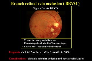
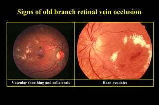
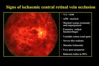
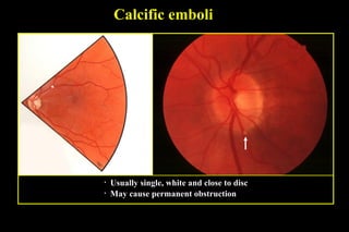
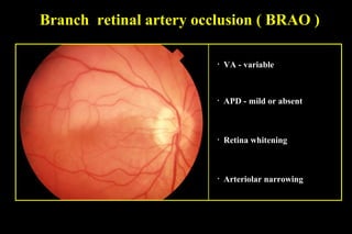
RETINAL VASCULAR OCCLUSIONS
Retinal vein occlusion
• Branch
• Central
• Hemi-retinal
Retinal artery occlusion
• Branch
• Cilio-retinal
• Hemi-retinal
• Central
Retinal vein occlusion - predisposing factors
1. Systemic
• Raised intraocular pressure
2. Ocular
• Periphlebitis
• Increasing age
• Hypertension
• Diabetes
• Abnormalities of coagulation
Patho-physiology of venous occlusion
Venous Occlusion
Stagnation
Increased extravascular
pressure
Hypoxia
Oedema and
haemorrhage
Branch retinal vein occlusion ( BRVO )
• Venous tortuosity and dilatation
• Flame-shaped and ‘dot-blot’ haemorrhages
• Cotton-wool spots and retinal oedema
Prognosis - VA 6/12 or better after 6 months in 50%
Complications - chronic macular oedema and neovascularization
Signs of acute BRVO
FA of branch retinal vein occlusion
Early - blocked background fluorescence due to
haemorrhage
Late - hyperfluorescence due
to diffuse oedema
Signs of old branch retinal vein occlusion
Vascular sheathing and collaterals Hard exudates
Management of chronic macular oedema
• Most common cause of persistent poor VA
• Wait 6-12 weeks and perform FA
Macular non-perfusion - no treatment Good macular perfusion and VA 6/18
or worse after 3 months - consider
laser photocoagulation
Management of neovascularization
• Perform laser photocoagulation to
involved segment• Most frequently after 6-12 months
• Occurs in about 30-50% of eyes
Central retinal vein occlusion ( CRVO )
• Chronic macular oedema
• Variable cotton-wool spots
• Mild to moderate disc oedema
• May subsequently convert
to ischaemic
• Guarded prognosis
• VA > CF
• APD - mild
• Mild venous tortuosity and
dilatation
• Mild to moderate retinal
haemorrhages
Signs of non-ischaemic CRVO
FA of non-ischaemic central retinal
vein occlusion
Good retinal capillary perfusion
Signs of ischaemic central retinal vein occlusion
• Variable cotton wool spots
• Severe disc oedema
• Very poor prognosis
• Macular ischaemia
• Rubeosis irides in 50%
• VA < 6/60
• APD - marked
• Marked venous tortuosity
and engorgement
• Extensive retinal
haemorrhages
FA of ischaemic central retinal vein occlusion
Extensive capillary non-perfusion
Management of ischaemic central
retinal vein occlusion
• Check every month for 6 months
• Look for rubeosis and angle new vessels
• Treat neovascularization by panretinal
photocoagulation
Hemi-retinal Vein Occlusion
• VA< reduction is variable
• Signs of BRVO
• Superior or inferior hemisphere
involve
FA Hemi-retinal Vein Occlusion
Management of CRVO
• Radial Optic Neurotomy
• A-V sheathotomy
• Chorio-retinal anastomosis
• Laser
• Surgical
• IVTA
• Anti-VEGF
RETINAL ARTERY
OCCLUSION
Causes of retinal artery occlusion
Embolism
Vaso-obliteration
Cholesterol emboli (Hollenhorst plaques)
• Multiple, bright, refractile crystals
• Often located at arteriolar bifurcations
• Frequently asymptomatic
Fibrinoplatelet emboli
• Multiple, dull grey particles
• Occasionally fill entire lumen
• May cause amaurosis fugax and occasionally permanent obstruction
Calcific emboli
• Usually single, white and close to disc
• May cause permanent obstruction
Branch retinal artery occlusion ( BRAO )
• VA - variable
• APD - mild or absent
• Retina whitening
• Arteriolar narrowing
FA of branch retinal artery occlusion
Early masking Extreme delay of arterial
phase
Late staining of arterial walls
Cilioretinal artery occlusion
• Present in about 30% of individuals
• In young individuals with
a systemic vasculitis
• Guarded prognosis
Combined with CRVO
• Usually good prognosis
• Elderly patients with
giant cell arteritis
• Very poor prognosis
Isolated
Combined with
anterior ischaemic
optic neuropathy
• Cilioretinal artery derived from posterior ciliary circulation
Central retinal artery occlusion ( CRAO )
• VA < 6/60
• ‘Cherry-red spot’ at macula
• Arteriolar and venular narrowing
• Very poor prognosis
• Sludging and segmentation of
blood column (cattle-trucking)
• APD - marked
• Retinal whitening
FA of central retinal artery occlusion
Early filling of cilioretinal
artery
Non-filling of other vessels Late staining of vessel walls
Treatment of central retinal artery
occlusion
• Ocular massage
• Sub-lingual Iso-sorbide di-nitrate
• Lowering of IOP
• AC paracentesis
• IV Streptokinase
Retinal Vascular occlusion

Retinal Vascular occlusion

  • 1.
    RETINAL VASCULAR OCCLUSIONS Retinalvein occlusion • Branch • Central • Hemi-retinal Retinal artery occlusion • Branch • Cilio-retinal • Hemi-retinal • Central
  • 2.
    Retinal vein occlusion- predisposing factors 1. Systemic • Raised intraocular pressure 2. Ocular • Periphlebitis • Increasing age • Hypertension • Diabetes • Abnormalities of coagulation
  • 3.
    Patho-physiology of venousocclusion Venous Occlusion Stagnation Increased extravascular pressure Hypoxia Oedema and haemorrhage
  • 4.
    Branch retinal veinocclusion ( BRVO ) • Venous tortuosity and dilatation • Flame-shaped and ‘dot-blot’ haemorrhages • Cotton-wool spots and retinal oedema Prognosis - VA 6/12 or better after 6 months in 50% Complications - chronic macular oedema and neovascularization Signs of acute BRVO
  • 5.
    FA of branchretinal vein occlusion Early - blocked background fluorescence due to haemorrhage Late - hyperfluorescence due to diffuse oedema
  • 6.
    Signs of oldbranch retinal vein occlusion Vascular sheathing and collaterals Hard exudates
  • 7.
    Management of chronicmacular oedema • Most common cause of persistent poor VA • Wait 6-12 weeks and perform FA Macular non-perfusion - no treatment Good macular perfusion and VA 6/18 or worse after 3 months - consider laser photocoagulation
  • 8.
    Management of neovascularization •Perform laser photocoagulation to involved segment• Most frequently after 6-12 months • Occurs in about 30-50% of eyes
  • 9.
    Central retinal veinocclusion ( CRVO ) • Chronic macular oedema • Variable cotton-wool spots • Mild to moderate disc oedema • May subsequently convert to ischaemic • Guarded prognosis • VA > CF • APD - mild • Mild venous tortuosity and dilatation • Mild to moderate retinal haemorrhages Signs of non-ischaemic CRVO
  • 10.
    FA of non-ischaemiccentral retinal vein occlusion Good retinal capillary perfusion
  • 11.
    Signs of ischaemiccentral retinal vein occlusion • Variable cotton wool spots • Severe disc oedema • Very poor prognosis • Macular ischaemia • Rubeosis irides in 50% • VA < 6/60 • APD - marked • Marked venous tortuosity and engorgement • Extensive retinal haemorrhages
  • 12.
    FA of ischaemiccentral retinal vein occlusion Extensive capillary non-perfusion
  • 13.
    Management of ischaemiccentral retinal vein occlusion • Check every month for 6 months • Look for rubeosis and angle new vessels • Treat neovascularization by panretinal photocoagulation
  • 14.
    Hemi-retinal Vein Occlusion •VA< reduction is variable • Signs of BRVO • Superior or inferior hemisphere involve
  • 15.
  • 16.
    Management of CRVO •Radial Optic Neurotomy • A-V sheathotomy • Chorio-retinal anastomosis • Laser • Surgical • IVTA • Anti-VEGF
  • 17.
  • 18.
    Causes of retinalartery occlusion Embolism Vaso-obliteration
  • 19.
    Cholesterol emboli (Hollenhorstplaques) • Multiple, bright, refractile crystals • Often located at arteriolar bifurcations • Frequently asymptomatic
  • 20.
    Fibrinoplatelet emboli • Multiple,dull grey particles • Occasionally fill entire lumen • May cause amaurosis fugax and occasionally permanent obstruction
  • 21.
    Calcific emboli • Usuallysingle, white and close to disc • May cause permanent obstruction
  • 22.
    Branch retinal arteryocclusion ( BRAO ) • VA - variable • APD - mild or absent • Retina whitening • Arteriolar narrowing
  • 23.
    FA of branchretinal artery occlusion Early masking Extreme delay of arterial phase Late staining of arterial walls
  • 24.
    Cilioretinal artery occlusion •Present in about 30% of individuals • In young individuals with a systemic vasculitis • Guarded prognosis Combined with CRVO • Usually good prognosis • Elderly patients with giant cell arteritis • Very poor prognosis Isolated Combined with anterior ischaemic optic neuropathy • Cilioretinal artery derived from posterior ciliary circulation
  • 25.
    Central retinal arteryocclusion ( CRAO ) • VA < 6/60 • ‘Cherry-red spot’ at macula • Arteriolar and venular narrowing • Very poor prognosis • Sludging and segmentation of blood column (cattle-trucking) • APD - marked • Retinal whitening
  • 26.
    FA of centralretinal artery occlusion Early filling of cilioretinal artery Non-filling of other vessels Late staining of vessel walls
  • 27.
    Treatment of centralretinal artery occlusion • Ocular massage • Sub-lingual Iso-sorbide di-nitrate • Lowering of IOP • AC paracentesis • IV Streptokinase