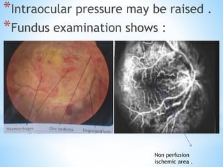
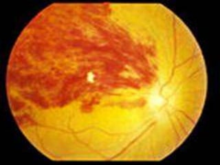
*
*Etiology .
*Clinical features .
*Symptoms .
*Signs .
*Investigation .
*Complication .
*Visual prognosis .
*Treatment :
*
*Definition:
*Central retinal vein occlusion (CRVO)
is a blockage of the main vein in the
retina .
*The central retinal vein is the venous
equivalent of the central retinal
artery and, like that blood vessel, it
can suffer from occlusion (central
retinal vein occlusion, also CRVO),
*similar to that seen in ocular
ischemic syndrome. Since the central
retinal artery and vein are the sole
source of blood supply and drainage
for the retina, such occlusion can
lead to severe damage to the retina
and blindness, due
to ischemia (restriction in blood
supply) and edema(swelling).
*Etiology :
*Age is the most important factor 50%
of cases occur after the age of 60y .
*External compression :
arteriosclerosis or athero sclerosis of
central retinal artery or its branch
compresses the vein at lamina cribosa
or its branch at arteriovenous
crossing because at these two sites
the artery and vein have common
facial sheath .
*Systemic Hypertension : it is the
most common cause of BRVO and is
present in up to 73% of cases .
*Diabetes mellitus : is present in
about 10% of cases .
*Raised Intraocular pressure : CRVO is
more common in primary open angle
glaucoma .
*Drugs : oral contraceptive (birth
controle pills are used to prevent
pregnancy )
*Hyperviscosity of blood
> Polycythemia ( An abnormally
increased concentration of
haemoglobin in the blood .
> Hyperlipidemia ( abnormally
elevated levels of any or all lipids and
or lipoproteins in the blood .
*Thrombophilic disorder .(
Thrombophilia is an abnormality of
blood coagulation that increases .
> Hyperhomocysteinurea ( is a medical
condition characterized by an abnormally high
level of homocysteine in the blood, conventionally
described as above 15 µmol/L.
* Inflammation .
> Eales disease (Eales disease is an idiopathic
obliterative vasculopathy that usually involves
the peripheral retina of young adults)
Eales disease ( periphlebitis )
> Saccoidosis (Sarcoidosis, also called sarcoid,
is a disease involving abnormal collections of
inflammatory cells that form lumps known as
granulomas)
*Local cause :
Orbital cellulitis
Cavernous sinus thrombosis .
*NON ISCHEMIC CRVO:
*Clinical Features :
*Is the most common clinical variety
75%
*Symptoms :
*Sudden onset of unilateral painless
deterioration of vision .
*Signs :
*Visual acuity : moderate to severe visual
loss .
*Afferent pupillary defect : is absent or
mild .
*IOP may be raised e.g in primary open
angle glaucoma .
*Fundus examination shows :
*Spontaneous venous pulsation is absent .
*Retinal veins are dilated, engorged and
tortuous (Paichida )
*Dot : blot and flamed- shaped
haemorrhages are present in all four
quadrants and most numerous in
periphery .
*Cotton whool : spots may be present
*Optic disc and macular oedema is
mild .
*Investigation :
*Are carried out to find out the cause
to prevent the similar attack in the
other eye . See fig ( 1.1 and 1.2 )
Fig 1.1 Fig 1.2
Mild disc
oedema .
Flamed
shaped
haemorrhages
,
Perfusion
area
*Blood pressure .
*ECG
*Blood ..
CP and ESR .
Blood glucose level .
Lipids .
plasma protein electrophoresis .
( Fibrinogen, a beta-2 protein) .
*Auto Antibodies : ANA (autoimmune
disorder, ) Anti DNA(Lupus erythematosus is
a name given to a collection of autoimmune
diseases ) and ANCA .(Anti-neutrophil
cytoplasmic antibodies (ANCAs) are a group
of autoantibodies, mainly of the IgG type, )
*Fluorescein angiography shows good
capillary perfusion .
*Optical coherence tomography OCT to
demonstrate the severity of macular
oedema and is useful to assess the
response of treatment .
*Fundus fluorescein angiography FFA is
useful to assess the perfusion of retina .
*Complication :
*Cystoid macular oedema is the main
complication .
*Conversion to ischemic type occurs in
about 15% cases which progress to
rubeosis in about 37% within 4 month.
*Visual prognosis :
*Heamorrhages resolve within 6 to
12 months .
*Visual prognosis is good with return
of vision nearly normal in about 50%
of cases .
*Visual loss in rest of cases is mainly
due to cystoid macular oedema .
*Treatment :
*Laser photocoagulation for macular
oedema is not effective .
*Consist of Anti VEGF ( drugs like lucentis
or
*Intravetral triamcinolone acetonide
(Triamcinolone acetonide is known as a
corticosteroid hormone (glucocorticoid).
It works by decreasing your body's
immune response to these diseases and
reduces symptoms such as swelling.)
*Oral steroid for 8 to 12 weeks may be
effective .
*Optic nerve sheathotomy via pars plana
approach for decomposing the central
retinal vein at lamina cribosa .
*ISCHEMIC CRVO:
*Clinical features :
*Presentation is sudden onset of severe
visual loss .
*Visual acuity is usually counting fingers
or worse .
*Afferent papillary defect is marked .
*Intraocular pressure may be raised .
*Fundus examination shows :
Non perfusion
ischemic area .
*Retinal veins are very tortuous and
engorged .
*Retinal haemorrhages dot : blot and
flamed shaped haemorrhages in all
four quadrants of retina and posterior
pole ( almost whole fundus is full of
heamorrhage giving tomato splashed
appearance .
*Disc odema and hyperemia is severe .
*Cotton whool spot may be present .
*Investigation:
*Blood pressure .
*ECG
*Blood ..
CP and ESR .
Blood glucose level .
Lipids .
plasma protein electrophoresis .
( Fibrinogen, a beta-2 protein) .
*Auto Antibodies : ANA (autoimmune disorder, )
Anti DNA(Lupus erythematosus is a name given
to a collection of autoimmune diseases ) and
ANCA .(Anti-neutrophil cytoplasmic
antibodies (ANCAs) are a group of
autoantibodies, mainly of the IgG type, )
*
*Fluorescein angiography shows good
capillary perfusion .
*Optical coherence tomography OCT to
demonstrate the severity of macular
oedema and is useful to assess the
response of treatment .
*Fundus fluorescein angiography FFA is
useful to assess the perfusion of retina.
*Prognosis :
*Prognosis is extremely poor due to
macular ischemia .
*Complication :
*Macular ischemia .
*Chronic cystoid macular oedema.
*Rubeois iridis , which causes neovascular
glaucoma NVG in more than 50% cases
between 2 to 4 months .
*Treatment:
*Panretinal photocoagulation PRP is
carried out in all eyes rubeosis iridis .
*Cryotherapy application when retinal
view is not possible due to hazy media .
*Branch retinal vein occlusion :
*Branch retinal vein occlusion (CRVO) is a
blockage of the Branch vein in the retina
.
*It is common than central retinal vein
occlusion .
*Types :
*Quadrant vein occlusion major branch
away from disc A-V crossing .
*Hemiretinal vein occlusion main branch
vein occlusion at disc margin .
*Clinical Features :
*The oedema and haemorrahages are
limited to the retinal area drained by
the affected vein .
*Vision is affected only when macular
area is involved .
*Cotton whool spots present .
*Secondary glaucoma occur rarely.
*Prognosis is reasonable good .
*Complication :
*Chronic macular oedema : is common cause
for poor vision .
*Neovascularization develops in 10% of cases.
*Treatment :
*Laser photocoagulation : for macular
oedema, when macular perfusion is good.
*Intravetral steroid triamcinolone acetonide
may improve vision .
*Intravetral anti VEGF showing good result .
Crvo vs brvo by Dr.kausar ali

Crvo vs brvo by Dr.kausar ali

  • 4.
    * *Etiology . *Clinical features. *Symptoms . *Signs . *Investigation . *Complication . *Visual prognosis . *Treatment :
  • 5.
    * *Definition: *Central retinal veinocclusion (CRVO) is a blockage of the main vein in the retina . *The central retinal vein is the venous equivalent of the central retinal artery and, like that blood vessel, it can suffer from occlusion (central retinal vein occlusion, also CRVO),
  • 6.
    *similar to thatseen in ocular ischemic syndrome. Since the central retinal artery and vein are the sole source of blood supply and drainage for the retina, such occlusion can lead to severe damage to the retina and blindness, due to ischemia (restriction in blood supply) and edema(swelling).
  • 7.
    *Etiology : *Age isthe most important factor 50% of cases occur after the age of 60y . *External compression : arteriosclerosis or athero sclerosis of central retinal artery or its branch compresses the vein at lamina cribosa or its branch at arteriovenous crossing because at these two sites the artery and vein have common facial sheath .
  • 8.
    *Systemic Hypertension :it is the most common cause of BRVO and is present in up to 73% of cases . *Diabetes mellitus : is present in about 10% of cases . *Raised Intraocular pressure : CRVO is more common in primary open angle glaucoma . *Drugs : oral contraceptive (birth controle pills are used to prevent pregnancy )
  • 9.
    *Hyperviscosity of blood >Polycythemia ( An abnormally increased concentration of haemoglobin in the blood . > Hyperlipidemia ( abnormally elevated levels of any or all lipids and or lipoproteins in the blood . *Thrombophilic disorder .( Thrombophilia is an abnormality of blood coagulation that increases .
  • 10.
    > Hyperhomocysteinurea (is a medical condition characterized by an abnormally high level of homocysteine in the blood, conventionally described as above 15 µmol/L. * Inflammation . > Eales disease (Eales disease is an idiopathic obliterative vasculopathy that usually involves the peripheral retina of young adults) Eales disease ( periphlebitis ) > Saccoidosis (Sarcoidosis, also called sarcoid, is a disease involving abnormal collections of inflammatory cells that form lumps known as granulomas)
  • 11.
    *Local cause : Orbitalcellulitis Cavernous sinus thrombosis . *NON ISCHEMIC CRVO: *Clinical Features : *Is the most common clinical variety 75% *Symptoms : *Sudden onset of unilateral painless deterioration of vision .
  • 12.
    *Signs : *Visual acuity: moderate to severe visual loss . *Afferent pupillary defect : is absent or mild . *IOP may be raised e.g in primary open angle glaucoma . *Fundus examination shows : *Spontaneous venous pulsation is absent . *Retinal veins are dilated, engorged and tortuous (Paichida )
  • 13.
    *Dot : blotand flamed- shaped haemorrhages are present in all four quadrants and most numerous in periphery . *Cotton whool : spots may be present *Optic disc and macular oedema is mild . *Investigation : *Are carried out to find out the cause to prevent the similar attack in the other eye . See fig ( 1.1 and 1.2 )
  • 14.
    Fig 1.1 Fig1.2 Mild disc oedema . Flamed shaped haemorrhages , Perfusion area
  • 15.
    *Blood pressure . *ECG *Blood.. CP and ESR . Blood glucose level . Lipids . plasma protein electrophoresis . ( Fibrinogen, a beta-2 protein) . *Auto Antibodies : ANA (autoimmune disorder, ) Anti DNA(Lupus erythematosus is a name given to a collection of autoimmune diseases ) and ANCA .(Anti-neutrophil cytoplasmic antibodies (ANCAs) are a group of autoantibodies, mainly of the IgG type, )
  • 16.
    *Fluorescein angiography showsgood capillary perfusion . *Optical coherence tomography OCT to demonstrate the severity of macular oedema and is useful to assess the response of treatment . *Fundus fluorescein angiography FFA is useful to assess the perfusion of retina . *Complication : *Cystoid macular oedema is the main complication .
  • 17.
    *Conversion to ischemictype occurs in about 15% cases which progress to rubeosis in about 37% within 4 month. *Visual prognosis : *Heamorrhages resolve within 6 to 12 months . *Visual prognosis is good with return of vision nearly normal in about 50% of cases . *Visual loss in rest of cases is mainly due to cystoid macular oedema .
  • 18.
    *Treatment : *Laser photocoagulationfor macular oedema is not effective . *Consist of Anti VEGF ( drugs like lucentis or *Intravetral triamcinolone acetonide (Triamcinolone acetonide is known as a corticosteroid hormone (glucocorticoid). It works by decreasing your body's immune response to these diseases and reduces symptoms such as swelling.) *Oral steroid for 8 to 12 weeks may be effective .
  • 19.
    *Optic nerve sheathotomyvia pars plana approach for decomposing the central retinal vein at lamina cribosa . *ISCHEMIC CRVO: *Clinical features : *Presentation is sudden onset of severe visual loss . *Visual acuity is usually counting fingers or worse . *Afferent papillary defect is marked .
  • 20.
    *Intraocular pressure maybe raised . *Fundus examination shows : Non perfusion ischemic area .
  • 21.
    *Retinal veins arevery tortuous and engorged . *Retinal haemorrhages dot : blot and flamed shaped haemorrhages in all four quadrants of retina and posterior pole ( almost whole fundus is full of heamorrhage giving tomato splashed appearance . *Disc odema and hyperemia is severe . *Cotton whool spot may be present .
  • 22.
    *Investigation: *Blood pressure . *ECG *Blood.. CP and ESR . Blood glucose level . Lipids . plasma protein electrophoresis . ( Fibrinogen, a beta-2 protein) . *Auto Antibodies : ANA (autoimmune disorder, ) Anti DNA(Lupus erythematosus is a name given to a collection of autoimmune diseases ) and ANCA .(Anti-neutrophil cytoplasmic antibodies (ANCAs) are a group of autoantibodies, mainly of the IgG type, ) *
  • 23.
    *Fluorescein angiography showsgood capillary perfusion . *Optical coherence tomography OCT to demonstrate the severity of macular oedema and is useful to assess the response of treatment . *Fundus fluorescein angiography FFA is useful to assess the perfusion of retina. *Prognosis : *Prognosis is extremely poor due to macular ischemia .
  • 24.
    *Complication : *Macular ischemia. *Chronic cystoid macular oedema. *Rubeois iridis , which causes neovascular glaucoma NVG in more than 50% cases between 2 to 4 months . *Treatment: *Panretinal photocoagulation PRP is carried out in all eyes rubeosis iridis . *Cryotherapy application when retinal view is not possible due to hazy media .
  • 25.
    *Branch retinal veinocclusion : *Branch retinal vein occlusion (CRVO) is a blockage of the Branch vein in the retina . *It is common than central retinal vein occlusion . *Types : *Quadrant vein occlusion major branch away from disc A-V crossing . *Hemiretinal vein occlusion main branch vein occlusion at disc margin .
  • 26.
    *Clinical Features : *Theoedema and haemorrahages are limited to the retinal area drained by the affected vein . *Vision is affected only when macular area is involved . *Cotton whool spots present . *Secondary glaucoma occur rarely. *Prognosis is reasonable good .
  • 28.
    *Complication : *Chronic macularoedema : is common cause for poor vision . *Neovascularization develops in 10% of cases. *Treatment : *Laser photocoagulation : for macular oedema, when macular perfusion is good. *Intravetral steroid triamcinolone acetonide may improve vision . *Intravetral anti VEGF showing good result .