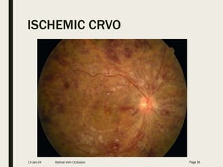
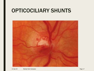
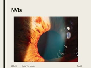
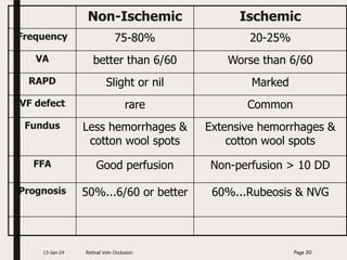
CENTRAL RETINAL
VEIN OCCLUSION
Dr. SALMAN AHMAD KHAN
FCPS Resident
Ophthalmology Unit-1
SIMS/SHL
13-Jan-24
Common mechanism
Venous blockage
back pressure on capillaries
endothelial junction dysfunction
leakage of fluid & blood
(edema / hemorrhages)
■ Severe nonperfusion leads to ischemia
13-Jan-24 Retinal Vein Occlusion Page 2
Predominant Associations
Patient
Group
Hypertension Hyperlipidemia Diabetes
Mellitus
No Obvious
Cause
Age<50
yrs
25% 35% 03% 40%
Age>50
yrs
64% 34% 4-15% 21%
Asian 64% 50% 29% 10.7%
West
Indian
83% 33% 38% 8.3%
Recurrent
cases
88% 47% 3% 6%
13-Jan-24 Retinal Vein Occlusion Page 3
Central retinal vein occlusion
■ Painless unilateral sudden loss of vision
■ Occlusion at or posterior to lamina cribrosa
■ Two clinical types
– Ischemic CRVO (I-CRVO)
– Non-ischemic (NI-CRVO)
13-Jan-24 Retinal Vein Occlusion Page 4
Pathogenesis
■ Virchow triad:
– Loss of vessel wall integrity
– Altered blood flow
– Hypercoagulable state
■ Disturbance leads to thrombus
formation & vessel occlusion
13-Jan-24 Retinal Vein Occlusion Page 5
■ Klein & Olwin postulated:
– Compression of vein by sclerotic central retinal
artery
– Occlusion by primary vessel wall disease
– Hemodynamic disturbance
Klein BA, Olwin JH. A survey of the pathogenesis of retinal venous occlusion. Arch Ophthalmol 1956;56:207.
13-Jan-24 Retinal Vein Occlusion Page 6
CRVO resistance to venous flow
blood stagnation & ischemia stimulates
production of VEGF (vascular endothelial growth factor)
neovascularization
capillary leakage (edema)
13-Jan-24 Retinal Vein Occlusion Page 7
Etiology
■ Any factor which directly or indirectly
activates virchow triad….
13-Jan-24 Retinal Vein Occlusion Page 8
External compression
– Arteriosclerosis of CRA (HTN, DM,
Hyperlipidemia)
– Glaucoma (5 times more likely to have CRVO)
– Papilledema
– Thyroid eye disease
– Orbital space occupying lesions
– Cavernous sinus thrombosis
– Closed-Head trauma
13-Jan-24 Retinal Vein Occlusion Page 9
Disease of vessel wall
– Systemic Vasculitis
■ TB
■ AIDS
■ Syphilis
■ SLE
– Localized inflammation
■ Sarcoidosis
13-Jan-24 Retinal Vein Occlusion Page 10
Hematological disorders
■ Clotting disorders
– Activated protein C
resistance
– Lupus anticoagulant
deficiency
– Anticardiolipin
antibodies
– Protein C & Protein S
deficiency
– Antithrombin III def
– Antiphospholipid
antibodies
■ Nephrotic syndrome
■ Paraproteinemia
– Multiple myeloma
■ Drugs
– Oral contraceptive
– Diuretics
■ Blood dyscrasia
– Lymphoma
– Leukemia
– Polycythemia vera
– Sickle cell disease
13-Jan-24 Retinal Vein Occlusion Page 11
MANAGEMENT
13-Jan-24 Retinal Vein Occlusion Page 12
History
■ Symptoms
– Painless loss of vision (mild to severe)
– Usually unilateral
■ Past & Personal Hx
– HTN, DM, smoking
– Hyperlipidemia
– Bleeding or clotting disorders
– Glaucoma
– Oral contraceptive use
– Head trauma / retrobulbar inj
13-Jan-24 Retinal Vein Occlusion Page 13
Examination
– VA & BCVA
– Pupillary reactions
– Congestion of conjunctiva or cornea
– Iris…neovessels
– AC angle…neovessels
– IOP
13-Jan-24 Retinal Vein Occlusion Page 14
■ Fundus findings
– Retinal hemorrhages in 4 quadrants
– Extensive hemorrhages…blood & thunder
appearance
– Dilated tortuous veins
– Cotton wool spots, macular edema
– Optic disc
■ Edema / optociliary shunts / atrophy
– Neovessels
■ NVD / NVE……vitreous hemorrhage
13-Jan-24 Retinal Vein Occlusion Page 15
ISCHEMIC CRVO
13-Jan-24 Retinal Vein Occlusion Page 16
OPTICOCILIARY SHUNTS
13-Jan-24 Retinal Vein Occlusion Page 17
NVIs
13-Jan-24 Retinal Vein Occlusion Page 18
■ Diagnosing CRVO is not difficult
■ Main task…differentiate btw ischemic &
non-ischemic CRVO
■ No single criterion is helpful
■ Various useful tools…
– Visual acuity, pupillary reflex
– Ocular neovascularization, Fundus findings
– ERG, FFA
13-Jan-24 Retinal Vein Occlusion Page 19
Non-Ischemic Ischemic
Frequency 75-80% 20-25%
VA better than 6/60 Worse than 6/60
RAPD Slight or nil Marked
VF defect rare Common
Fundus Less hemorrhages &
cotton wool spots
Extensive hemorrhages &
cotton wool spots
FFA Good perfusion Non-perfusion > 10 DD
Prognosis 50%...6/60 or better 60%...Rubeosis & NVG
13-Jan-24 Retinal Vein Occlusion Page 20
NON ISCHEMIC CRVO
13-Jan-24 Retinal Vein Occlusion Page 21
Complications
■ Principle causes of visual morbidity
– Macular edema (ME)
– Neovascularization (NVI>NVD>NVE) &
Neovascular glaucoma (100 days)
– Vitreous hemorrhage
– Optic atrophy
13-Jan-24 Retinal Vein Occlusion Page 22
Differential diagnosis
■ Ocular ischemic syndrome:dilated veins
without tortuosity,no disc edema,patients
have history of visual loss.
■ Diabetic retinopathy:hemorrhages on
posterior pole and bilateral.
■ Papilledema:bilateral disc swelling with
hemorrhages surrounding disc.
■ Radiation retinopathy:history of
radiation,disc swelling,more cotton wool
spots than hemorrhages.
13-Jan-24 Retinal Vein Occlusion Page 23
Ocular Investigations
■ ERG
– Is depressed
– Extent of this is used to assess neovascular
risk
■ OCT
– For macular thickness.Enables quantification
of cystoid macular edema.
13-Jan-24 Retinal Vein Occlusion Page 24
■ Fluorescein angiography
– Very useful for detecting…
■ Capillary nonperfusion
■ Neovascularization
■ Macular edema
– Reliable to differentiate btw I-CRVO & NI-CRVO
– >10 DD retinal nonperfusion is termed as I-CRVO*
– Limitations
■ It provides little information in early stages bcz of
extensive hemorrhages
13-Jan-24 Retinal Vein Occlusion Page 25
■ FFA findings
– Delayed arteriovenous transit
– Macular edema
– Staining along the retinal veins
– Micro aneurysms, Arteriovenous collaterals
– NVD, NVE
– Dilated optic nerve head capillaries
– Nonperfusion…hypofluorescence
13-Jan-24 Retinal Vein Occlusion Page 26
Extensive hypofluorescence due
to capillary non-perfusion On
FFA
13-Jan-24 Retinal Vein Occlusion Page 27
Wide-field FA showing
Extensive Peripheral
Ischaemia
13-Jan-24 Retinal Vein Occlusion Page 28
TREATMENT
■ Treat the underline cause
■ It is the responsibility of the diagnosing
physician or ophthalmologist to:
– Investigate and interpret results.
– Refer the patient for appropriate medical
advice with urgency according to the severity of
underlying risk factor(s).
– Ensure that initiation of medical management
occurs within 2 months of diagnosis
Royal college of ophthalmologists guidelines: July, 2015
13-Jan-24 Retinal Vein Occlusion Page 29
Initial medical investigations
■ ALL PATIENTS
– FBC & ESR
– Renal function tests
– Random blood
glucose
– Lipid profile
– Thyroid function
– ECG
Royal college of ophthalmologists guidelines: JULY,
2015
■ ACCORDING TO
CLINICAL INDICATION
– Thrombophilia screen
– CRP
– Serum ACE
– Autoantibodies
– CXR
– Fasting homocystine
levels
13-Jan-24 Retinal Vein Occlusion Page 30
Natural history of CRVO
■ NI-CRVO
– Completely resolution…10% a
– ME resolves…30% in 6-15 months b
– About 50%...VA is 6/60 or worse a
– 1/3rd progress to I-CRVO in 6-12 months a
– Neovessels develop…33% in 12-15 months b
a Central Vein Occlusion Study Group. Baseline and early natural history report. Arch Ophthalmol. Aug 1993;111(8):1087-95
b McIntosh RL et al. Natural History of Central Retinal Vein Occlusion: An Evidence-Based Systematic Review. Ophthalmology
2010;117:1113–1123
13-Jan-24 Retinal Vein Occlusion Page 31
■ I-CRVO
– >90%...VA is 6/60 or worse a
– ME resolves…73% in 15 months b
– NVG…>60% in 1-2 yrs a
– About 10% develop RVO in same or fellow eye in 2 yrs
■ Vitreous hemorrhage…10 % of CRVO by 9 months
b
a Central Vein Occlusion Study Group. Baseline and early natural history report. Arch Ophthalmol. Aug 1993;111(8):1087-95
b McIntosh RL et al. Natural History of Central Retinal Vein Occlusion: An Evidence-Based Systematic Review. Ophthalmology
2010;117:1113–1123
13-Jan-24 Retinal Vein Occlusion Page 32
Treatment
■ Systemic treatment a
– Anticoagulants…Heparin, warfarin
– Fibrinolytic agents…Streptokinase, tissue
plasminogen activator
– Antiplatelets…Aspirin, prostacyclin
– Hemodilution
a Mahmood T. CRVO: current management options. Pak J Ophthalmol 2009. 25(1):56-9.
13-Jan-24 Retinal Vein Occlusion Page 33
■ Ocular treatment
– Pharmacotherapy
– Photocoagulation
– New techniques (Surgical)
■ Certain clinical trials needs attention
13-Jan-24 Retinal Vein Occlusion Page 34
Central Vein Occlusion Study (CVOS
■ Results
– Group M--Macular Edema: Macular grid
photocoagulation was effective in reducing
angiographic evidence of macular edema but
did not improve visual acuity.
– Group N--PRP for Ischemic CVO: Prophylactic
PRP did not prevent the development of NVI in
eyes with >10 disc areas of retinal capillary
nonperfusion confirmed by FFA
13-Jan-24 Retinal Vein Occlusion Page 35
THE SCORE STUDY
13-Jan-24 Retinal Vein Occlusion Page 36
■ The SCORE study showed an improvement
of three or more lines of vision at one year
in over 25% of patients (versus 7% of
controls) treated with an average of two
injections of 1 mg triamcinolone, using a
preservative-free preparation developed for
intraocular use. There was a slightly higher
rate of IOP elevation and cataract than with
observation.
CRUISE Study
■ In June 2010, the FDA approved a new
indication for Ranibizumab(0.5 mg)
intravitreal injection…for the treatment of
macular edema after retinal vein occlusion.
■ Ranibizumab may initially be given monthly
for 6 months and subsequently less
intensively, with typically a two- to three-
line gain in VA.
13-Jan-24 Retinal Vein Occlusion Page 37
The GENEVA study
■ Evaluated safety and efficacy of an intravitreal
implant of dexamethasone (Ozurdex; Allergan Inc.,
Irvine, California, USA).
13-Jan-24 Retinal Vein Occlusion Page 38
The Royal College of Ophthalmologists
Guidelines
■ Treatment Algorithm: Published in JULY 2015.
■ NON-ISCHAEMIC CRVO
■ If no iris or angle NV and there is OCT evidence of MO:
 If visual acuity is 6/96 or better Anti VEGF or OZURDEX
 If visual acuity is less than 6/96, potential ↑ in VA is low
and high risk of developing NVI/NVA… Treatment can be
offered.
 If visual acuity is better than 6/12…. Observation
*
13-Jan-24 Retinal Vein Occlusion Page 39
ISCHAEMIC CRVO
■ If iris or angle neovascularisation occurs and the
anterior chamber angle is open:
■ Urgent PRP is recommended and with review at two
weeks initially and then less frequently as regression
occurs ± Bevacizumab.
■ If iris or angle NV are present with a closed angle and
raised intraocular pressure
■ Urgent PRP is recommended with cyclodiode laser
therapy / tube shunt surgery ± Bevacizumab.
13-Jan-24 Retinal Vein Occlusion Page 40
If an ischaemic CRVO is present without
NVI/NVG
■ Patient should be seen monthly for 6
months.Prophylactic PRP is generally not
recommended even with marked ischaemia
unless iris new vessels develop, though may
be considered in patients unlikely to attend
scheduled review.
■ FFA shows > 30 DD non-perfusion.
13-Jan-24 Retinal Vein Occlusion Page 41
Recommendations for further follow-up
■ Follow-up after 6 months for ischemia should
be every 3 months for 1 year
■ Non-ischemic eyes…every 3 months for 6
months.
■ Subsequent follow-up will depend on laser Tx
& complications.
■ Development of disc collaterals +/- resolution
of CRVO should lead to discharge from clinical
supervision
13-Jan-24 Retinal Vein Occlusion Page 42
Experimental treatments
– Chorio-retinal anastomosis
– Radial optic neurotomy with PPV
■ Currently…these are not recommended
except as part of clinical trials.
a
13-Jan-24 Retinal Vein Occlusion Page 43
Take home message
■ Emphasis should be on:
– Differentiating ischemic & Nonischemic CRVO
– Exploring the risk factors (local & systemic)
– Treating CRVO and Referral to physician for
risk factors
– Proper follow-up
13-Jan-24 Retinal Vein Occlusion Page 44
13-Jan-24 Retinal Vein Occlusion Page 45
THANKS

CRVO final.ppt

  • 1.
    CENTRAL RETINAL VEIN OCCLUSION Dr.SALMAN AHMAD KHAN FCPS Resident Ophthalmology Unit-1 SIMS/SHL 13-Jan-24
  • 2.
    Common mechanism Venous blockage backpressure on capillaries endothelial junction dysfunction leakage of fluid & blood (edema / hemorrhages) ■ Severe nonperfusion leads to ischemia 13-Jan-24 Retinal Vein Occlusion Page 2
  • 3.
    Predominant Associations Patient Group Hypertension HyperlipidemiaDiabetes Mellitus No Obvious Cause Age<50 yrs 25% 35% 03% 40% Age>50 yrs 64% 34% 4-15% 21% Asian 64% 50% 29% 10.7% West Indian 83% 33% 38% 8.3% Recurrent cases 88% 47% 3% 6% 13-Jan-24 Retinal Vein Occlusion Page 3
  • 4.
    Central retinal veinocclusion ■ Painless unilateral sudden loss of vision ■ Occlusion at or posterior to lamina cribrosa ■ Two clinical types – Ischemic CRVO (I-CRVO) – Non-ischemic (NI-CRVO) 13-Jan-24 Retinal Vein Occlusion Page 4
  • 5.
    Pathogenesis ■ Virchow triad: –Loss of vessel wall integrity – Altered blood flow – Hypercoagulable state ■ Disturbance leads to thrombus formation & vessel occlusion 13-Jan-24 Retinal Vein Occlusion Page 5
  • 6.
    ■ Klein &Olwin postulated: – Compression of vein by sclerotic central retinal artery – Occlusion by primary vessel wall disease – Hemodynamic disturbance Klein BA, Olwin JH. A survey of the pathogenesis of retinal venous occlusion. Arch Ophthalmol 1956;56:207. 13-Jan-24 Retinal Vein Occlusion Page 6
  • 7.
    CRVO resistance tovenous flow blood stagnation & ischemia stimulates production of VEGF (vascular endothelial growth factor) neovascularization capillary leakage (edema) 13-Jan-24 Retinal Vein Occlusion Page 7
  • 8.
    Etiology ■ Any factorwhich directly or indirectly activates virchow triad…. 13-Jan-24 Retinal Vein Occlusion Page 8
  • 9.
    External compression – Arteriosclerosisof CRA (HTN, DM, Hyperlipidemia) – Glaucoma (5 times more likely to have CRVO) – Papilledema – Thyroid eye disease – Orbital space occupying lesions – Cavernous sinus thrombosis – Closed-Head trauma 13-Jan-24 Retinal Vein Occlusion Page 9
  • 10.
    Disease of vesselwall – Systemic Vasculitis ■ TB ■ AIDS ■ Syphilis ■ SLE – Localized inflammation ■ Sarcoidosis 13-Jan-24 Retinal Vein Occlusion Page 10
  • 11.
    Hematological disorders ■ Clottingdisorders – Activated protein C resistance – Lupus anticoagulant deficiency – Anticardiolipin antibodies – Protein C & Protein S deficiency – Antithrombin III def – Antiphospholipid antibodies ■ Nephrotic syndrome ■ Paraproteinemia – Multiple myeloma ■ Drugs – Oral contraceptive – Diuretics ■ Blood dyscrasia – Lymphoma – Leukemia – Polycythemia vera – Sickle cell disease 13-Jan-24 Retinal Vein Occlusion Page 11
  • 12.
  • 13.
    History ■ Symptoms – Painlessloss of vision (mild to severe) – Usually unilateral ■ Past & Personal Hx – HTN, DM, smoking – Hyperlipidemia – Bleeding or clotting disorders – Glaucoma – Oral contraceptive use – Head trauma / retrobulbar inj 13-Jan-24 Retinal Vein Occlusion Page 13
  • 14.
    Examination – VA &BCVA – Pupillary reactions – Congestion of conjunctiva or cornea – Iris…neovessels – AC angle…neovessels – IOP 13-Jan-24 Retinal Vein Occlusion Page 14
  • 15.
    ■ Fundus findings –Retinal hemorrhages in 4 quadrants – Extensive hemorrhages…blood & thunder appearance – Dilated tortuous veins – Cotton wool spots, macular edema – Optic disc ■ Edema / optociliary shunts / atrophy – Neovessels ■ NVD / NVE……vitreous hemorrhage 13-Jan-24 Retinal Vein Occlusion Page 15
  • 16.
    ISCHEMIC CRVO 13-Jan-24 RetinalVein Occlusion Page 16
  • 17.
  • 18.
    NVIs 13-Jan-24 Retinal VeinOcclusion Page 18
  • 19.
    ■ Diagnosing CRVOis not difficult ■ Main task…differentiate btw ischemic & non-ischemic CRVO ■ No single criterion is helpful ■ Various useful tools… – Visual acuity, pupillary reflex – Ocular neovascularization, Fundus findings – ERG, FFA 13-Jan-24 Retinal Vein Occlusion Page 19
  • 20.
    Non-Ischemic Ischemic Frequency 75-80%20-25% VA better than 6/60 Worse than 6/60 RAPD Slight or nil Marked VF defect rare Common Fundus Less hemorrhages & cotton wool spots Extensive hemorrhages & cotton wool spots FFA Good perfusion Non-perfusion > 10 DD Prognosis 50%...6/60 or better 60%...Rubeosis & NVG 13-Jan-24 Retinal Vein Occlusion Page 20
  • 21.
    NON ISCHEMIC CRVO 13-Jan-24Retinal Vein Occlusion Page 21
  • 22.
    Complications ■ Principle causesof visual morbidity – Macular edema (ME) – Neovascularization (NVI>NVD>NVE) & Neovascular glaucoma (100 days) – Vitreous hemorrhage – Optic atrophy 13-Jan-24 Retinal Vein Occlusion Page 22
  • 23.
    Differential diagnosis ■ Ocularischemic syndrome:dilated veins without tortuosity,no disc edema,patients have history of visual loss. ■ Diabetic retinopathy:hemorrhages on posterior pole and bilateral. ■ Papilledema:bilateral disc swelling with hemorrhages surrounding disc. ■ Radiation retinopathy:history of radiation,disc swelling,more cotton wool spots than hemorrhages. 13-Jan-24 Retinal Vein Occlusion Page 23
  • 24.
    Ocular Investigations ■ ERG –Is depressed – Extent of this is used to assess neovascular risk ■ OCT – For macular thickness.Enables quantification of cystoid macular edema. 13-Jan-24 Retinal Vein Occlusion Page 24
  • 25.
    ■ Fluorescein angiography –Very useful for detecting… ■ Capillary nonperfusion ■ Neovascularization ■ Macular edema – Reliable to differentiate btw I-CRVO & NI-CRVO – >10 DD retinal nonperfusion is termed as I-CRVO* – Limitations ■ It provides little information in early stages bcz of extensive hemorrhages 13-Jan-24 Retinal Vein Occlusion Page 25
  • 26.
    ■ FFA findings –Delayed arteriovenous transit – Macular edema – Staining along the retinal veins – Micro aneurysms, Arteriovenous collaterals – NVD, NVE – Dilated optic nerve head capillaries – Nonperfusion…hypofluorescence 13-Jan-24 Retinal Vein Occlusion Page 26
  • 27.
    Extensive hypofluorescence due tocapillary non-perfusion On FFA 13-Jan-24 Retinal Vein Occlusion Page 27
  • 28.
    Wide-field FA showing ExtensivePeripheral Ischaemia 13-Jan-24 Retinal Vein Occlusion Page 28
  • 29.
    TREATMENT ■ Treat theunderline cause ■ It is the responsibility of the diagnosing physician or ophthalmologist to: – Investigate and interpret results. – Refer the patient for appropriate medical advice with urgency according to the severity of underlying risk factor(s). – Ensure that initiation of medical management occurs within 2 months of diagnosis Royal college of ophthalmologists guidelines: July, 2015 13-Jan-24 Retinal Vein Occlusion Page 29
  • 30.
    Initial medical investigations ■ALL PATIENTS – FBC & ESR – Renal function tests – Random blood glucose – Lipid profile – Thyroid function – ECG Royal college of ophthalmologists guidelines: JULY, 2015 ■ ACCORDING TO CLINICAL INDICATION – Thrombophilia screen – CRP – Serum ACE – Autoantibodies – CXR – Fasting homocystine levels 13-Jan-24 Retinal Vein Occlusion Page 30
  • 31.
    Natural history ofCRVO ■ NI-CRVO – Completely resolution…10% a – ME resolves…30% in 6-15 months b – About 50%...VA is 6/60 or worse a – 1/3rd progress to I-CRVO in 6-12 months a – Neovessels develop…33% in 12-15 months b a Central Vein Occlusion Study Group. Baseline and early natural history report. Arch Ophthalmol. Aug 1993;111(8):1087-95 b McIntosh RL et al. Natural History of Central Retinal Vein Occlusion: An Evidence-Based Systematic Review. Ophthalmology 2010;117:1113–1123 13-Jan-24 Retinal Vein Occlusion Page 31
  • 32.
    ■ I-CRVO – >90%...VAis 6/60 or worse a – ME resolves…73% in 15 months b – NVG…>60% in 1-2 yrs a – About 10% develop RVO in same or fellow eye in 2 yrs ■ Vitreous hemorrhage…10 % of CRVO by 9 months b a Central Vein Occlusion Study Group. Baseline and early natural history report. Arch Ophthalmol. Aug 1993;111(8):1087-95 b McIntosh RL et al. Natural History of Central Retinal Vein Occlusion: An Evidence-Based Systematic Review. Ophthalmology 2010;117:1113–1123 13-Jan-24 Retinal Vein Occlusion Page 32
  • 33.
    Treatment ■ Systemic treatmenta – Anticoagulants…Heparin, warfarin – Fibrinolytic agents…Streptokinase, tissue plasminogen activator – Antiplatelets…Aspirin, prostacyclin – Hemodilution a Mahmood T. CRVO: current management options. Pak J Ophthalmol 2009. 25(1):56-9. 13-Jan-24 Retinal Vein Occlusion Page 33
  • 34.
    ■ Ocular treatment –Pharmacotherapy – Photocoagulation – New techniques (Surgical) ■ Certain clinical trials needs attention 13-Jan-24 Retinal Vein Occlusion Page 34
  • 35.
    Central Vein OcclusionStudy (CVOS ■ Results – Group M--Macular Edema: Macular grid photocoagulation was effective in reducing angiographic evidence of macular edema but did not improve visual acuity. – Group N--PRP for Ischemic CVO: Prophylactic PRP did not prevent the development of NVI in eyes with >10 disc areas of retinal capillary nonperfusion confirmed by FFA 13-Jan-24 Retinal Vein Occlusion Page 35
  • 36.
    THE SCORE STUDY 13-Jan-24Retinal Vein Occlusion Page 36 ■ The SCORE study showed an improvement of three or more lines of vision at one year in over 25% of patients (versus 7% of controls) treated with an average of two injections of 1 mg triamcinolone, using a preservative-free preparation developed for intraocular use. There was a slightly higher rate of IOP elevation and cataract than with observation.
  • 37.
    CRUISE Study ■ InJune 2010, the FDA approved a new indication for Ranibizumab(0.5 mg) intravitreal injection…for the treatment of macular edema after retinal vein occlusion. ■ Ranibizumab may initially be given monthly for 6 months and subsequently less intensively, with typically a two- to three- line gain in VA. 13-Jan-24 Retinal Vein Occlusion Page 37
  • 38.
    The GENEVA study ■Evaluated safety and efficacy of an intravitreal implant of dexamethasone (Ozurdex; Allergan Inc., Irvine, California, USA). 13-Jan-24 Retinal Vein Occlusion Page 38
  • 39.
    The Royal Collegeof Ophthalmologists Guidelines ■ Treatment Algorithm: Published in JULY 2015. ■ NON-ISCHAEMIC CRVO ■ If no iris or angle NV and there is OCT evidence of MO:  If visual acuity is 6/96 or better Anti VEGF or OZURDEX  If visual acuity is less than 6/96, potential ↑ in VA is low and high risk of developing NVI/NVA… Treatment can be offered.  If visual acuity is better than 6/12…. Observation * 13-Jan-24 Retinal Vein Occlusion Page 39
  • 40.
    ISCHAEMIC CRVO ■ Ifiris or angle neovascularisation occurs and the anterior chamber angle is open: ■ Urgent PRP is recommended and with review at two weeks initially and then less frequently as regression occurs ± Bevacizumab. ■ If iris or angle NV are present with a closed angle and raised intraocular pressure ■ Urgent PRP is recommended with cyclodiode laser therapy / tube shunt surgery ± Bevacizumab. 13-Jan-24 Retinal Vein Occlusion Page 40
  • 41.
    If an ischaemicCRVO is present without NVI/NVG ■ Patient should be seen monthly for 6 months.Prophylactic PRP is generally not recommended even with marked ischaemia unless iris new vessels develop, though may be considered in patients unlikely to attend scheduled review. ■ FFA shows > 30 DD non-perfusion. 13-Jan-24 Retinal Vein Occlusion Page 41
  • 42.
    Recommendations for furtherfollow-up ■ Follow-up after 6 months for ischemia should be every 3 months for 1 year ■ Non-ischemic eyes…every 3 months for 6 months. ■ Subsequent follow-up will depend on laser Tx & complications. ■ Development of disc collaterals +/- resolution of CRVO should lead to discharge from clinical supervision 13-Jan-24 Retinal Vein Occlusion Page 42
  • 43.
    Experimental treatments – Chorio-retinalanastomosis – Radial optic neurotomy with PPV ■ Currently…these are not recommended except as part of clinical trials. a 13-Jan-24 Retinal Vein Occlusion Page 43
  • 44.
    Take home message ■Emphasis should be on: – Differentiating ischemic & Nonischemic CRVO – Exploring the risk factors (local & systemic) – Treating CRVO and Referral to physician for risk factors – Proper follow-up 13-Jan-24 Retinal Vein Occlusion Page 44
  • 45.
    13-Jan-24 Retinal VeinOcclusion Page 45 THANKS