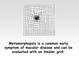
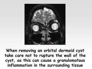
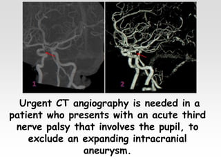
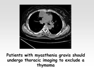
Black & White !
By
Ahmed Alsherbeny
MD, CABOphth, MRCSEd Ophth, FRCS Ophth (Glasg), FICO
A missed intraocular foreign body with
a ferrous component can result in late
visual loss secondary to siderosis.
In optic neuritis secondary to multiple
sclerosis the vision usually improves
without treatment over a period of
several months.
Stargardt disease is the most commonly
inherited macular dystrophy and is usually
associated with mutations in the ABCA4
gene.
In a patient with AMD who has not
responded to anti-VEGF treatment after
3 months, undertake ICG angiography to
check for polypoidal choroidal
vasculopathy
Patients with a large pigment epithelial
detachment should be warned that there
is a risk of inducing a pigment epithelial
tear with anti-VEGF injection
Metamorphopsia is a common early
symptom of macular disease and can be
evaluated with an Amsler grid.
When removing an orbital dermoid cyst
take care not to rupture the wall of the
cyst, as this can cause a granulomatous
inflammation in the surrounding tissue
In thyroid eye disease, compression
of the optic nerve secondary to
enlarged and congested rectus muscles
may occur in the absence of proptosis
The most common tumour associated with
opticociliary shunt vessels on the disc is an
optic nerve sheath meningioma
Because orbital cellulitis often occurs as a
consequence of a sinus infection, urgent
radiological investigation of the sinuses is
mandatory.
Urgent CT angiography is needed in a
patient who presents with an acute third
nerve palsy that involves the pupil, to
exclude an expanding intracranial
aneurysm.
Hearing and corneal sensation should be tested
in patients with a sixth nerve palsy, to exclude
a vestibular schwannoma (acoustic neuroma).
Patients with myasthenia gravis should
undergo thoracic imaging to exclude a
thymoma
All patients with papilloedema should
undergo urgent neuroradiological
investigation to exclude intracranial
pathology.
Neurological lesions causing optic nerve or chiasmal
compression can produce visual field defects that may be
misinterpreted as glaucomatous, and neuroimaging should
be performed if there is any suspicion; some practitioners
routinely perform a cranial MRI in all cases of NTG.
MRI should never be performed if
a metallic foreign body is suspected
In pleomorphic lacrimal gland adenoma
, it is wise to avoid prior biopsy, to
avoid tumour seeding into adjacent
orbital tissue, with recurrences which
may later turn malignant
Glaucoma with deep A.C:
1- Plateau iris syndrome
2- pigment dispersion syndrome
3- Buphthalmos
In Sturge-Weber syndrome , Ipsilateral parietal
or occipital leptomeningeal haemangioma may
cause contralateral or generalized focal or
generalized seizers, hemiparesis or hemianopia
In acoustic neuroma, the first symptom
is hearing loss and the first sign is
diminished corneal sensitivity
In Kerns-Sayer syndrome,ECG
demonstrates cardiac conduction defects
and should be performed periodically;
pacemaker implantation may be required.
Malignancies with calcifications related to
the eye :
1- lacrimal gland carcinoma
2-retinoblastoma
3- optic nerve sheath meningioma
4- osseous choristoma
A small child with characteristic
ophthalmic features of abusive head
trauma (especially unexplained retinal
haemorrhages) should be examined in
conjunction with a specialist paediatrician.
Thank You

Black & White !

  • 1.
    Black & White! By Ahmed Alsherbeny MD, CABOphth, MRCSEd Ophth, FRCS Ophth (Glasg), FICO
  • 2.
    A missed intraocularforeign body with a ferrous component can result in late visual loss secondary to siderosis.
  • 3.
    In optic neuritissecondary to multiple sclerosis the vision usually improves without treatment over a period of several months.
  • 4.
    Stargardt disease isthe most commonly inherited macular dystrophy and is usually associated with mutations in the ABCA4 gene.
  • 5.
    In a patientwith AMD who has not responded to anti-VEGF treatment after 3 months, undertake ICG angiography to check for polypoidal choroidal vasculopathy
  • 6.
    Patients with alarge pigment epithelial detachment should be warned that there is a risk of inducing a pigment epithelial tear with anti-VEGF injection
  • 7.
    Metamorphopsia is acommon early symptom of macular disease and can be evaluated with an Amsler grid.
  • 8.
    When removing anorbital dermoid cyst take care not to rupture the wall of the cyst, as this can cause a granulomatous inflammation in the surrounding tissue
  • 9.
    In thyroid eyedisease, compression of the optic nerve secondary to enlarged and congested rectus muscles may occur in the absence of proptosis
  • 10.
    The most commontumour associated with opticociliary shunt vessels on the disc is an optic nerve sheath meningioma
  • 11.
    Because orbital cellulitisoften occurs as a consequence of a sinus infection, urgent radiological investigation of the sinuses is mandatory.
  • 12.
    Urgent CT angiographyis needed in a patient who presents with an acute third nerve palsy that involves the pupil, to exclude an expanding intracranial aneurysm.
  • 13.
    Hearing and cornealsensation should be tested in patients with a sixth nerve palsy, to exclude a vestibular schwannoma (acoustic neuroma).
  • 14.
    Patients with myastheniagravis should undergo thoracic imaging to exclude a thymoma
  • 15.
    All patients withpapilloedema should undergo urgent neuroradiological investigation to exclude intracranial pathology.
  • 16.
    Neurological lesions causingoptic nerve or chiasmal compression can produce visual field defects that may be misinterpreted as glaucomatous, and neuroimaging should be performed if there is any suspicion; some practitioners routinely perform a cranial MRI in all cases of NTG.
  • 17.
    MRI should neverbe performed if a metallic foreign body is suspected
  • 18.
    In pleomorphic lacrimalgland adenoma , it is wise to avoid prior biopsy, to avoid tumour seeding into adjacent orbital tissue, with recurrences which may later turn malignant
  • 19.
    Glaucoma with deepA.C: 1- Plateau iris syndrome 2- pigment dispersion syndrome 3- Buphthalmos
  • 20.
    In Sturge-Weber syndrome, Ipsilateral parietal or occipital leptomeningeal haemangioma may cause contralateral or generalized focal or generalized seizers, hemiparesis or hemianopia
  • 21.
    In acoustic neuroma,the first symptom is hearing loss and the first sign is diminished corneal sensitivity
  • 22.
    In Kerns-Sayer syndrome,ECG demonstratescardiac conduction defects and should be performed periodically; pacemaker implantation may be required.
  • 23.
    Malignancies with calcificationsrelated to the eye : 1- lacrimal gland carcinoma 2-retinoblastoma 3- optic nerve sheath meningioma 4- osseous choristoma
  • 24.
    A small childwith characteristic ophthalmic features of abusive head trauma (especially unexplained retinal haemorrhages) should be examined in conjunction with a specialist paediatrician.
  • 25.