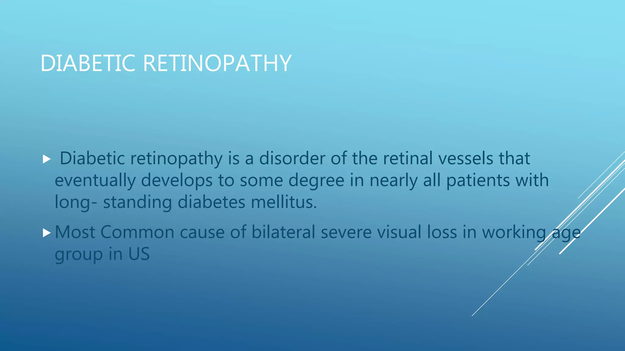
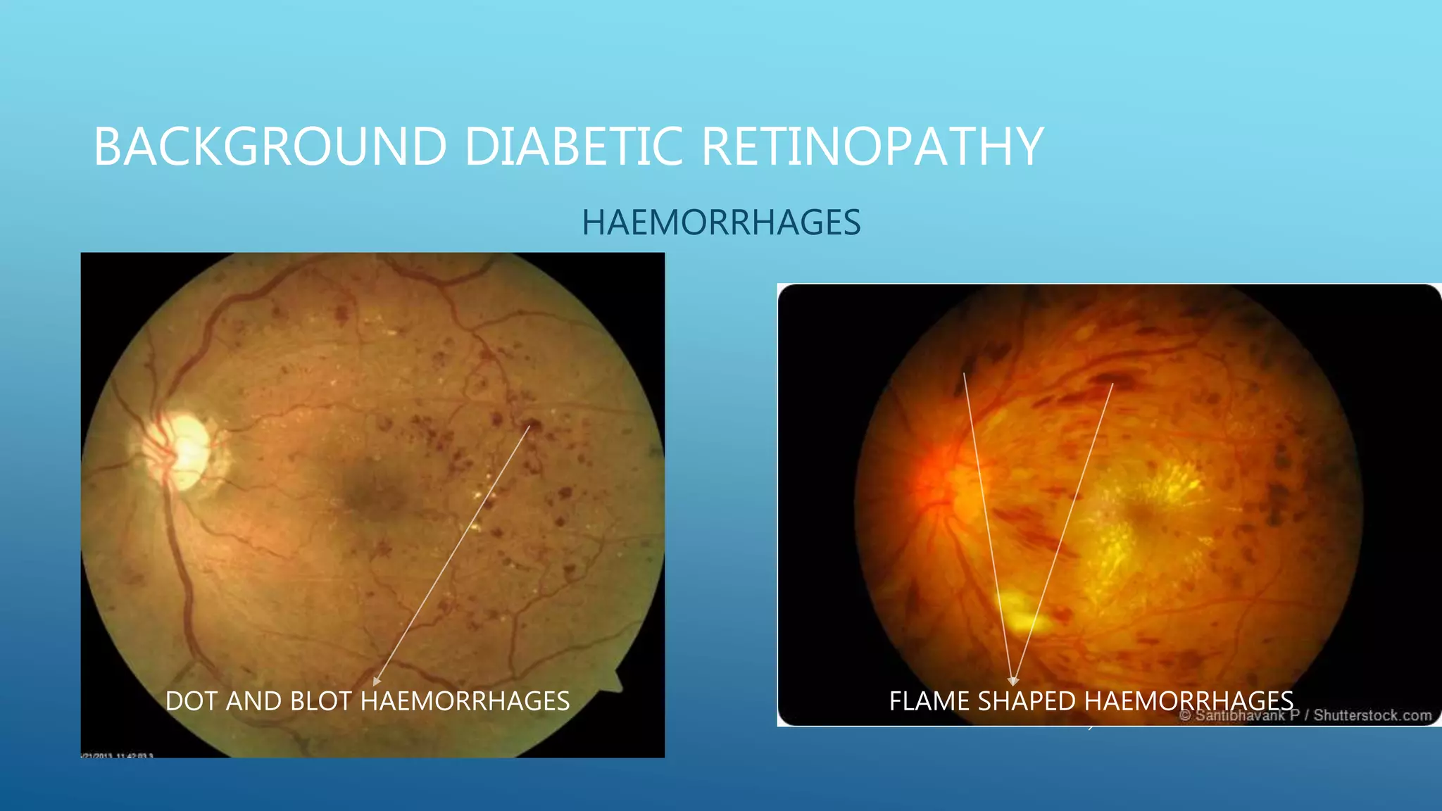
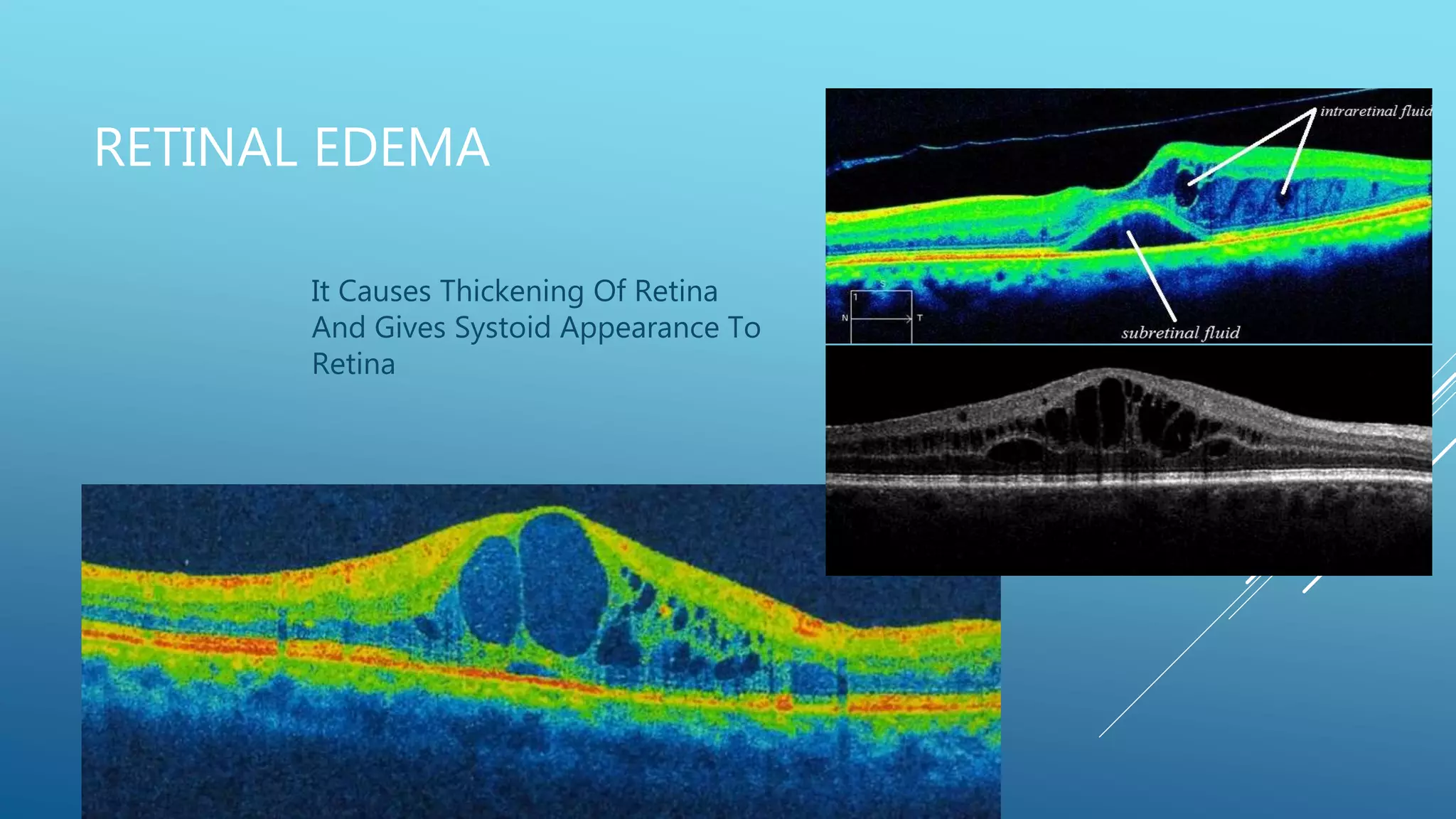
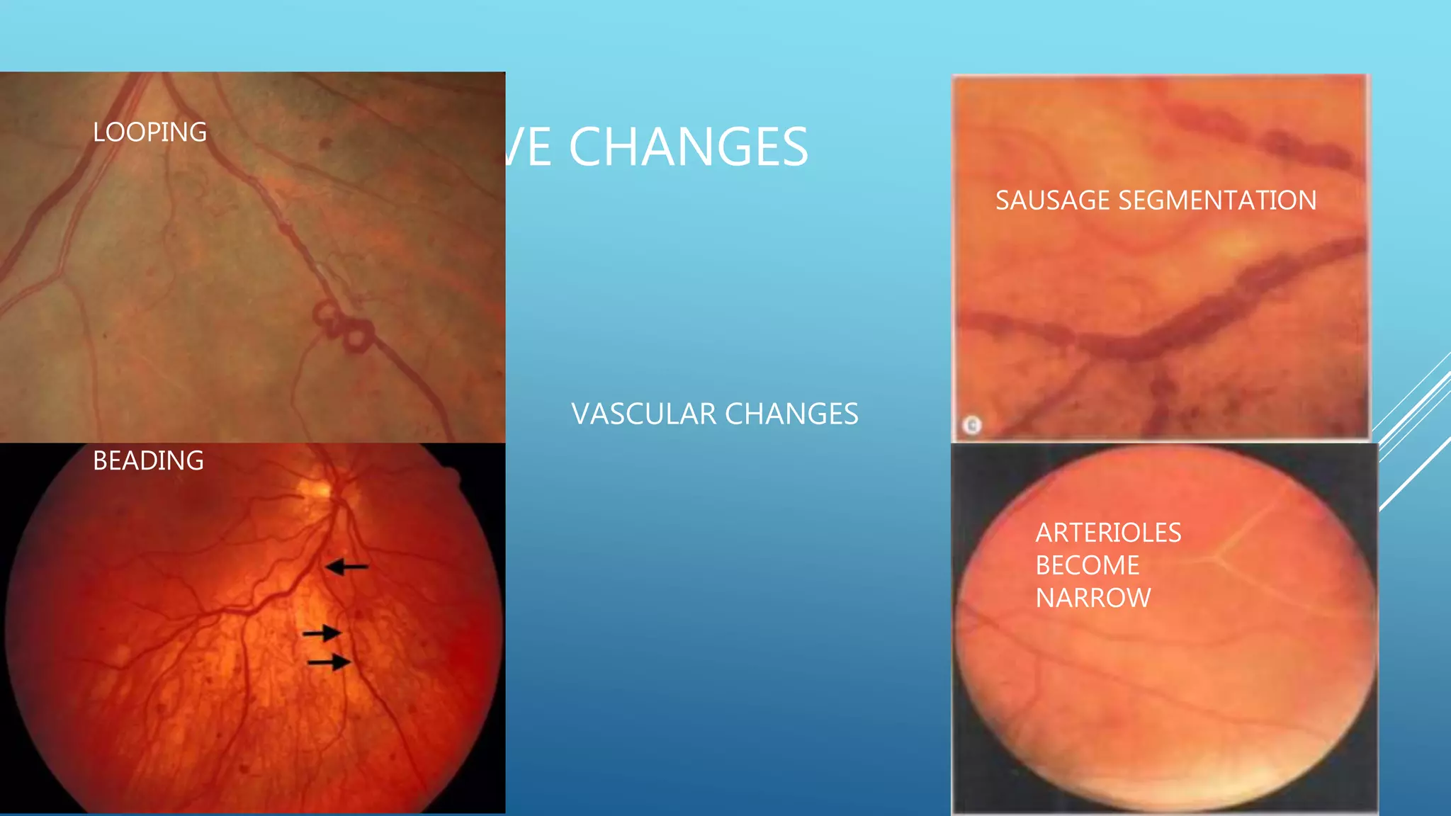
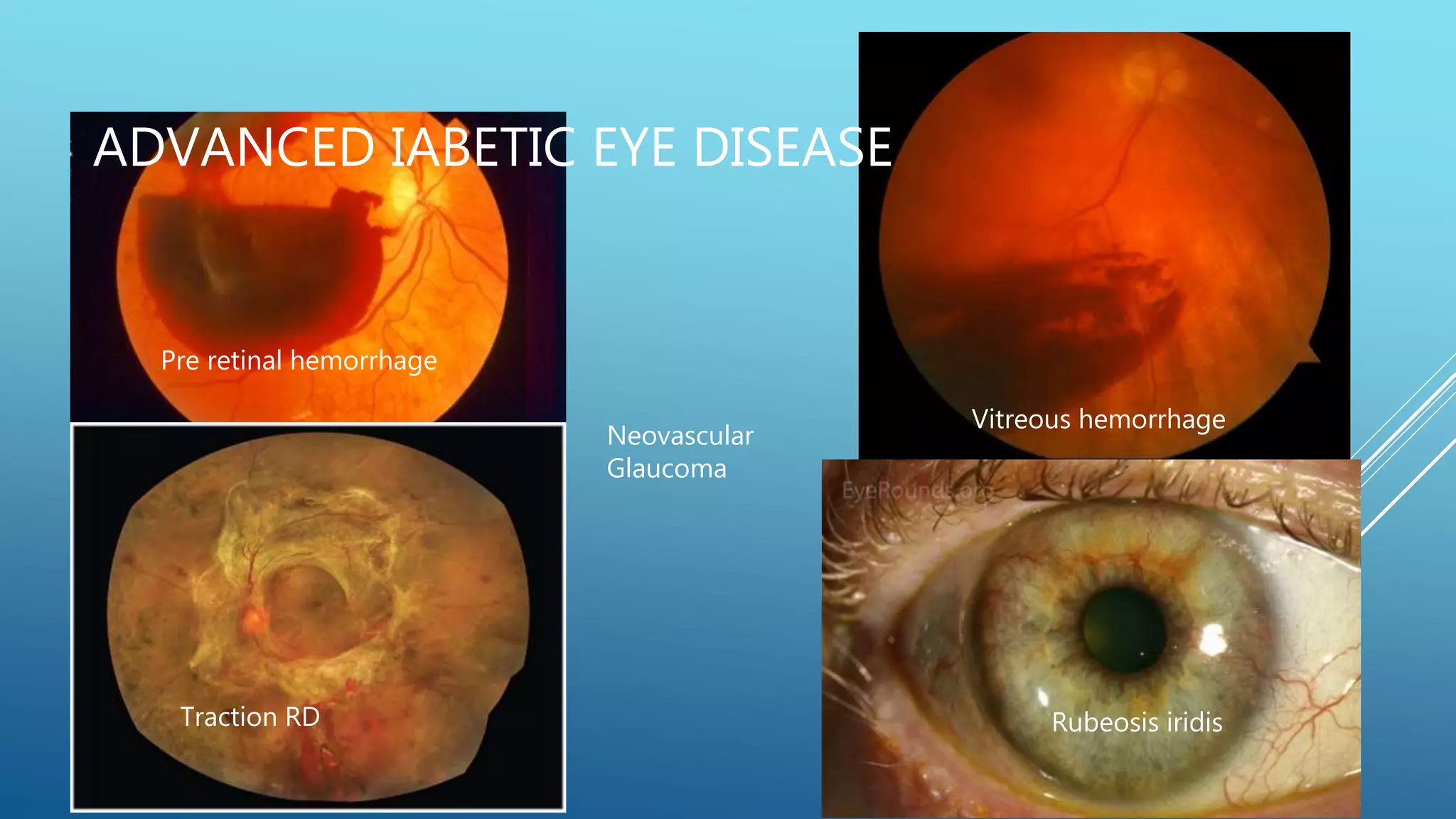
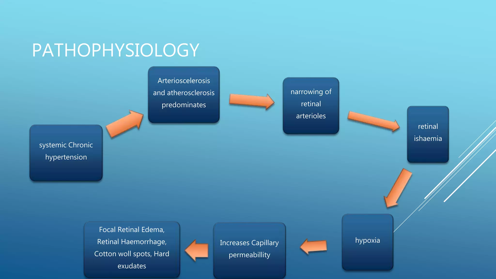
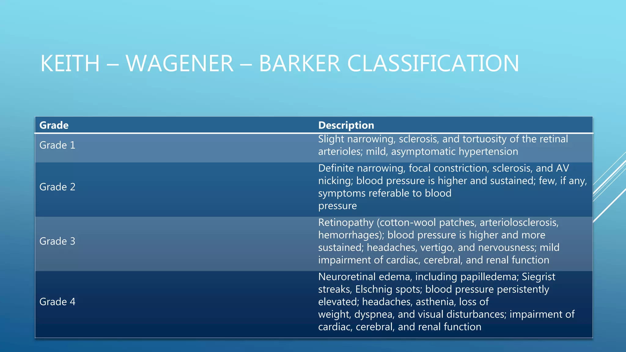
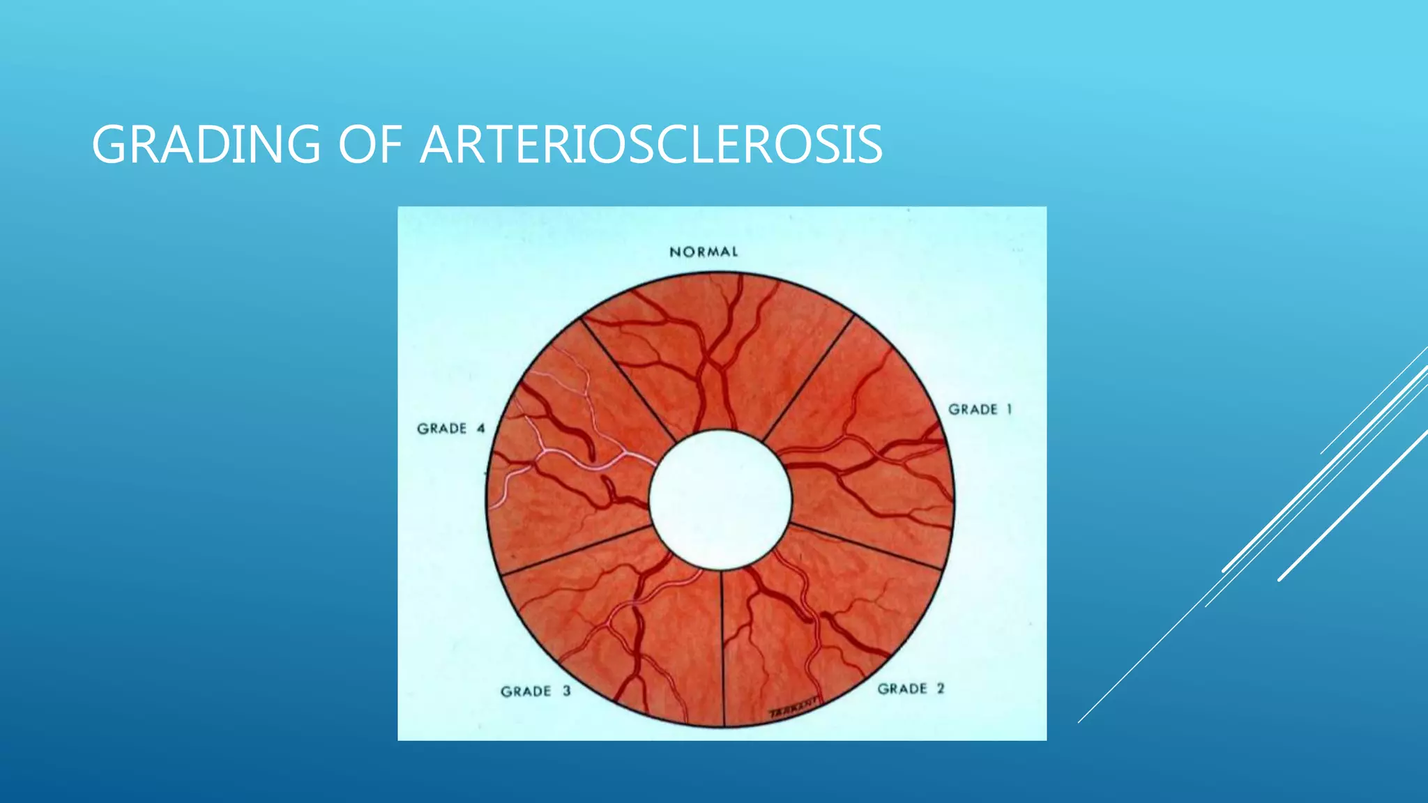
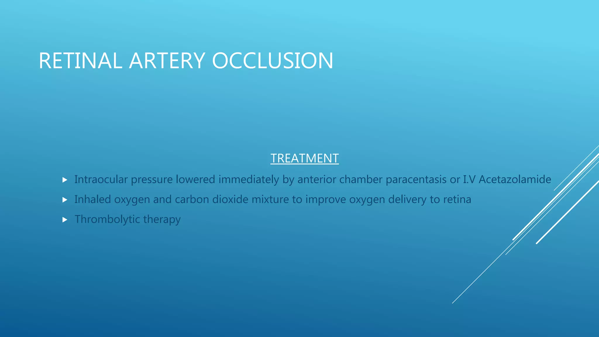
Vascular disorders of the retina can cause vision loss or blindness if not properly treated. The document discusses several types of vascular disorders:
1. Diabetic retinopathy, which affects nearly all patients with long-standing diabetes and is a leading cause of vision loss. It involves microaneurysms, hemorrhages, hard exudates, and new blood vessel growth if proliferative. Laser treatment and anti-VEGF injections can help treat it.
2. Hypertensive retinopathy results from high blood pressure and involves arteriolar narrowing, hemorrhages, cotton wool spots, and exudates. Grading systems describe the severity.
3. Retinal vein oc
This document discusses cystoid macular edema (CME), including its pathogenesis, etiology, associated ocular conditions, manifestations, diagnosis and testing. Specifically, it focuses on pseudophakic or Irvine-Gass syndrome CME, which can occur after cataract surgery. The summary discusses how CME results from fluid accumulation in the retina, its appearance on fluorescein angiography, risk factors for pseudophakic CME like vitreous loss during surgery, and how it is diagnosed using techniques like optical coherence tomography.
This document provides information on vascular disorders of the retina. It begins with the anatomy and blood supply of the retina. It then discusses diabetic retinopathy in detail, including pathogenesis, signs, diagnosis and treatment. It also briefly covers hypertensive retinopathy and retinal vein occlusion, describing their pathophysiology, classification systems, clinical presentation and management. The document focuses primarily on providing an overview of diabetic retinopathy and its various stages and complications.
This document provides an overview of keratoconus, including its etiology, signs and symptoms, classification, and management. Keratoconus is a non-inflammatory thinning of the cornea that results in a cone-shaped protrusion and irregular astigmatism. It typically onset in teenagers and progresses over time. Management includes rigid gas permeable contact lenses, collagen cross-linking to halt progression, and keratoplasty for advanced cases.
This document discusses low vision aids and their use for people with low vision. It defines low vision as visual acuity between 6/18 and 3/60 in the better eye after correction, or a field of vision between 20 to 30 degrees. Common causes of low vision include macular degeneration, glaucoma, and diabetic retinopathy. Optical low vision aids like magnifying spectacles, hand magnifiers, and telescopes use magnification to improve vision. Non-optical aids include increased lighting, contrast enhancement, and electronic magnifiers. Proper evaluation and prescribing of low vision aids depends on the patient's needs, vision status, and motivation. The goal is to prescribe simple, portable devices to help low vision
1) VKH disease is a multisystem disorder characterized by granulomatous panuveitis with exudative retinal detachments and associated neurologic and cutaneous manifestations.
2) It was first described separately by three doctors (Vogt, Koyanagi, and Harada) in the early 20th century before being recognized as a single disease.
3) It has distinct prodromal, uveitic, chronic, and chronic recurrent stages with characteristic ocular and systemic findings at each stage. International criteria have been developed to classify its complete, incomplete, and probable forms.
This document discusses various types of ischemic optic neuropathy (ION). It begins by introducing ION as a major cause of vision loss and outlines its classification into anterior and posterior forms. It then details the anatomy and vascular supply of the optic nerve, risk factors for ION such as nocturnal blood pressure changes, and the pathogenesis involving hypoperfusion and axonal swelling. Non-arteritic anterior ION is described as the most common type, while posterior ION and arteritic forms are less prevalent but can involve vascular inflammation. The document outlines signs, investigations, management approaches including steroids, and variable prognoses for the different ION types.
Vision therapy is an individualized treatment program designed to correct visual and perceptual deficiencies and improve vision skills through activities like eye tracking and coordination exercises. It can help with conditions like amblyopia, strabismus, and convergence insufficiency by strengthening the visual system and improving connections in the brain. Research studies have found vision therapy to be an effective treatment for improving visual acuity and stereopsis in patients with refractive amblyopia and for establishing sensory fusion in patients with strabismus.
This document discusses the effects of diabetes on the eye. It begins with an introduction to diabetes mellitus and its long-term damaging effects on organs. It then covers the two main types of diabetes and their characteristics. The document discusses the pathogenesis of diabetic retinopathy and how high blood glucose damages blood vessels in the eye. It provides a detailed overview of the stages of diabetic retinopathy from mild non-proliferative to proliferative and potential vision loss outcomes. Management strategies like glucose control, laser therapy, anti-VEGF drugs, and vitrectomy are summarized.
Retinal diseases can affect vision and cause blindness. Common retinal conditions include floaters, macular degeneration, diabetic eye disease, retinal detachment, and retinitis pigmentosa. Floaters are spots in vision caused by age-related changes in the vitreous jelly inside the eyes. Macular degeneration is deterioration of the macula and is diagnosed as dry or wet. Diabetics are susceptible to retinal damage from conditions like diabetic eye disease. Retinal detachment occurs when fluid builds behind the retina, causing separation. Retinitis pigmentosa describes genetic conditions that cause retinal degeneration over time.
This document provides information on hereditary macular dystrophies and macular function tests. It begins by describing the anatomical landmarks of the macula, including the fovea and foveola. It then discusses psychophysical macular function tests such as visual acuity testing, color vision testing, photostress testing, and Amsler grid testing. The document also covers electrophysiological tests like electroretinography (ERG) which objectively measures retinal electrical activity in response to light. ERG testing analyzes the a-wave from photoreceptors and b-wave from bipolar cells.
Optic atrophy is the degeneration of the optic nerve that occurs when the ganglion cells and axons in the retina are damaged. It can be classified clinically, etiologically, and anatomically. Clinically, it is classified as primary, secondary, consecutive, cavernous, or segmental based on ophthalmoscopic features. Causes include diseases of the visual pathways, swelling/congestion of the optic nerve head, destruction of ganglion cells, or axonal degeneration without glial proliferation. Etiologically, it can be hereditary, consecutive, vascular, toxic, metabolic, demyelinating, pressure-related, post-inflammatory, or traumatic. Anatomically it is classified
Cystoid macular edema is a pathological accumulation of fluid in the macula that can be caused by conditions like diabetic retinopathy, retinal vein occlusions, or following cataract surgery. It is diagnosed using optical coherence tomography or fluorescein angiography and treated initially with anti-inflammatory eye drops, corticosteroid injections, or anti-VEGF drugs to reduce fluid accumulation. Laser therapy or vitrectomy may also be used in some cases to treat underlying causes like vitreomacular traction.
Contact lens for congenital aphakia and other eye conditions for infants and toddlers. The slide presentation encompasses indications for CL fitting in paediatric, contact lens options, fitting techniques, challenges and contact lens as myopia control.
This document discusses different types of low vision devices used to help people with low vision. It describes optical devices like telescopes, magnifying spectacles, and magnifiers that use lenses to improve vision. It also discusses non-optical devices like illumination aids, reading stands, and software that help without using lenses. A variety of telescopes, magnifiers, and other low vision devices are presented along with their uses, advantages, and disadvantages. The document provides an overview of the options available to enhance vision for those with low vision.
This document discusses low vision and provides definitions, classifications, common causes, and management strategies.
[1] Low vision is defined as visual impairment even after treatment that results in visual acuity worse than 6/18 but ability to use vision. It can be caused by conditions like macular degeneration, retinitis pigmentosa, cataract, and glaucoma.
[2] Low vision affects people's ability to perform visual tasks and can cause blurry or decreased vision, loss of peripheral vision, and light sensitivity. Evaluation involves assessing vision and goals, while management includes low vision devices and counseling.
[3] Common low vision devices include telescopes, magnifiers, and electronic
This document discusses branch retinal vein occlusion (BRVO), including its pathogenesis, clinical features, complications, investigations, and management. BRVO is caused by obstruction of one of the retinal veins, usually at the arteriovenous crossing point. It can lead to macular edema, neovascularization, vitreous hemorrhage, and retinal detachment. Treatment involves anti-VEGF injections, steroids, laser photocoagulation, and occasionally surgery. Several clinical trials have evaluated therapies for BRVO, finding that anti-VEGF drugs and steroids reduce macular edema but laser provides little additional benefit when combined with anti-VEGF treatment.
Keratoconus is a degenerative eye condition where the cornea thins and changes shape, causing vision problems. The cause is unknown but risk factors include eye rubbing and genetics. Symptoms include progressively worsening vision not fully corrected by glasses. Diagnosis involves examining the cornea shape using keratometry and topography to detect thinning, steepening, and irregular astigmatism. Mild cases may need no treatment, while progressive cases can be managed with contact lenses or corneal cross-linking depending on severity.
This document discusses low vision aids and their use for people with visual impairments. It defines low vision according to the WHO and describes common causes of visual dysfunction like macular degeneration and glaucoma. The goals of low vision rehabilitation are to maintain and improve visual function through clinical assessment and optometric intervention. Low vision aids can be optical devices like magnifying glasses, telescopes, or non-optical devices that alter lighting, contrast and size of objects. Common optical devices discussed include magnifying spectacles, hand magnifiers, stand magnifiers, and telescopes.
1. The extraocular muscles develop from three masses of cranial mesoderm that receive innervation from cranial nerves as early as the first month of gestation.
2. By week 7, the four recti muscles have differentiated and the superior oblique differentiates from the superior rectus by week 8.
3. The extraocular muscles are innervated by the oculomotor, trochlear, and abducens cranial nerves and are supplied by branches of the ophthalmic artery.
GENERAL INFORMATION ABOUT DIABETIC MACULAR EDEMA WITH 2 PATIENT CASES, TREATED WITH 2 DIFFERENT TREATMENT TECHNIQUES.
CLASSIFICATION (CSME)
RISK FACTORS
CAUSES
SIGNS AND SYMPTOMS
MANAGEMENT AND TREATMENT OPTIONS
DIAGNISTIC TESTS, BLOOD AND URINE TEST
SCORING SYSTEM
PATHOLOGY
DIFFERENTIAL DIAGNOSIS
PROGNOSIS
EPIDEMIOLOGY
DESCRIPTION OF 2 CASES, THEIR DIAGNOSTIC RESULT AND DETAILS ABOUT TREATMENTS PERFORMED.
The document discusses the major causes of low vision in adults, including age-related macular degeneration, cataracts, diabetic retinopathy, multiple sclerosis, myopic degeneration, retinal detachment, and glaucoma. For each condition, it describes how visual acuity and visual fields are typically affected and recommends approaches to low vision management such as magnification, filters, prisms, lighting aids, and mobility training. Overall, the document provides an overview of the leading causes and treatment considerations for low vision in the adult population.
Dry eye is a disease of the tear film and ocular surface caused by reduced tear production or increased tear evaporation. It results in eye discomfort, visual disturbance, and potential ocular surface damage. Dry eye can be caused by problems with the lacrimal functional unit such as aging, autoimmune disease like Sjogren's syndrome, or environmental factors. Diagnosis involves evaluating tear production via tests like Schirmer's test and tear breakup time, and assessing ocular surface staining. Treatment depends on dry eye severity and may include artificial tears, anti-inflammatories, punctal plugs, and management of underlying conditions. The goal is to supplement tears, reduce evaporation, stimulate natural tear production, and minimize
Duane's retraction syndrome involves congenital miswiring of the medial and lateral rectus muscles, causing limited eye movement. There are typically four types based on the pattern of limited adduction and/or abduction. Treatment may involve glasses, prisms, botulinum toxin injections, or surgery such as recession of the medial or lateral rectus muscles to improve eye alignment and positioning. Brown syndrome similarly involves a congenital or acquired restriction of eye elevation in adduction, believed to be caused by an abnormality of the superior oblique tendon. It is characterized by limited elevation in adduction and downshoot, and may cause a vertical eye misalignment.
This document discusses contact lens wear and corneal health. It outlines the key factors for successful contact lens wear, including 12 hours of comfortable wear and good end-of-day comfort. It also discusses consumers' priorities around contact lens health and definitions of a "healthier contact lens." Silicone hydrogel lenses are highlighted as improving oxygen transmission and reducing complications compared to conventional lenses. Proper lens care, handling, materials selection and follow-up care are emphasized to prevent complications and support corneal health with contact lens wear.
Presbyopia is the loss of accommodation that occurs with aging. It results in a decreased ability to focus on near objects and is caused by lenticular and extralenticular changes within the eye. Symptoms typically begin around age 40 and accommodation is completely lost by ages 50-60, affecting 100% of the population. Treatment options include reading glasses, bifocal and multifocal contact lenses, refractive surgery such as LASIK, and intraocular lens implants. Newer treatments being researched include corneal inlays and injectable accommodating intraocular lenses.
This document discusses low vision in childhood, including various pathologies that can cause low vision such as Leber's amaurosis, optic atrophy, and retinitis pigmentosa. It outlines the visual prognosis and visual field defects associated with each condition. The document also discusses the use of low vision aids in children, noting that children are more accepting of aids and that aids should be introduced early. Various types of aids are described, from magnifiers to closed-circuit television. The document concludes with references.
1) Hypertensive retinopathy involves vasoconstrictive and vasospastic responses in the retinal arterioles that can be classified into increasing stages of severity based on observed retinal changes.
2) As hypertension progresses, it can cause narrowing and sclerosis of retinal arterioles, hemorrhages, exudates, microaneurysms, and cotton wool spots in more severe cases.
3) Evaluation of hypertensive retinopathy provides insight into the severity of hypertension and risk for end organ damage, with more advanced changes indicating poorly controlled blood pressure that threatens cardiac, cerebral and renal function.
Retinal diseases can have extra-mural, mural, or intra-mural causes. Central retinal vein occlusion is common in middle-aged, arteriosclerotic patients and causes sudden vision loss and dark patches in the vision. Signs include hemorrhages radiating from the optic disc. Prolonged hypertension can cause hypertensive retinopathy with arteriolar narrowing, disc edema, and hemorrhages. Diabetic retinopathy is caused by microvascular occlusion and leakage, initially appearing as microaneurysms and hemorrhages, and can progress to proliferative retinopathy with neovascularization. Retinal detachment occurs when fluid accumulates under the retina through breaks
Hypertensive retinopathy consists of retinal vascular changes caused by elevated blood pressure. It progresses through vasoconstrictive, exudative, and sclerotic phases. Key features include arteriolar narrowing, AV nicking, hemorrhages, and cotton wool spots. Left untreated, malignant hypertension can lead to hypertensive choroidopathy characterized by Elsching spots and Siegrist streaks, as well as hypertensive optic neuropathy. Strict blood pressure control and regular screening can help prevent progression of hypertensive eye disease.
This document summarizes diabetic retinopathy and hypertensive retinopathy. It discusses the pathogenesis, risk factors, classification, clinical features and treatment of diabetic retinopathy. It describes how diabetes can damage retinal blood vessels and lead to proliferative changes. It also outlines the screening guidelines for diabetic retinopathy. The document also summarizes hypertensive retinopathy, describing how high blood pressure can cause vascular changes in the retina. It discusses the different grades of hypertensive retinopathy and the aim of treatment to control high blood pressure.
This document discusses hypertensive retinopathy, which is caused by high blood pressure damaging the retina. It begins by describing the anatomy of the eye and retina. It then discusses the prevalence, risk factors, stages, signs, and classification of hypertensive retinopathy. The stages involve changes from arteriolar narrowing to hemorrhages and exudates. Key signs include arteriovenous nicking, cotton wool spots, and flame-shaped hemorrhages. Treatment involves monitoring blood pressure and controlling risk factors, with more urgent treatment needed for malignant cases. In summary, this document covers the pathogenesis, stages, signs, classification and clinical management of hypertensive retinopathy.
Central retinal vein occlusion (CRVO) occurs when the central retinal vein that drains blood from the retina becomes blocked. This causes blood and fluid to spill into the retina, which can lead to swelling of the macula and loss of central vision. CRVO is classified as either ischemic or non-ischemic. Treatment aims to prevent further swelling and seal leaking blood vessels through medications, laser treatment, or injections into the eye. While some vision can be regained, CRVO often results in long-term vision loss or legal blindness without prompt treatment.
The document discusses fundus examination and various findings that may be observed during examination. It describes how to perform direct and indirect opthalmoscopy and what structures should be evaluated such as the optic disc, blood vessels, macula, and peripheral retina. It then lists and describes various abnormalities that may be seen, including hemorrhages, exudates, retinal detachment, neovascularization, and signs of various systemic diseases.
This document discusses hypertensive retinopathy, which refers to fundus changes that occur in patients with systemic hypertension. Uncontrolled high blood pressure can lead to non-perfusion in the retina and neuronal loss. The main risk factors include age, family history, obesity, smoking, and lack of exercise. Chronic hypertension can cause arteriolar narrowing, arteriovenous nicking, hemorrhages, exudates, and cotton wool spots in the retina. Malignant hypertension is characterized by severe arteriolar narrowing and papilledema. Treatment involves controlling blood pressure through lifestyle changes and medication, as well as laser therapy or injections for vision-threatening complications.
This document provides information on diabetic retinopathy including:
1. It defines diabetic retinopathy as progressive damage to the retinal vasculature caused by chronic hyperglycemia, affecting up to 40% of diabetics. Proliferative diabetic retinopathy affects 5-10% and can lead to vision loss.
2. Risk factors include duration of diabetes, poor blood sugar control, pregnancy, hypertension, and nephropathy. The pathogenesis involves cellular damage from hyperglycemia and changes to the capillaries leading to leakage, occlusion, and neovascularization.
3. Treatment depends on the severity and includes laser photocoagulation, anti-VEGF injections, and vitrect
This document discusses various retinal vascular diseases and associated findings. It covers central retinal vein occlusion and the associated findings except for neovascular glaucoma. It notes that the most common cause of neovascular glaucoma is ischemic central retinal vein occlusion. The document also discusses ophthalmic artery occlusion findings compared to central retinal artery occlusion. Additional topics covered include hypertensive retinopathy, sickle cell retinopathy, Coats disease, retinal artery macroaneurysms, and other retinal conditions like radiation retinopathy. Treatment options are provided for several of the conditions.
Hypertensive Retinopathy (HTN-R) for undergraduate MBBS Students.
Covers the basics of Aetiology, pathophysiology, clinical features, Classification and management of HTN-R.
Also encompasses salient points for PGMEE
1. Hypertensive retinopathy refers to changes in the retina due to high blood pressure and includes narrowing of retinal arterioles, arteriovenous nicking, and hemorrhages.
2. It affects around 30% of people with hypertension. Major risk factors include age, family history, obesity, smoking, and stress.
3. Changes are caused by vasoconstriction, arteriosclerotic changes, and increased vascular permeability from hypertension.
Hypertensive retinopathy is typically asymptomatic but some people experience blurry vision. Grading systems describe levels of changes from mild to severe involving hemorrhages, exudates, and optic disc swelling. Treatment focuses on blood
The document summarizes various causes of sudden vision loss including retinal artery obstruction, retinal vein obstruction, ischemic optic neuropathy, optic neuritis, vitreous hemorrhage, and acute glaucoma. It then describes the blood supply of the eye from the central retinal artery, anterior ciliary arteries, and posterior ciliary arteries. It provides details on retinal vein occlusion including presentation, risk factors, and treatments such as anti-VEGF injections. It also discusses retinal artery occlusion presenting with sudden severe vision loss and visible emboli, as well as treatments focused on underlying causes.
This document discusses the pathophysiology of diabetic retinopathy. It notes that the earliest response to hyperglycemia is changes in blood vessel dilation and blood flow. Over time, this can lead to microvascular changes like loss of pericytes and endothelial cells. Chronic hyperglycemia also causes inflammation through increased cytokines and chemokines. This inflammation, along with other factors like oxidative stress, can eventually lead to neurodegeneration in the retina. Proliferative diabetic retinopathy develops as the retina responds to hypoxia through growth of new blood vessels. This angiogenesis can cause complications like vitreous hemorrhage or tractional retinal detachment that threaten vision.
This document discusses the pathogenesis and progression of diabetic retinopathy. It notes that early responses to hyperglycemia include blood vessel dilation and changes in blood flow. Over time, this can lead to microvascular damage, inflammation, neurodegeneration, and structural changes in the retina. Chronic hyperglycemia can also cause hypoxia, increased vascular permeability, and new vessel growth. Proliferative diabetic retinopathy develops as the disease progresses, characterized by the growth of new abnormal blood vessels which can lead to vision loss. The document outlines various clinical findings and complications of diabetic retinopathy.
This document summarizes key aspects of diabetic retinopathy including:
1. Microvascular changes like pericyte loss, endothelial cell dysfunction and basement membrane thickening occur early in diabetic retinopathy. Inflammation also plays a role in the pathogenesis.
2. As diabetic retinopathy progresses, there is increased capillary non-perfusion leading to ischemia. This drives pathological angiogenesis resulting in new vessel formation. Proliferative diabetic retinopathy and its complications can develop, including vitreous hemorrhage.
3. Tight glycemic control is important for preventing and slowing the progression of diabetic retinopathy. Laser photocoagulation reduces the risk of vision loss for patients with
This document provides information on various types of corneal degeneration. It discusses age-related degenerations like arcus senilis and Vogt's white limbal girdle. Pathological degenerations described include fatty degeneration, hyaline degeneration, amyloidosis, and calcific degeneration (band keratopathy). Specific conditions like Salzmann's nodular degeneration, furrow degeneration, pellucid marginal degeneration, and Terrien's marginal degeneration are also summarized. The classifications, clinical features, etiology, and treatment approaches for different corneal degenerations are concisely outlined.
This document discusses central retinal vein occlusion (CRVO), a retinal vascular disorder where the central retinal vein becomes blocked. It has a prevalence of 0.4% and can be caused by compression of the vein, intraluminal thrombosis, or inflammation. CRVO is classified as perfused or non-perfused based on fluorescein angiography and the degree of retinal capillary non-perfusion. Treatment focuses on managing macular edema and neovascularization, common complications. While no treatment reverses CRVO itself, risk factor modification and therapies targeting edema and neovascularization can help preserve vision.
This document discusses diabetic retinopathy, which is a microvascular complication of longstanding diabetes mellitus that can cause retinal damage and vision loss. It defines the different classifications of diabetic retinopathy from non-proliferative to proliferative stages. Signs and symptoms, risk factors, pathogenesis, differential diagnoses, screening recommendations, and treatment options such as laser photocoagulation are described in detail. Case studies are suggested to apply the concepts.
Travel Clinic Cardiff: Health Advice for International TravelersNX Healthcare
Travel Clinic Cardiff offers comprehensive travel health services, including vaccinations, travel advice, and preventive care for international travelers. Our expert team ensures you are well-prepared and protected for your journey, providing personalized consultations tailored to your destination. Conveniently located in Cardiff, we help you travel with confidence and peace of mind. Visit us: www.nxhealthcare.co.uk
Osteoporosis - Definition , Evaluation and Management .pdfJim Jacob Roy
Osteoporosis is an increasing cause of morbidity among the elderly.
In this document , a brief outline of osteoporosis is given , including the risk factors of osteoporosis fractures , the indications for testing bone mineral density and the management of osteoporosis
These lecture slides, by Dr Sidra Arshad, offer a simplified look into the mechanisms involved in the regulation of respiration:
Learning objectives:
1. Describe the organisation of respiratory center
2. Describe the nervous control of inspiration and respiratory rhythm
3. Describe the functions of the dorsal and respiratory groups of neurons
4. Describe the influences of the Pneumotaxic and Apneustic centers
5. Explain the role of Hering-Breur inflation reflex in regulation of inspiration
6. Explain the role of central chemoreceptors in regulation of respiration
7. Explain the role of peripheral chemoreceptors in regulation of respiration
8. Explain the regulation of respiration during exercise
9. Integrate the respiratory regulatory mechanisms
10. Describe the Cheyne-Stokes breathing
Study Resources:
1. Chapter 42, Guyton and Hall Textbook of Medical Physiology, 14th edition
2. Chapter 36, Ganong’s Review of Medical Physiology, 26th edition
3. Chapter 13, Human Physiology by Lauralee Sherwood, 9th edition
Travel vaccination in Manchester offers comprehensive immunization services for individuals planning international trips. Expert healthcare providers administer vaccines tailored to your destination, ensuring you stay protected against various diseases. Conveniently located clinics and flexible appointment options make it easy to get the necessary shots before your journey. Stay healthy and travel with confidence by getting vaccinated in Manchester. Visit us: www.nxhealthcare.co.uk
Promoting Wellbeing - Applied Social Psychology - Psychology SuperNotesPsychoTech Services
A proprietary approach developed by bringing together the best of learning theories from Psychology, design principles from the world of visualization, and pedagogical methods from over a decade of training experience, that enables you to: Learn better, faster!
10 Benefits an EPCR Software should Bring to EMS Organizations Traumasoft LLC
The benefits of an ePCR solution should extend to the whole EMS organization, not just certain groups of people or certain departments. It should provide more than just a form for entering and a database for storing information. It should also include a workflow of how information is communicated, used and stored across the entire organization.
8 Surprising Reasons To Meditate 40 Minutes A Day That Can Change Your Life.pptxHolistified Wellness
We’re talking about Vedic Meditation, a form of meditation that has been around for at least 5,000 years. Back then, the people who lived in the Indus Valley, now known as India and Pakistan, practised meditation as a fundamental part of daily life. This knowledge that has given us yoga and Ayurveda, was known as Veda, hence the name Vedic. And though there are some written records, the practice has been passed down verbally from generation to generation.
Kosmoderma Academy, a leading institution in the field of dermatology and aesthetics, offers comprehensive courses in cosmetology and trichology. Our specialized courses on PRP (Hair), DR+Growth Factor, GFC, and Qr678 are designed to equip practitioners with advanced skills and knowledge to excel in hair restoration and growth treatments.
Mercurius is named after the roman god mercurius, the god of trade and science. The planet mercurius is named after the same god. Mercurius is sometimes called hydrargyrum, means ‘watery silver’. Its shine and colour are very similar to silver, but mercury is a fluid at room temperatures. The name quick silver is a translation of hydrargyrum, where the word quick describes its tendency to scatter away in all directions.
The droplets have a tendency to conglomerate to one big mass, but on being shaken they fall apart into countless little droplets again. It is used to ignite explosives, like mercury fulminate, the explosive character is one of its general themes.
2. ANATOMY OF RETINA
The retina is a thin, semitransparent, multilayered sheet of
neural tissue that lines the inner aspect of the posterior
two-thirds of the wall of the globe
3. LANDMARKS OF RETINA
1. Optic Disc
2. Retinal Blood Vessels
3. Area Centralis With Fovea And
Foveola
4. Peripheral Retina And Ora Serrata
5. Thickest Near The Optic Disc
6. Thin Towards The Peripheral
5. BLOOD SUPPLY OF THE RETINA
Outer 4 layers of retina is supplied by (till outer nuclear layer) choriocapillaries.
The inner six layers gets its supply from central retinal artery which is a branch of ophthalmic
artery.
The outer plexiform layer gets partly by both the above arteries. •The fovea is avascular and is
mainly supplied by choriocapillaries.
The inner portion of the retina is perfused by branches of the central retinal artery.
In 30% of eyes ,a cilioretinal artery,branching from the ciliary circulation ,supplies part of
inner retina mainly The Macula Region.
The retinal blood vessels maintain the inner blood-retinal barrier.This physiological barrier is
due to single layer of non-fenestrated endothelial cells,whose tight junctions are impervious
to tracer substances such as fluorescein
7. BLOOD SUPPLY OF THE RETINA
Retinal blood vessels lack an internal elastic lamina & a continuous layer of smooth muscle
cells.
The retinal arteries are end arteries & have no anastomoses.The only place where the retinal
system anastomoses is in the neighborhood of lamina cribrosa.
The veins of the retina unite to form Central retinal vein at the disc, which follows the
corresponding artery.
The terminal fundus arterioles bend sharply and dip almost vertically into the retina.
In most of the Extramacular fundus- two retinal capillary networks- a superficial and a deep.
In parafoveal zone it is well developed and in 3 layers.
A capillary free zone of 500 micro meter diameter in foveal zone- FAZ.
10. DIABETIC RETINOPATHY
Diabetic retinopathy is a disorder of the retinal vessels that
eventually develops to some degree in nearly all patients with
long- standing diabetes mellitus.
Most Common cause of bilateral severe visual loss in working age
group in US
11. RISK FACTORS
Age at diagnosis of diabetes
Duration
Poor control of diabetes
Pregnancy
Hypertension
Nephropathy
Hyperlipidemia
Obesity
Anemia
Smoking
Cataract surgery
12. PATHOGENESIS
MICROVASCULAR LEAKAGE
1. Loss Of Pericytes
2. Micro Aneurysn
3. Blood Retinal Barrier Breakdown
IT CAUSES
1. Retinal Edema
2. Hard Exudate
3. Retinal Hemorrhages
1. Superficial (Flame Shaped)
2. Deep (Dot & Blot )
MICROVASCULAR OCCLUSION
1. Thickening Of Basement Membrane
2. Endothelial Damage
3. Stickiness & Aggregation Of Platelets
4. Fibrinolytic System Defective
5. Red Cell Aggregation
6. Defective Oxygen Transport
IT CAUSES
1. Retinal Ischaemia (Cotton Wool Spots)
2. Neovasculariization On The Surface Of
Retina, Optic Nerve Head And Iris
(Rubeosis Iridis )
3. Arterio Venous Shunts
16. HARD EXUDATES
located between inner plexiform and
inner layer of retina
Composed of plasma proteins and lipid
Yellow waxy appearance with distinct
margins
23. PROLIFERATIVE DIABETIC RETINOPATHY
NEO VASCULARIZATION : ELSEWHERE
New vessels proliferate along the
course of internal temporal vascular
arcades or tother areas of retina
26. DIAGNOSIS
1. FUNDUS EXAMINATION WITH
Direct Ophthalmoscope
Indirect Ophthalmoscope
Slit Lamp Bimicroscopy With Contact Lens And Non Contact Lens
2. FUNDUS FLUORESCEIN ANGIOGRAPHY FOR ASSESSMENT OF:
Leaking Areas
Occlusion Areas
3. OPTICAL COHERENCE TOMOGRAPHY IS USEFUL TO ASSESS RETINAL OEDEMA
27. TREATMENT
1. Control Of Risk Factors
Diabetes Mellitus
Hypertension
Anaemia
Nephropathy
Hyperlipidemia
2. Antivascular Endothelial Growth Factor
3. Intravitreal Steroids Are Useful To Reduce The Macular Oedema (Intravitreal Injection Of
Triamcinolone
MEDICAL
28. LASER PHOTOCOAGULATION
OBJECTIVES
To destroy the hypoxic retina, stop the
release of vasoformative substance and
cause involution of new vessels
To destroy the leakage areas and
enhance the absorption of edema and
exudate
TYPES
Focal treatment for focal macular edema
Grid treatment for diffuse macular
edema
Panretinal photocoagulation
34. KEITH – WAGENER – BARKER CLASSIFICATION
Grade Description
Grade 1
Slight narrowing, sclerosis, and tortuosity of the retinal
arterioles; mild, asymptomatic hypertension
Grade 2
Definite narrowing, focal constriction, sclerosis, and AV
nicking; blood pressure is higher and sustained; few, if any,
symptoms referable to blood
pressure
Grade 3
Retinopathy (cotton-wool patches, arteriolosclerosis,
hemorrhages); blood pressure is higher and more
sustained; headaches, vertigo, and nervousness; mild
impairment of cardiac, cerebral, and renal function
Grade 4
Neuroretinal edema, including papilledema; Siegrist
streaks, Elschnig spots; blood pressure persistently
elevated; headaches, asthenia, loss of
weight, dyspnea, and visual disturbances; impairment of
cardiac, cerebral, and renal function
35. MODIFIED SCHEIE CLASSIFICATION
Grade 0 No changes
Grade 1 Barely detectable arterial narrowing
Grade 2
Obvious arterial narrowing with
focal irregularities
Grade 3
Grade 2 plus retinal hemorrhages
and/or exudates
Grade 4 Grade 3 plus disc swelling
38. RETINAL VEIN OCCLUSION
Definition
It is a common vascular disorder characterized by retinal vein occlusion resulting in
edema and haemorrhages on retina in the affected region with potential blinding
complications
Types
central retinal vein occlusion o Branch Retinal vein occlusion
Aetiology and Risk Factors
Age of age of above 50 years
Systemic diseases like hyperlipidaemia, Diabetes, Chronic Renal Failure
Chronic Open Angle Glaucoma
39. RETINAL VEIN OCCLUSION
Clinical Presentation
Sudden painless loss of vision
Persistent decreased central vision
Clinical Examination
Visual Acuity- Severe visual loss, up to 20/200
Intra Ocular Pressure- Raised
Fundus Examination- dilated, tortuous veins, retinal and macular edema, flame shaped
haemorrhages, and cotton wool spots
41. CENTRAL RETINAL VEIN OCCLUSION
Non-ischemic type
Mild fundus change
retinal hemorrhage and tortuous vein
Mild VA decrease
capillary nonperfusion rare
Visual field defect (retinal hemorrhage)
42. CENTRAL RETINAL VEIN OCCLUSION
Ischemic type
More Common
Extensive Retinal Haemorrhage And
Tortuous Vein, Multiple Cotton-wool Spots
Severe VA Decrease
Widespread Capillary Nonperfusion, 60%
Cases Present Iridal Neovascularization.
43. BRANCH RETINAL VEIN
OCCLUSION
More Common Than CRVO
Edema And Haemorrhage Limited To The
Affected Vein
Vision Affected Only Macular Area Is Involved
Secondary Glaucoma Occurs Rarely
Prognosis Is Reasonably Good
47. RETINAL ARTERY OCCLUSION
Definition
Vascular disorder of retina resulting in sudden painless loss of vision, with antecedent transient
visual loss
Types
Central Retinal Artery Occlusion o Branch Retinal Artery Occlusion
Etiology
Thrombosis due to atherosclerosis
Embolism
Raised Intra Ocular Pressure o Giant Cell Arteritis
Angiospasm- Retinal Migraine
48. CLINICAL MANIFESTATION
Symptoms
Sudden painless vision loss of one eye
Signs
Direct light reflex disappear
Indirect light reflex normal
Retinal edema – cherry red spot
Retinal artery narrow, mild hemorrhage
49. CENTRAL RETINAL ARTERY OCCLUSION
Retinal Oedema
Cherry Red Spots
Retinal Artery Narrow
Mild Haemorrhage
Whitish Appearance Of Retina
50. BRANCH RETINAL ARTERY OCCLUSION
Due To Lodgement Of Emboli At
Bifurcation Of Retinal Artery Retinal
Distal To Occlusion Becomes
Oedematous With Narrowed
Arterioles
Involved Area Atrophied Causing
Sectorial Visual Field Defect
Permanently
51. RETINAL ARTERY OCCLUSION
TREATMENT
Intraocular pressure lowered immediately by anterior chamber paracentasis or I.V Acetazolamide
Inhaled oxygen and carbon dioxide mixture to improve oxygen delivery to retina
Thrombolytic therapy
53. RETINOPATHY OF PREMATURITY
Definition
It Is A Bilateral Vasoproliferative Retinopathy Occurring In Premature Infants With Low Birth Weight And
Exposed To High Concentration Of Oxygen
Etiology And Risk Factors
Low Birth Weight
Exposure To High Concentrations Of Oxygen O Premature Birth
Pathology
THE TEMPORAL RETINAL VASCULARIZATION IS COMPLETED 1 MONTH AFTER BIRTH
TOXIC LEVEL OF OXYGEN INTERFERES WITH REVASCULARIZATION BY DAMAGING THE ENDOTHELIUM
AND OBLITERATING NEWLY FORMED CAPILLARIES.