
More Related Content
What's hot
What's hot (20)
Similar to Basic opthalmoscopy findings - presentation at www.eyenirvaan.com
Similar to Basic opthalmoscopy findings - presentation at www.eyenirvaan.com (20)
More from Eyenirvaan
More from Eyenirvaan (20)
Recently uploaded
Recently uploaded (20)
Basic opthalmoscopy findings - presentation at www.eyenirvaan.com
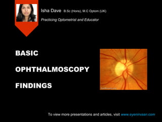
- 1. BASIC OPHTHALMOSCOPY FINDINGS Isha Dave B.Sc (Hons), M.C Optom (UK) Practicing Optometrist and Educator To view more presentations and articles, visit www.eyenirvaan.com
- 2. The Normal Fundus • Definition of Fundus: – The interior of the globe as seen with an opthalmoscope – Limit is the orra serrata – Direct ophthalmoscopy allows visualisation of approx 60-70% of fundus To view more presentations and articles, visit www.eyenirvaan.com
- 3. To view more presentations and articles, visit www.eyenirvaan.com
- 4. Fundus Background • What colour is the retina???? To view more presentations and articles, visit www.eyenirvaan.com
- 5. Fundus Background • Orange/red background comes from: – Light directly reflected from choroidal blood vessels – Light reflected from sclera and transmitted through choroidal blood vessels • Amount of light reflected depends on: – Degree of pigmentation of retina – Degree of pigmentation of choroid • Most fundus layers are transparent and do not contribute to appearance of fundus To view more presentations and articles, visit www.eyenirvaan.com
- 6. Types of fundus 1. Dark/Negroid 2. Medium/Caucasian 3. Light/Blonde 4. Tesselated 5. Albinotic To view more presentations and articles, visit www.eyenirvaan.com
- 7. 1. Dark/Negroid • Heavily pigmented RPE • Heavily pigmented choroid To view more presentations and articles, visit www.eyenirvaan.com
- 8. 2. Medium/Caucasian • Normally pigmented RPE • Normally pigmented choroid To view more presentations and articles, visit www.eyenirvaan.com
- 9. 3. Light/Blonde • Lightly pigmented RPE • Lightly pigmented choroid To view more presentations and articles, visit www.eyenirvaan.com
- 10. 4. Tessilated • Lightly pigmented RPE • Normal/heavily pigmented choroid To view more presentations and articles, visit www.eyenirvaan.com
- 11. 5. Albinotic • Virtually no pigment in RPE or choroid To view more presentations and articles, visit www.eyenirvaan.com
- 12. Why are some fundi more pigmented than others? • Age • Race • Hereditary • Metabolism To view more presentations and articles, visit www.eyenirvaan.com
- 13. Optic Disc • Definition: – The ophthalmoscopic view of the optic nerve head • Features: – Size – Shape – Colour (NRR) – Margins – Cupping – Vessels To view more presentations and articles, visit www.eyenirvaan.com
- 14. To view more presentations and articles, visit www.eyenirvaan.com
- 15. Optic Disc Cupping • No detectable cup in 15% of eyes • Can be measured by recording vertical C/D ratio • >0.2 difference between the eyes is suspicious of glaucoma To view more presentations and articles, visit www.eyenirvaan.com
- 16. Optic Disc Vessels • Central Retinal Artery – Becomes and arteriole after branching • Central Retinal Vein – Becomes a venule after branching • Cilio-retinal arteries – Not branches of CRA, derived from blood vessels supplying the choroid To view more presentations and articles, visit www.eyenirvaan.com
- 17. Optic Disc surroundings • Elschnig’s scleral ring – RPE and choroid stop short of the disc • Choroidal crescent – RPE stops short of the disc • Peri-peripheral pigment
- 18. Retinal Blood Vessels • Two layers of capillaries: – Superficial network in nerve fibre layer – Deep network at junction of inner nuclear and outer plexiform layer – No capillaries at the fovea • Walls of BVs are transparent • Linear light reflex notable feature of arterioles • Purpose of vessels • Ocular vessel health reflects health of circulation throughout body
- 19. A/V and V/A crossing phenomena • Generally arterioles cross venules • Venule dips to avoid arteriole • Disease induced changes are most noticeable at the crossings To view more presentations and articles, visit www.eyenirvaan.com
- 20. Vessel Calibre • Pathological changes cause arteriole narrowing or venule thickening • A/V ratio good indication of patholgy – Normal = 2/3
- 21. Macula • Macula is 1.5 to 2 disc diameters temporal to optic disc • Horizontally oval • Roughly same size as the optic disc • Foveal reflex – Bright spot of light at centre of macula – Only real landmark on healthy macula
- 23. Peripheral Retina • Peripheral retinal lesions: – Size (in mm or DD) – Location – Shape – Colour – Flat/raised?? To view more presentations and articles, visit www.eyenirvaan.com
- 24. Direct Vs. Indirect Opthalmoscopy • Direct – Good magnification – Good for macular assessment – V. useful is assessment of media opacities – Real image – right way up! – Light and portable • Indirect – Stereopsis!! – Good FOV – Good for optic disc and peripheral retina assessment To view more presentations and articles, visit www.eyenirvaan.com
- 25. THANK YOU To view more presentations and articles, visit www.eyenirvaan.com