PRESENTER: DR CHETHANA G R
MODERATOR: DR N.VIDHYA
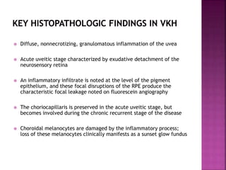
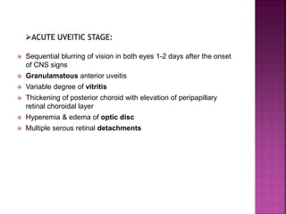
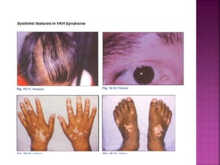
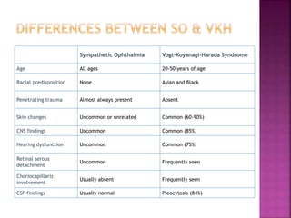
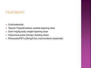
Vogt-Koyanagi-Harada (VKH) disease is a multisystemic
disorder characterized by granulomatous panuveitis with
exudative retinal detachments that is often associated
with neurologic and cutaneous manifestations.
 Commonly affects darkly pigmented ethnic groups
 Uncommon among whites
 Women more commonly affected than Men Except in Japanese
populations
 Most common in second to fouth decade of life
 Unknown
 Experimental evidence suggests Cell mediated autoimmune
process against Melanocytes of all organ systems(genetically
susceptible individuals)
 T helper-1 cells & upregulation of associated cytokines(IL-2,IL-6
&INF-gamma) also plays a role
 Recenty study suggests that IL-23(differentiation of IL-17
producing CD4 helper T lymphocytes) responsible for development
& maintanence of autoimmune process
 Sensitisation to melanocyte antigenic peptides by cutaneous
injury/viral infections-possible trigger
 Tyrosinase/Tyrosinase related protiens(75 Kda protein & S-100
protein targets melaocytes
 HLA-DR4 in Japanese population
 HLA DRB1 *0405,HLA DRB1*0410 haplotypes-stongly associated
risk
 84% Hispanic patients from Southern California found to have high
relative risk with HLA-DR1 than HLADR4
 Diffuse, nonnecrotizing, granulomatous inflammation of the uvea
 Acute uveitic stage characterized by exudative detachment of the
neurosensory retina
 An inflammatory infiltrate is noted at the level of the pigment
epithelium, and these focal disruptions of the RPE produce the
characteristic focal leakage noted on fluorescein angiography
 The choriocapillaris is preserved in the acute uveitic stage, but
becomes involved during the chronic recurrent stage of the disease
 Choroidal melanocytes are damaged by the inflammatory process;
loss of these melanocytes clinically manifests as a sunset glow fundus
 Flu like symptoms – 3-5 days
 Headache,neck stiffness,confusion ,nausea,fever,
dysacusia,tinnitus,orbital pain,photophobia Hypersensitivity of
skin & hair
 Focal Neurological signs:Cranial
neuropathies,Hemiparesis,Aphasia,Transverse myelitis &
ganglionitis
 CSF Analysis:lymphocytic pleocytosis,Normal level of glucose>80%
of patients(may persist up to 8 wks)
 Sequential blurring of vision in both eyes 1-2 days after the onset
of CNS signs
 Granulamatous anterior uveitis
 Variable degree of vitritis
 Thickening of posterior choroid with elevation of peripapillary
retinal choroidal layer
 Hyperemia & edema of optic disc
 Multiple serous retinal detachments
 B/L multiple serous RD in uveitic stage
 Multiple, focal areas of
leakage are noted at the
level of the RPE, and
subretinal fluid
accumulation may be
seen
 Right eye, early
arteriovenous phase of
fluorescein angiogram
exhibiting multifocal
areas of
hyperfluorescence at the
level of the RPE along
with pooling of dye in
the subretinal space
 (a) Multiple serous retinal detachments in the acute
uveitic stage of VKH. Also note marked optic disk
hyperemia.
 (b) OCT of the same eye confirms exudative retinal
detachment as well as retinal edema.
 Focal serous RD often shallow(clover leaf pattern)
coalasce to form large bullous exudative RD-profound visual
loss
 Less commonly,mutton fat KP’s,iris nodules at pupillary
margin are observed
 AC may be shallow due to forward displacement of lens-iris
diaphragm(ciliary body edema & annular choroidal
detachment)
 IOP may be elevated or low secondary to ciliary body shut
down
 Several weeks later to several months
 Cutaneous symptoms
 Resolution of exudative RD
 Gradual depigmentation of choroid leads to classic
orange-red discolouration(Sunset glow fundus)
 In addition,small,round discrete depigmented lesions
–inferior mid peripheral fundus (nummular scars )
 Juxta papillary depigmentation (pale disc) may also
occur
 Perilimbal vitiligo(Sugiura sign)-85% of japanese
patients,not in whites
 Integumentary changes:Vitiligo,poliosis,alopecia corresponds to
fundus depigmentation occurs in 30% of patients
 Skin & hair changes usually occur weeks – months after onset of
ocular inflamation but it may occur simultaneously
 10-63% develops vitiligo on ethnic background
 A smoldering panuveitis with acute episodic exacerbations of
granulomatous anterior uveitis
 Iris nodules may appear as round, whitish, well-circumscribed
lesions on a background of atrophic iris stroma
 Posterior segment recurrences associated with
vitritis,papillitis,multifocal choroiditis,exudative RD- rare
 Anterior segment recurrence coincides with subclinical choroidal
inflammation requires systemic therapy
 Sequelae of chronic inflammation leads to
PSCC,glaucoma,CNV,sub retinal fibrosis
1. No history of penetrating ocular trauma or surgery
preceding the initial onset of uveitis
2. No clinical or laboratory evidence suggestive of other
ocular disease entities
3. Bilateral ocular involvement (early n late - must be met,
depending on the stage of disease when the patient is
examined)
4. Neurological/auditory findings (may resolve by time of
evaluation)
5. Intergumentory finding (not preceding onset of central
nevous system or ocular disease)
Complete
VKH
Requires criteria 1to5
Incomplete
VHK
Requires Criteria 1–3 and
either 4 or 5 must be
present
Probable
VKH
(isolated ocular disease):
Criteria 1–3 must be
present
 Usually clinical
 Characterised by Exudative RD in acute stage,Sunset glow fundus
in chronic recurrent stage
 CBC,Mantoux test,TPHA-To rule out infectious cause
 FFA,ICG Angiography,OCT,USG,Lumbar puncture help in
confirming diagnisis
 Acute uveitic stage:
 Numerous hyperfluorescent foci at level of RPE in early stage
followed by pooling of dye in sub-retinal space in areas of
Neurosensory dtachment
 Majority shows disc leakage,CME & retinal vascular leakage are
uncommon
 Convalescent & Chronic recurrent stage:
 Focal RPE loss and atrophy produce multiple hyperfluorescent
window defects without progressive staining
 Highlights choroidal pathology
 Shows delay in choriocapillaries & choroidal vessel perfusion
 Early choroidal vessel stromal hyperfluorescence & leakage
 Disc hyperfluorescence
 Multiple hypofluorescent spots throughout the fundus indicates
foci of lymphocytic infiltration
 Hyperfluorescent pinpoint changes with in areas of exudative RD
 Hypofluorescent spots-sensitive marker and follow up of sub
clinical choroidal inflammation(when fundoscopic & FFA findings
are unremarkable)
ICG photographs of the right eye of a patient in the acute phase of VKHD
showing (A) patchy hypofluorescence during the early angiographic phase,
(B) large choroidal stromal vessel hyperfluorescence with fuzzy choroidal
vessels in the early phase, (C) hypofluorescent dark dots during the
intermediate phase of angiography, and (D) diffuse choroidal
hyperfluorescence in the late phase.
 Helpful in diagnosis in presence of media opacity
 Shows diffuse,low to medium reflective thickening of posterior
choroid ,most prominent in peripapillary area with extension to
equatorial region
 Exudative RD
 Vitreous opacification
 Posterior thickening of sclera
 Helps in diagnosis &
monitoring of
 Serous macular
detachment
 CME
 CNVM
 Done in atypical cases who presented early with neurological signs
 Shows lymphocytic pleocytosis
 Sympathetic ophthalmia
 Uveal effusion syndrome
 Posterior scleritis
 Primary intra ocular lymphoma
 Uveal lymphoid infiltration
 APMPPE
 Sarcoidosis
 Syphilis
 Lyme disease
Sympathetic Ophthalmia Vogt-Koyanagi-Harada Syndrome
Age All ages 20-50 years of age
Racial predisposition None Asian and Black
Penetrating trauma Almost always present Absent
Skin changes Uncommon or unrelated Common (60-90%)
CNS findings Uncommon Common (85%)
Hearing dysfunction Uncommon Common (75%)
Retinal serous
detachment
Uncommon Frequently seen
Choriocapillaris
involvement
Usually absent Frequently seen
CSF findings Usually normal Pleocytosis (84%)
 Corticosteroids:
 Topical-1%prednisolone acetate-tapering dose
 Oral-1mg/kg body weight-tapering dose
 Intavenous-pulse therapy (loading dose)
 Periocular(PST)-(20mg/0.5cc triamcinolone acetonide)
 Immunosupprasants
 Cyclosporine, 5 mg/kg/ day
 Tacrolimus 0.1–0.15 mg /kg /day
 Cytotoxic Agents[10,42]
 Azathioprine, 1–2.5 mg/kg/ day
 Mycophenylate mofetil, 1-3 g/day
 Cyclophosphamide 1–2 mg/ kg/ day
 Chlorambucil 0.1 mg/kg/day with adjustment of dose every 3
weeks up to a maximum of 18 mg/day
 Good with prompt and agressive therapy
 In addition to cataract and glaucoma,subretinal fibrosis and
choroidal neovascular membrances may occur
 Albert and jacobie 3rd edition vol 1.page chapter 97
 Uveitis fundamentals and clinical practice – robert &
scott:4th edition page no 303 -315
Thank you

Vkh

  • 1.
    PRESENTER: DR CHETHANAG R MODERATOR: DR N.VIDHYA
  • 2.
    Vogt-Koyanagi-Harada (VKH) diseaseis a multisystemic disorder characterized by granulomatous panuveitis with exudative retinal detachments that is often associated with neurologic and cutaneous manifestations.
  • 3.
     Commonly affectsdarkly pigmented ethnic groups  Uncommon among whites  Women more commonly affected than Men Except in Japanese populations  Most common in second to fouth decade of life
  • 4.
     Unknown  Experimentalevidence suggests Cell mediated autoimmune process against Melanocytes of all organ systems(genetically susceptible individuals)  T helper-1 cells & upregulation of associated cytokines(IL-2,IL-6 &INF-gamma) also plays a role  Recenty study suggests that IL-23(differentiation of IL-17 producing CD4 helper T lymphocytes) responsible for development & maintanence of autoimmune process
  • 5.
     Sensitisation tomelanocyte antigenic peptides by cutaneous injury/viral infections-possible trigger  Tyrosinase/Tyrosinase related protiens(75 Kda protein & S-100 protein targets melaocytes
  • 6.
     HLA-DR4 inJapanese population  HLA DRB1 *0405,HLA DRB1*0410 haplotypes-stongly associated risk  84% Hispanic patients from Southern California found to have high relative risk with HLA-DR1 than HLADR4
  • 7.
     Diffuse, nonnecrotizing,granulomatous inflammation of the uvea  Acute uveitic stage characterized by exudative detachment of the neurosensory retina  An inflammatory infiltrate is noted at the level of the pigment epithelium, and these focal disruptions of the RPE produce the characteristic focal leakage noted on fluorescein angiography  The choriocapillaris is preserved in the acute uveitic stage, but becomes involved during the chronic recurrent stage of the disease  Choroidal melanocytes are damaged by the inflammatory process; loss of these melanocytes clinically manifests as a sunset glow fundus
  • 8.
     Flu likesymptoms – 3-5 days  Headache,neck stiffness,confusion ,nausea,fever, dysacusia,tinnitus,orbital pain,photophobia Hypersensitivity of skin & hair  Focal Neurological signs:Cranial neuropathies,Hemiparesis,Aphasia,Transverse myelitis & ganglionitis  CSF Analysis:lymphocytic pleocytosis,Normal level of glucose>80% of patients(may persist up to 8 wks)
  • 9.
     Sequential blurringof vision in both eyes 1-2 days after the onset of CNS signs  Granulamatous anterior uveitis  Variable degree of vitritis  Thickening of posterior choroid with elevation of peripapillary retinal choroidal layer  Hyperemia & edema of optic disc  Multiple serous retinal detachments
  • 10.
     B/L multipleserous RD in uveitic stage
  • 11.
     Multiple, focalareas of leakage are noted at the level of the RPE, and subretinal fluid accumulation may be seen  Right eye, early arteriovenous phase of fluorescein angiogram exhibiting multifocal areas of hyperfluorescence at the level of the RPE along with pooling of dye in the subretinal space
  • 12.
     (a) Multipleserous retinal detachments in the acute uveitic stage of VKH. Also note marked optic disk hyperemia.  (b) OCT of the same eye confirms exudative retinal detachment as well as retinal edema.
  • 14.
     Focal serousRD often shallow(clover leaf pattern) coalasce to form large bullous exudative RD-profound visual loss  Less commonly,mutton fat KP’s,iris nodules at pupillary margin are observed  AC may be shallow due to forward displacement of lens-iris diaphragm(ciliary body edema & annular choroidal detachment)  IOP may be elevated or low secondary to ciliary body shut down
  • 15.
     Several weekslater to several months  Cutaneous symptoms  Resolution of exudative RD  Gradual depigmentation of choroid leads to classic orange-red discolouration(Sunset glow fundus)  In addition,small,round discrete depigmented lesions –inferior mid peripheral fundus (nummular scars )  Juxta papillary depigmentation (pale disc) may also occur  Perilimbal vitiligo(Sugiura sign)-85% of japanese patients,not in whites
  • 18.
     Integumentary changes:Vitiligo,poliosis,alopeciacorresponds to fundus depigmentation occurs in 30% of patients  Skin & hair changes usually occur weeks – months after onset of ocular inflamation but it may occur simultaneously  10-63% develops vitiligo on ethnic background
  • 19.
     A smolderingpanuveitis with acute episodic exacerbations of granulomatous anterior uveitis  Iris nodules may appear as round, whitish, well-circumscribed lesions on a background of atrophic iris stroma  Posterior segment recurrences associated with vitritis,papillitis,multifocal choroiditis,exudative RD- rare  Anterior segment recurrence coincides with subclinical choroidal inflammation requires systemic therapy  Sequelae of chronic inflammation leads to PSCC,glaucoma,CNV,sub retinal fibrosis
  • 21.
    1. No historyof penetrating ocular trauma or surgery preceding the initial onset of uveitis 2. No clinical or laboratory evidence suggestive of other ocular disease entities 3. Bilateral ocular involvement (early n late - must be met, depending on the stage of disease when the patient is examined) 4. Neurological/auditory findings (may resolve by time of evaluation) 5. Intergumentory finding (not preceding onset of central nevous system or ocular disease)
  • 22.
    Complete VKH Requires criteria 1to5 Incomplete VHK RequiresCriteria 1–3 and either 4 or 5 must be present Probable VKH (isolated ocular disease): Criteria 1–3 must be present
  • 24.
     Usually clinical Characterised by Exudative RD in acute stage,Sunset glow fundus in chronic recurrent stage  CBC,Mantoux test,TPHA-To rule out infectious cause  FFA,ICG Angiography,OCT,USG,Lumbar puncture help in confirming diagnisis
  • 25.
     Acute uveiticstage:  Numerous hyperfluorescent foci at level of RPE in early stage followed by pooling of dye in sub-retinal space in areas of Neurosensory dtachment  Majority shows disc leakage,CME & retinal vascular leakage are uncommon  Convalescent & Chronic recurrent stage:  Focal RPE loss and atrophy produce multiple hyperfluorescent window defects without progressive staining
  • 27.
     Highlights choroidalpathology  Shows delay in choriocapillaries & choroidal vessel perfusion  Early choroidal vessel stromal hyperfluorescence & leakage  Disc hyperfluorescence  Multiple hypofluorescent spots throughout the fundus indicates foci of lymphocytic infiltration  Hyperfluorescent pinpoint changes with in areas of exudative RD  Hypofluorescent spots-sensitive marker and follow up of sub clinical choroidal inflammation(when fundoscopic & FFA findings are unremarkable)
  • 28.
    ICG photographs ofthe right eye of a patient in the acute phase of VKHD showing (A) patchy hypofluorescence during the early angiographic phase, (B) large choroidal stromal vessel hyperfluorescence with fuzzy choroidal vessels in the early phase, (C) hypofluorescent dark dots during the intermediate phase of angiography, and (D) diffuse choroidal hyperfluorescence in the late phase.
  • 29.
     Helpful indiagnosis in presence of media opacity  Shows diffuse,low to medium reflective thickening of posterior choroid ,most prominent in peripapillary area with extension to equatorial region  Exudative RD  Vitreous opacification  Posterior thickening of sclera
  • 31.
     Helps indiagnosis & monitoring of  Serous macular detachment  CME  CNVM
  • 32.
     Done inatypical cases who presented early with neurological signs  Shows lymphocytic pleocytosis
  • 33.
     Sympathetic ophthalmia Uveal effusion syndrome  Posterior scleritis  Primary intra ocular lymphoma  Uveal lymphoid infiltration  APMPPE  Sarcoidosis  Syphilis  Lyme disease
  • 34.
    Sympathetic Ophthalmia Vogt-Koyanagi-HaradaSyndrome Age All ages 20-50 years of age Racial predisposition None Asian and Black Penetrating trauma Almost always present Absent Skin changes Uncommon or unrelated Common (60-90%) CNS findings Uncommon Common (85%) Hearing dysfunction Uncommon Common (75%) Retinal serous detachment Uncommon Frequently seen Choriocapillaris involvement Usually absent Frequently seen CSF findings Usually normal Pleocytosis (84%)
  • 35.
     Corticosteroids:  Topical-1%prednisoloneacetate-tapering dose  Oral-1mg/kg body weight-tapering dose  Intavenous-pulse therapy (loading dose)  Periocular(PST)-(20mg/0.5cc triamcinolone acetonide)
  • 36.
     Immunosupprasants  Cyclosporine,5 mg/kg/ day  Tacrolimus 0.1–0.15 mg /kg /day  Cytotoxic Agents[10,42]  Azathioprine, 1–2.5 mg/kg/ day  Mycophenylate mofetil, 1-3 g/day  Cyclophosphamide 1–2 mg/ kg/ day  Chlorambucil 0.1 mg/kg/day with adjustment of dose every 3 weeks up to a maximum of 18 mg/day
  • 37.
     Good withprompt and agressive therapy  In addition to cataract and glaucoma,subretinal fibrosis and choroidal neovascular membrances may occur
  • 38.
     Albert andjacobie 3rd edition vol 1.page chapter 97  Uveitis fundamentals and clinical practice – robert & scott:4th edition page no 303 -315
  • 39.