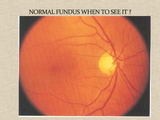
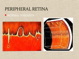
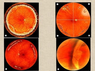
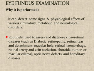
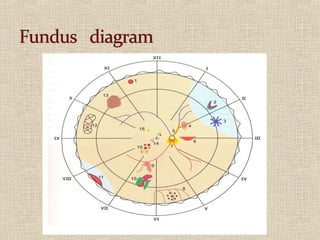
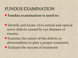
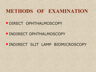
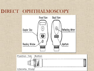
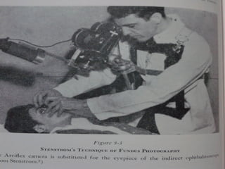
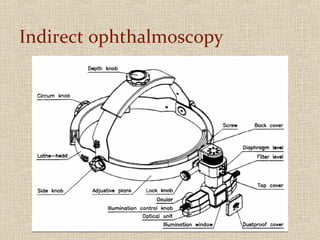
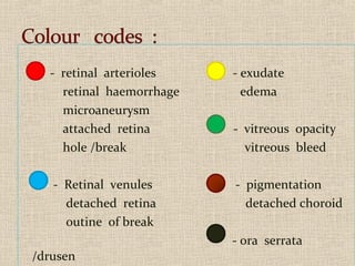
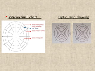
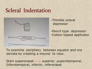
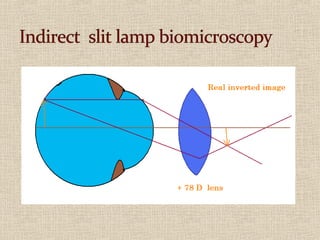
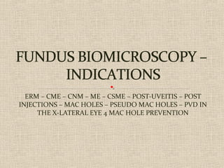
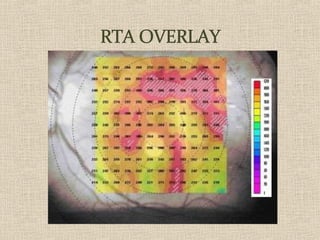
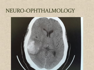
10. Why it is performed:
It can detect some signs & physiological effects of
various circulatory, metabolic and neurological
disorders.
Routinely used to assess and diagnose vitro-retinal
diseases (such as Diabetic retinopathy, retinal tear
and detachment, macular hole, retinal haemorrhage,
retinal artery and vein occlusion, choroidal tumor, or
macular edema), optic nerve defects, and hereditary
diseases.
11.
12.
13. Fundus examination is used to:
Identify and locate vitro-retinal and optical
nerve defects caused by eye diseases or
trauma.
Examine the extent of the defects or
abnormalities to plan a proper treatment.
Evaluate the success of treatment.
28. ERM – CME – CNM – ME – CSME – POST-UVEITIS – POST
INJECTIONS – MAC HOLES – PSEUDO MAC HOLES
29. -Thimble scleral
depressor
-Pencil type depressor
-Cotton tipped applicator
To examine periphery between equator and ora
serrata by creating a mound to view.
Start superonasal superior ,superotemporal,
Inferotemporal, inferior, inferonasal
30.
31.
32.
33.
34.
35. ERM – CME – CNM – ME – CSME – POST-UVEITIS – POST
INJECTIONS – MAC HOLES – PSEUDO MAC HOLES – PVD IN
THE X-LATERAL EYE 4 MAC HOLE PREVENTION
36. ERM – CME – CNM – ME – CSME – POST-UVEITIS – POST
INJECTIONS – MAC HOLES – PSEUDO MAC HOLES
45. B-scan ultrasound is most useful when direct
visualization of intraocular structures is
difficult or impossible. Situations that
prevent normal examination include lid
problems (eg, severe edema, partial or total
tarsorrhaphy), keratoprosthesis, corneal
opacities (eg, scars, severe edema), hyphema,
hypopyon, miosis, pupillary membranes,
dense cataracts, or vitreous opacities (eg,
hemorrhage, inflammatory debris).
47. In such cases, diagnostic B-scan ultrasound can accurately
image intraocular structures and give valuable information
on the status of the lens, vitreous, retina, choroid, and
sclera. However, in many instances, ultrasound is used for
diagnostic purposes even though pathology is clinically
visible. Such instances include differentiating iris or
ciliary body lesions; ruling out ciliary body detachments; and di
intraocular tumors, serous versus hemorrhagic
choroidal detachments, rhegmatogenous versus
exudative retinal detachments, and disc drusen versus
papilledema.
48.
49. VISUAL EVOKED
POTENTIAL - VEP
The visually evoked potential (VEP)
measures the electrical response of
the brain's primary visual cortex to
a visual stimulus.
NEURO-OPHTH DISORDERS
OPTIC PATHWAY DISORDERS
EXCLUDING CRITERIA
MEDICAL LEGAL
OTHERS
50.
51.
52.
53.
54.
55.
56. Preemie Eye test (ROP)...
how bad is it?
I made the mistake of
reading up about the
ROP eye test that they
are giving my baby early
next week. It sounds
absolutely terrible. How
did your babies do
afterwards? The nurses
have warned that he may
not eat well that day.
57.
58. Your Baby’s Eye Test
Why your baby needs an eye test
All babies on the neonatal unit are screened for an
eye condition called retinopathy of prematurity
(ROP) if they are born before 32 weeks of
pregnancy or if they weighed 1500 grams or less
at birth.
59.
60. ERM – CME – CNM – ME – CSME – POST-UVEITIS – POST
INJECTIONS – MAC HOLES – PSEUDO MAC HOLES
61.
62. NO DIABETIC RETINOPATHY WHAT FOR ???
NO POSTERIOR POLE CHANGES WHAT FOR ???
NO KNOWN ALLERGIES AND THE TWO ABOVE
WHAT FOR ???
NO CHANGES SO MEDICAL LEGAL ASPECT WHAT
FOR ???
THINK ABOUT