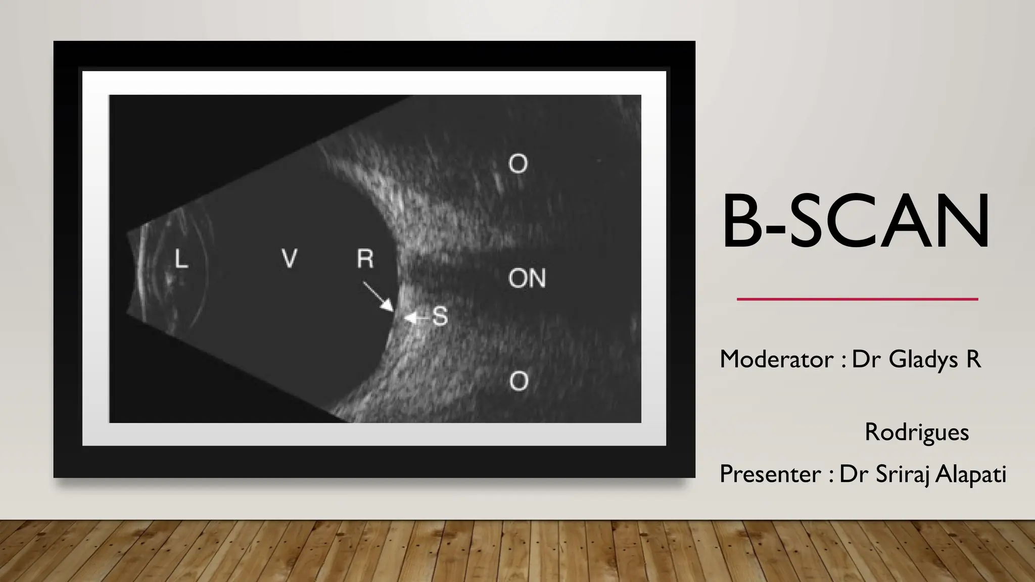
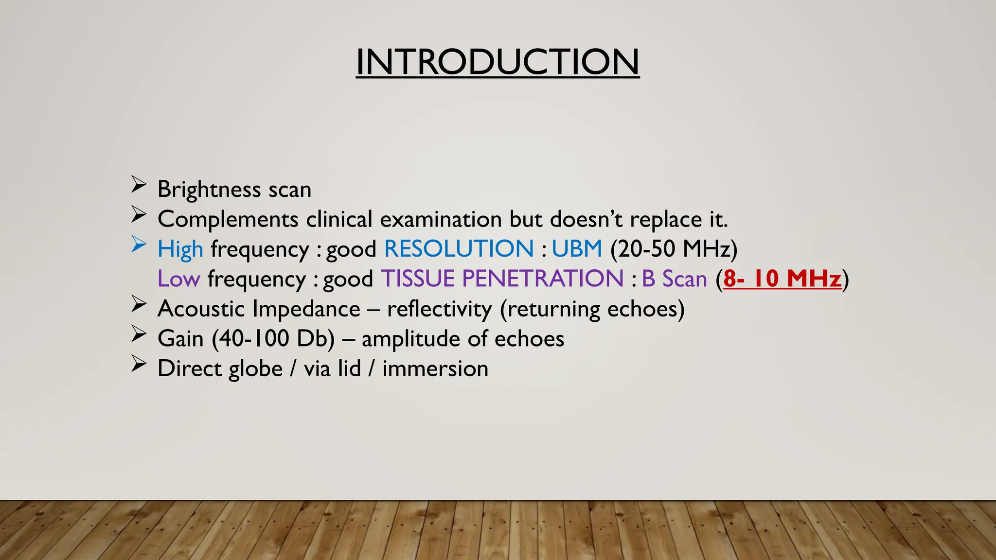
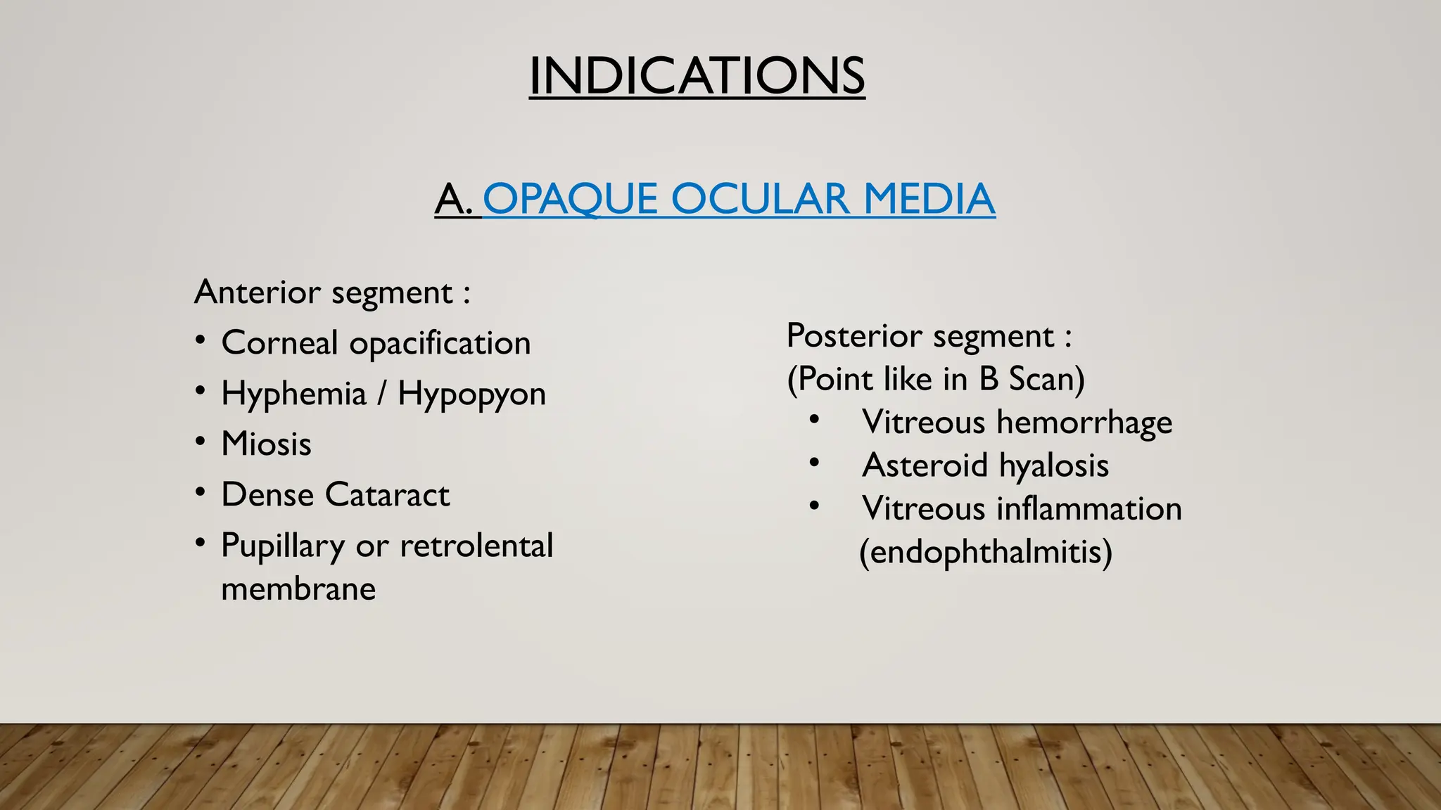
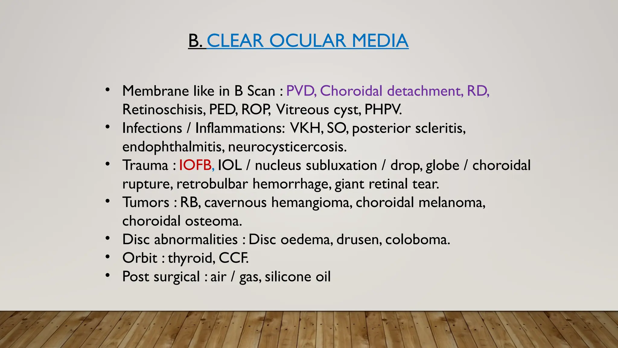
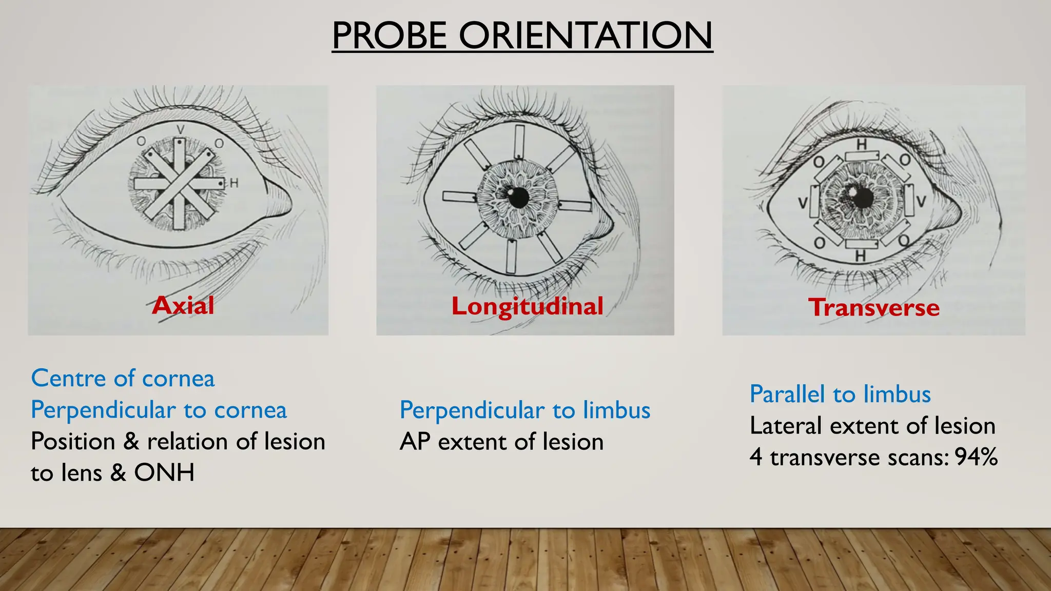
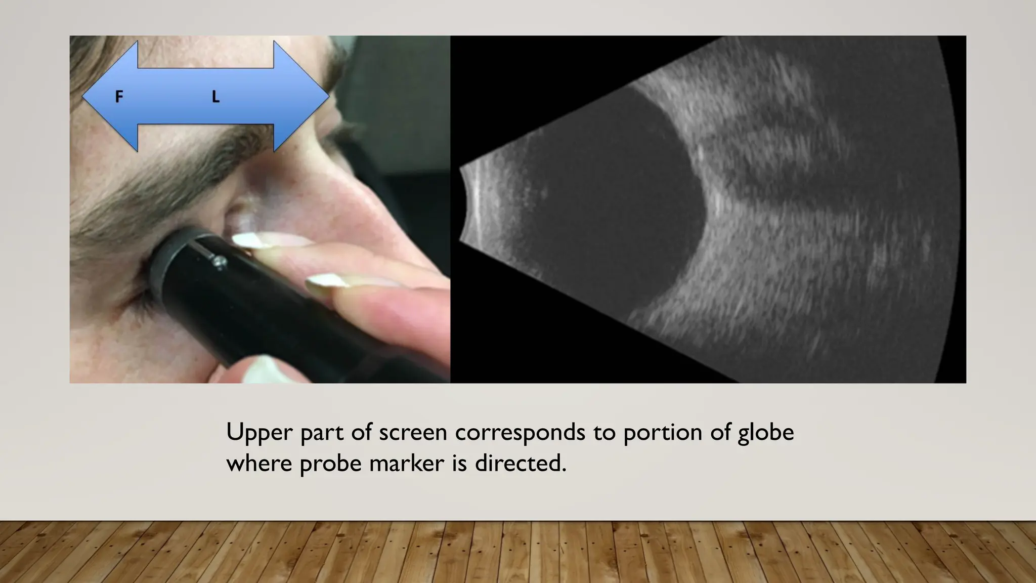
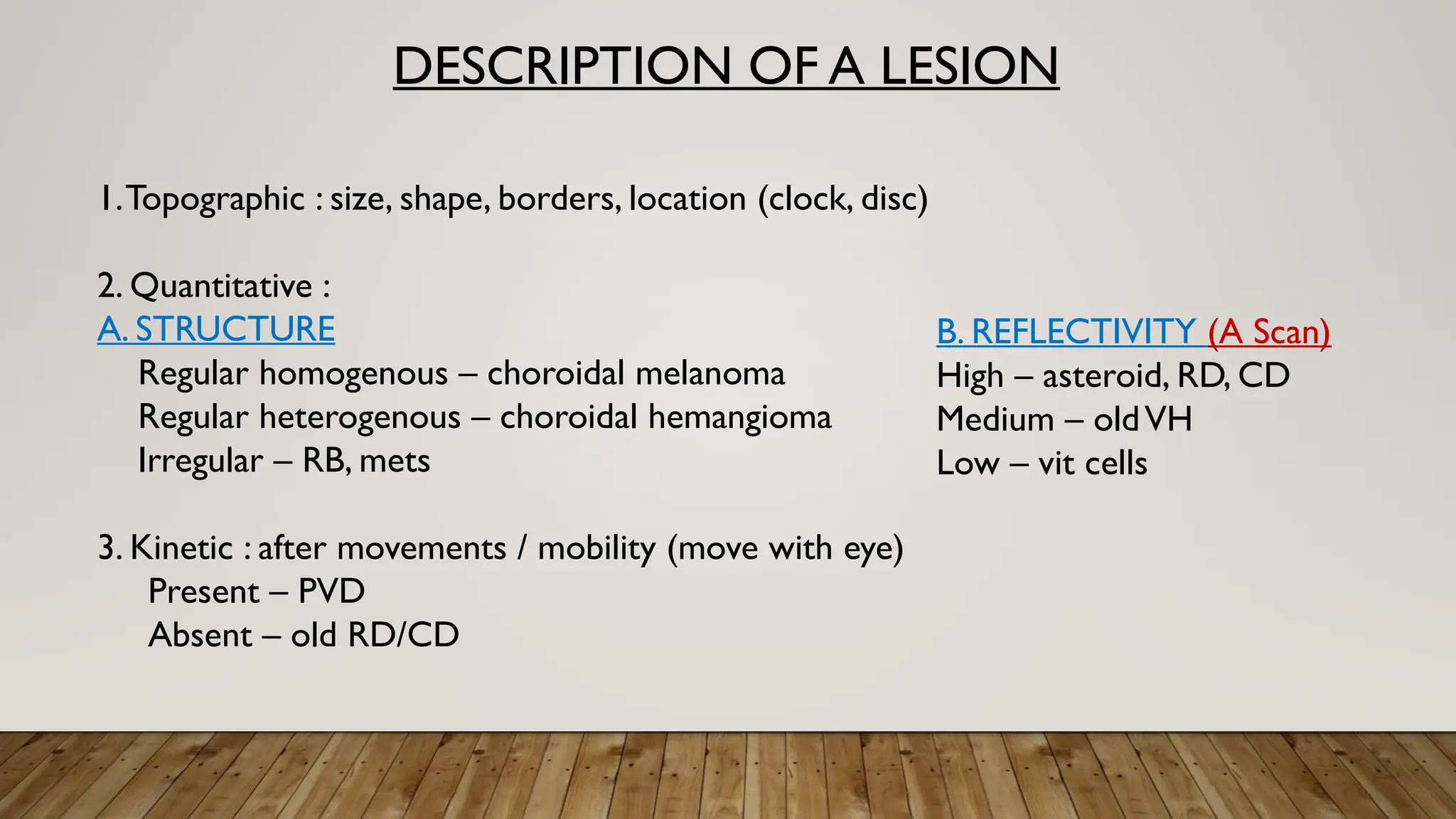
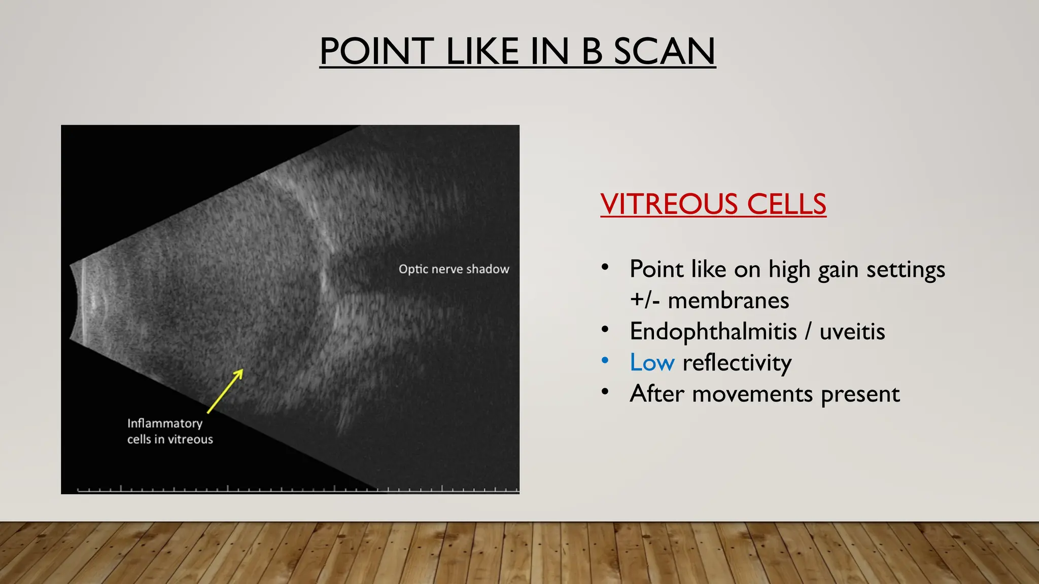
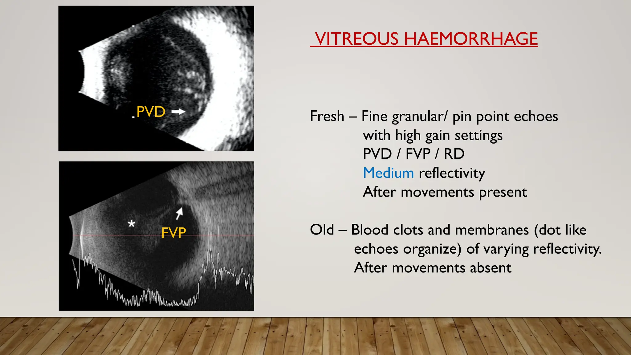
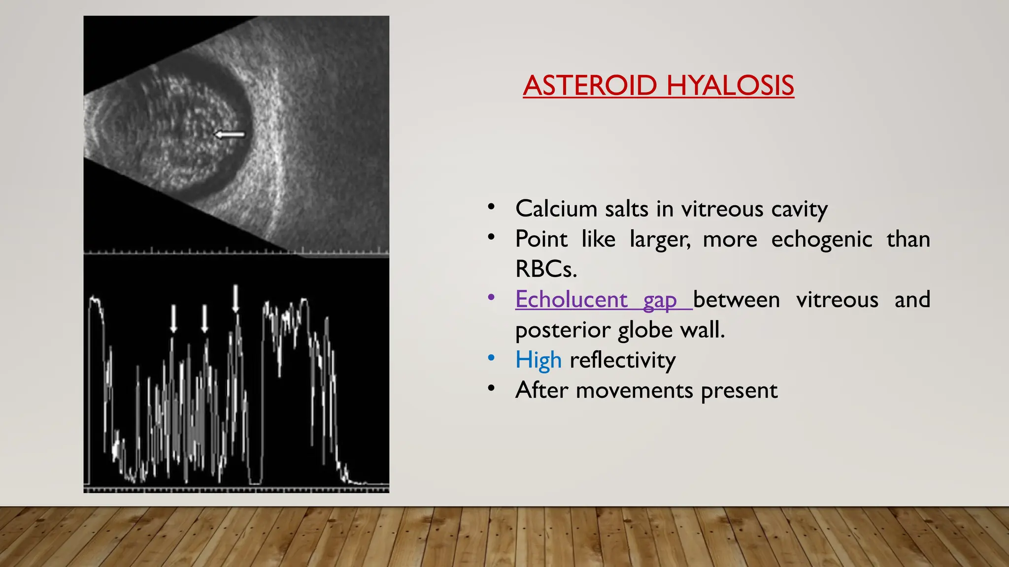
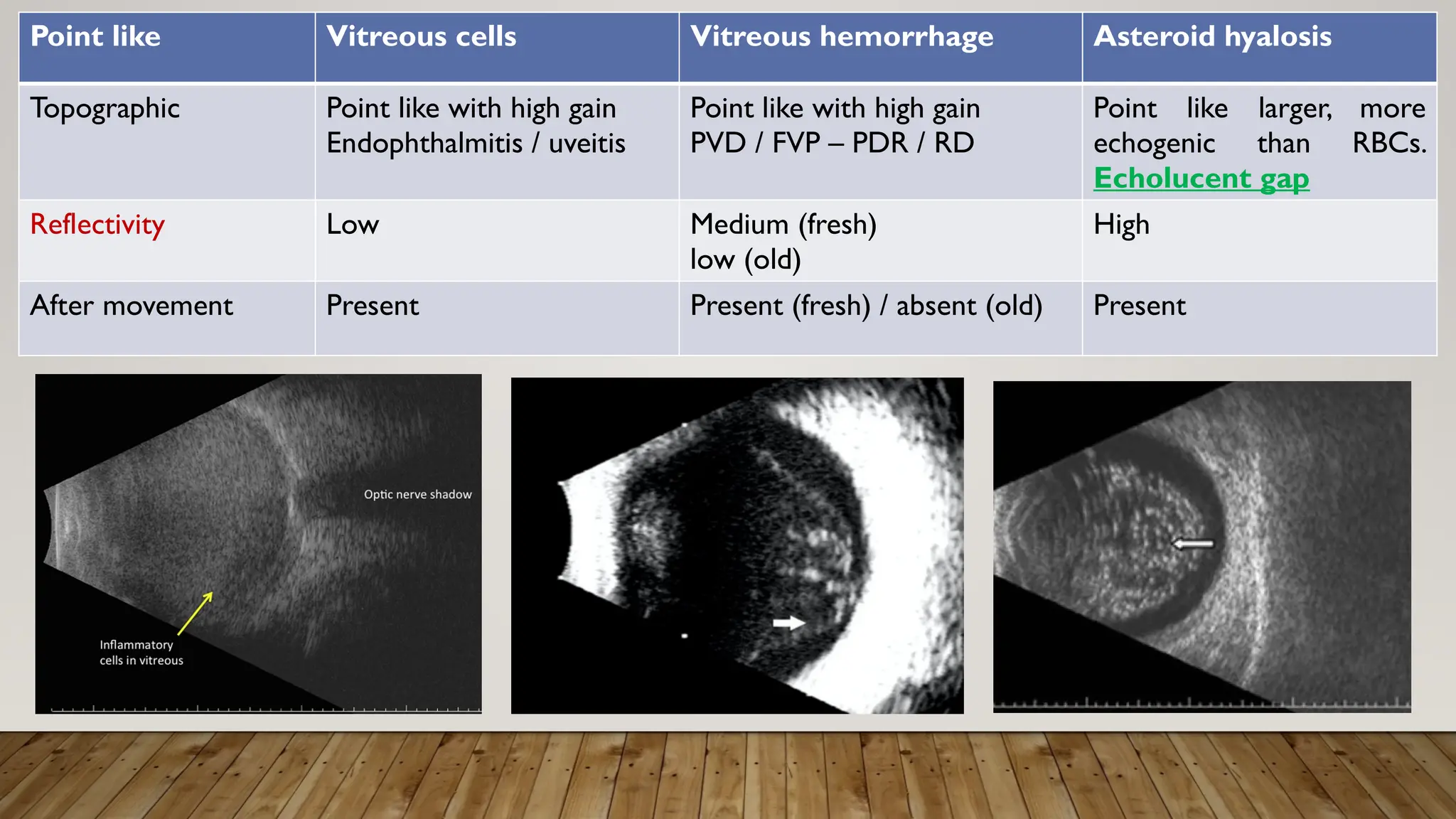
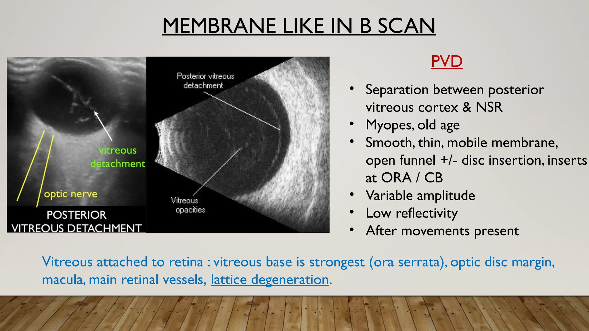
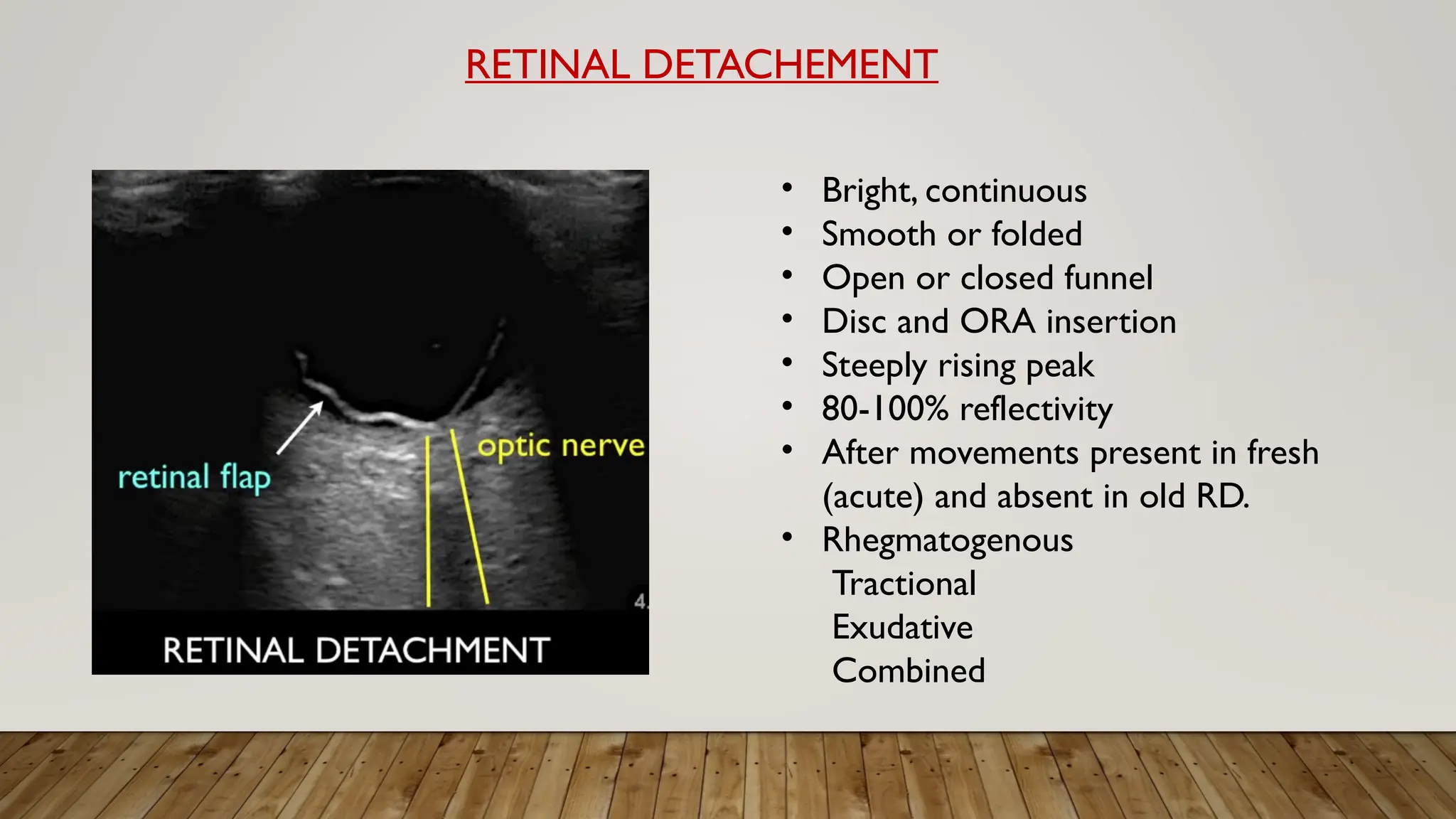
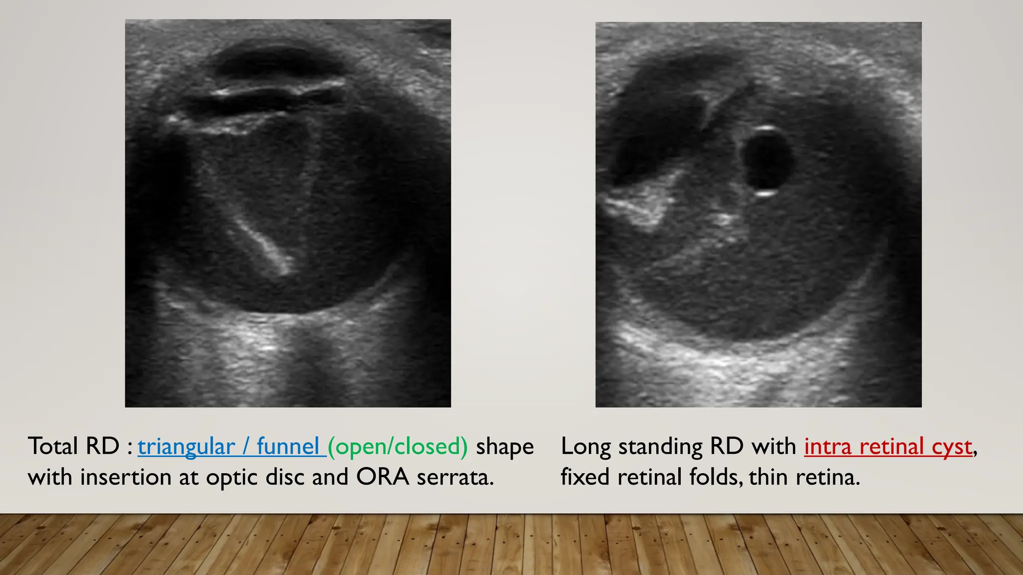
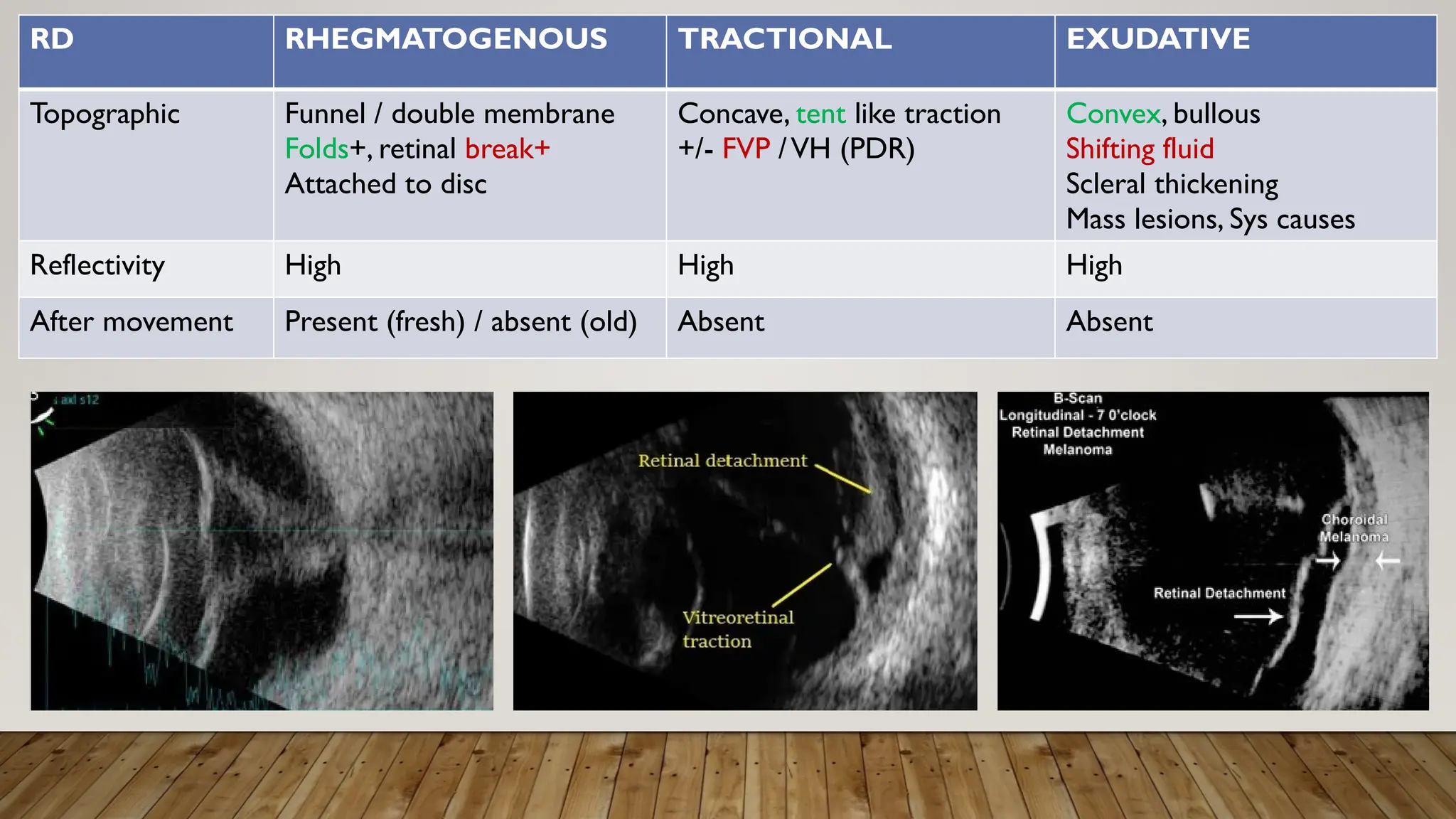
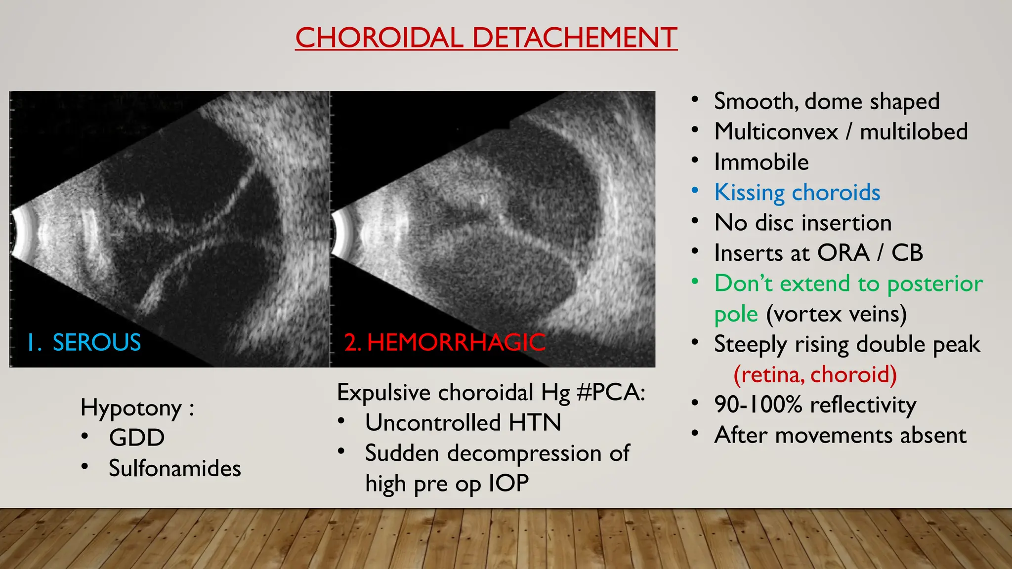
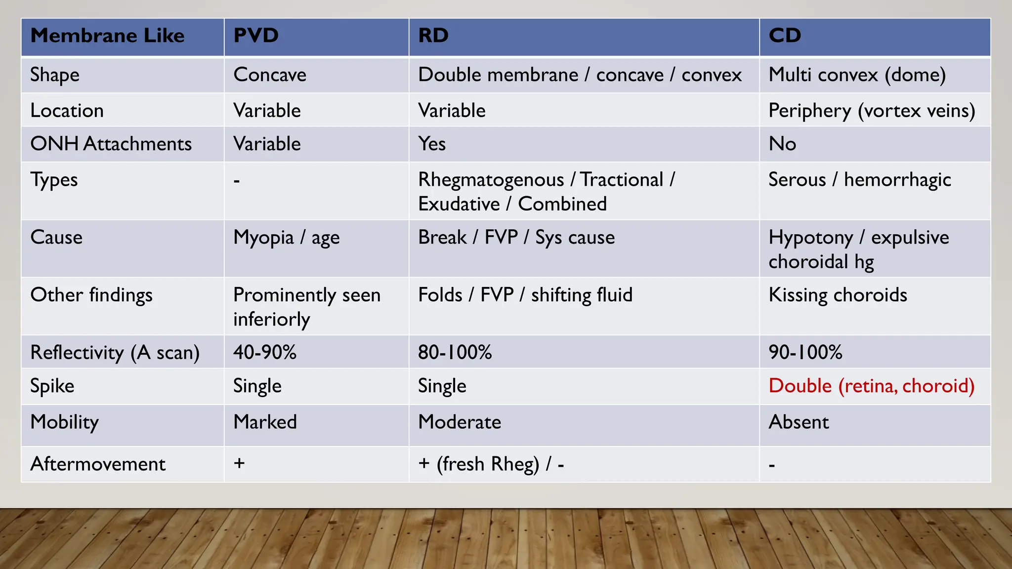
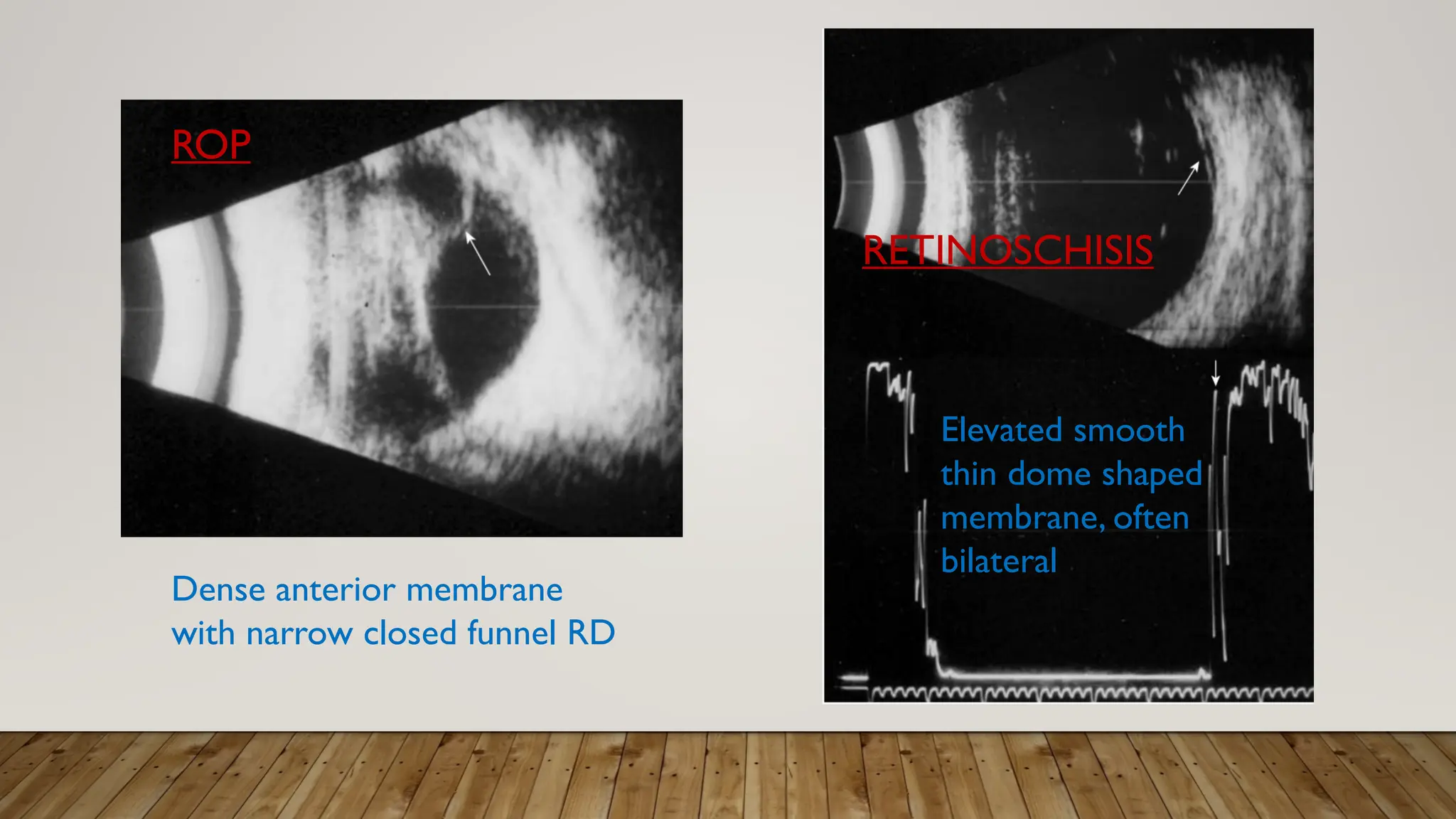
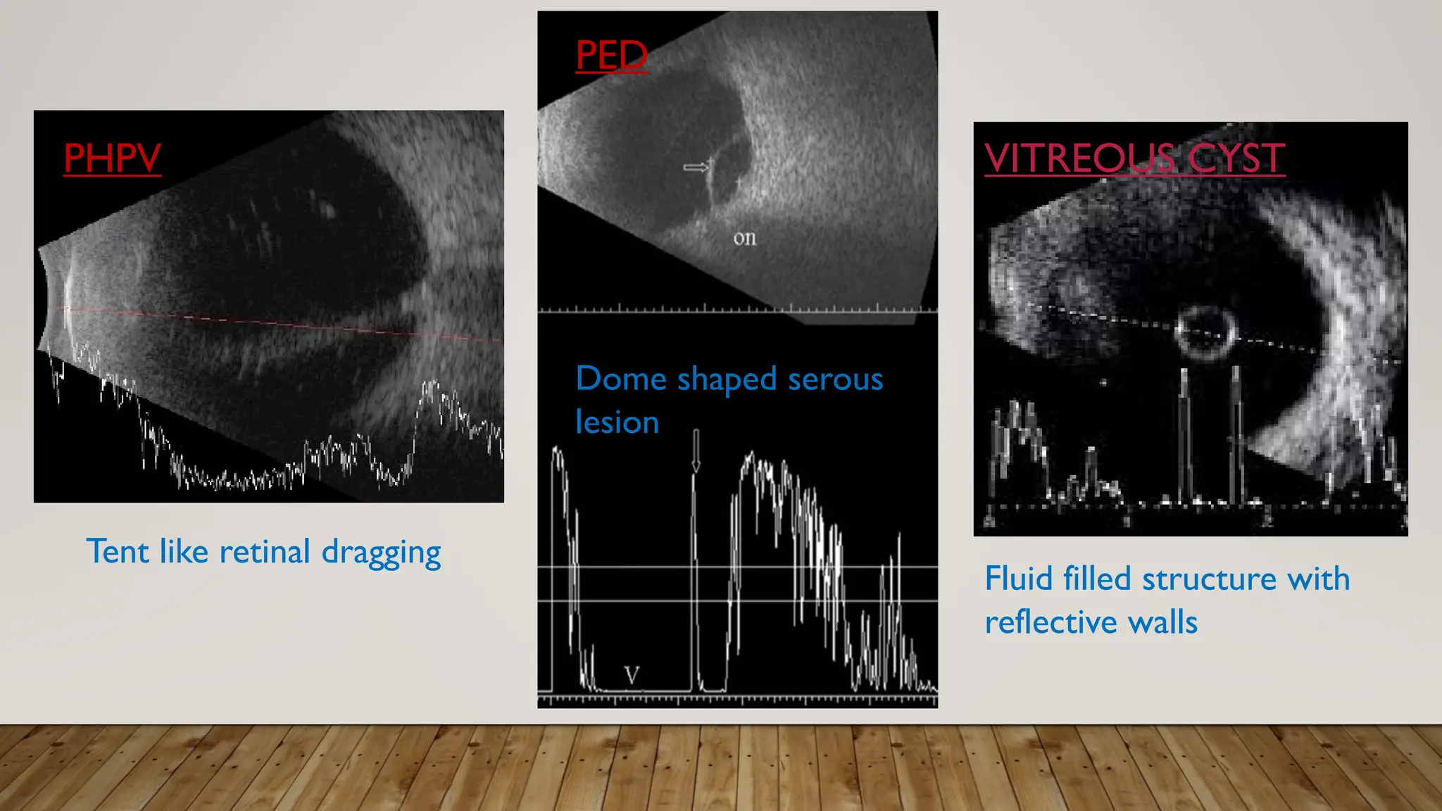
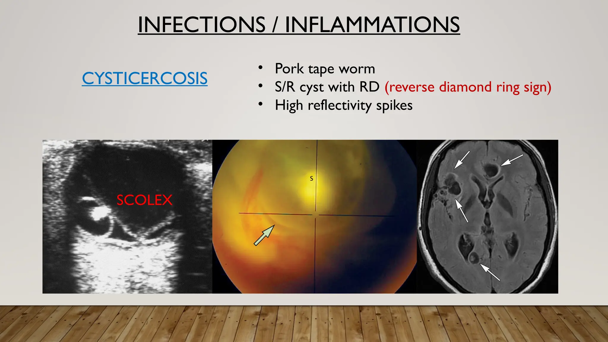
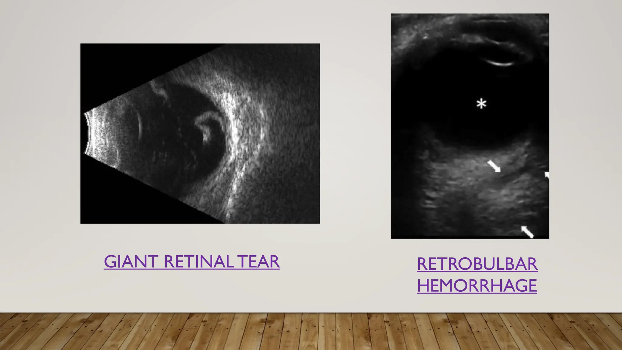
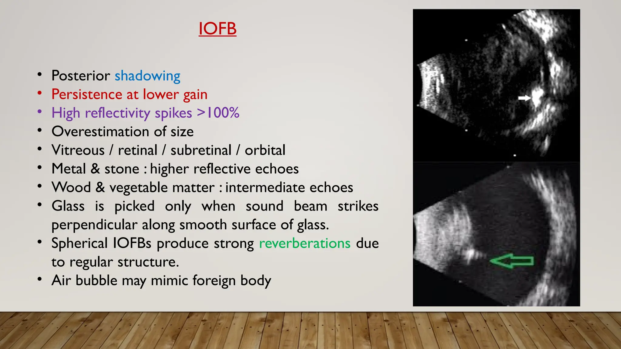
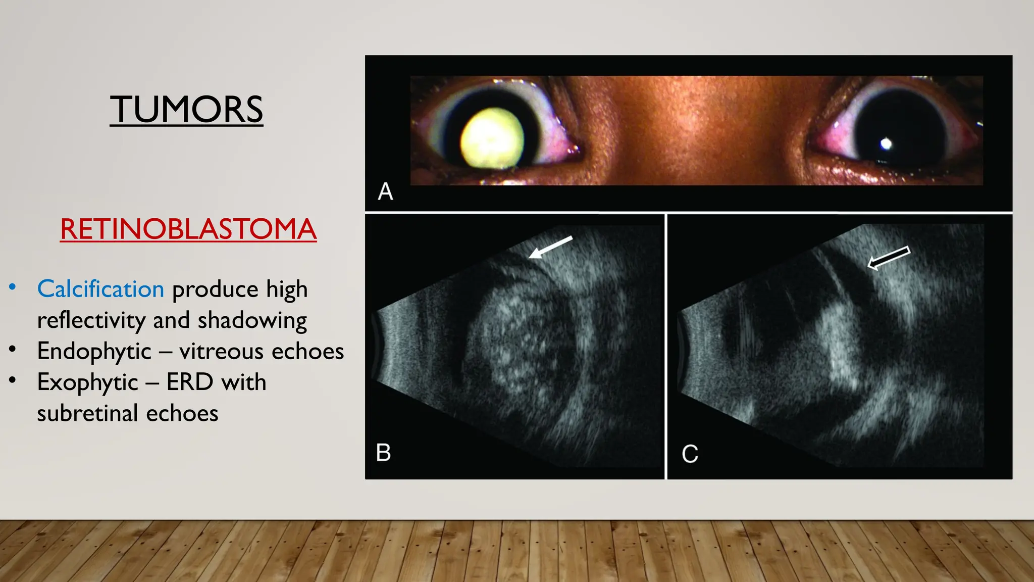
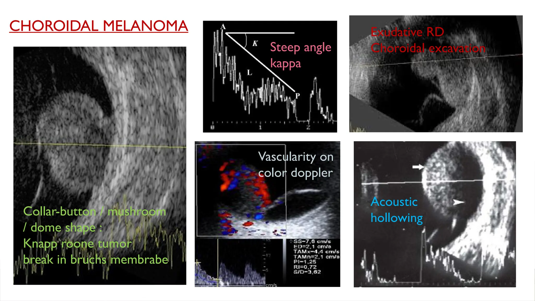
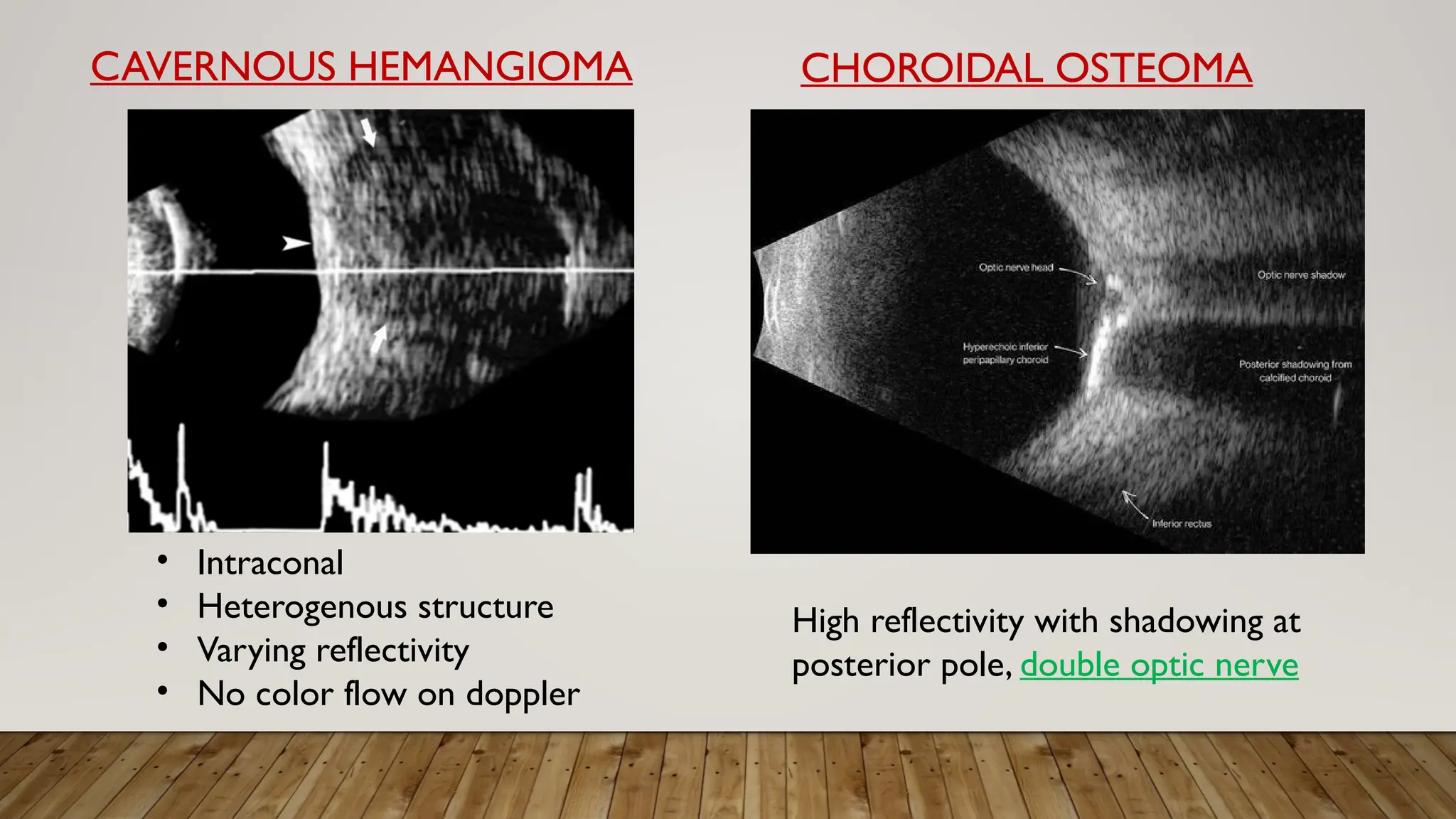
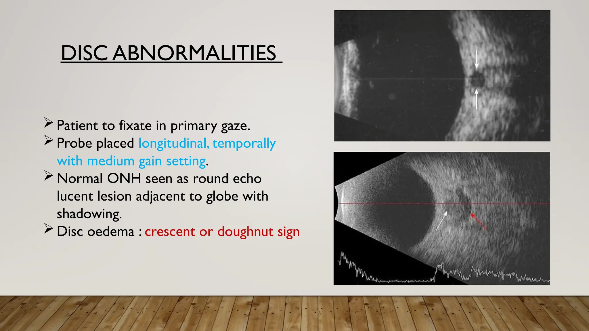
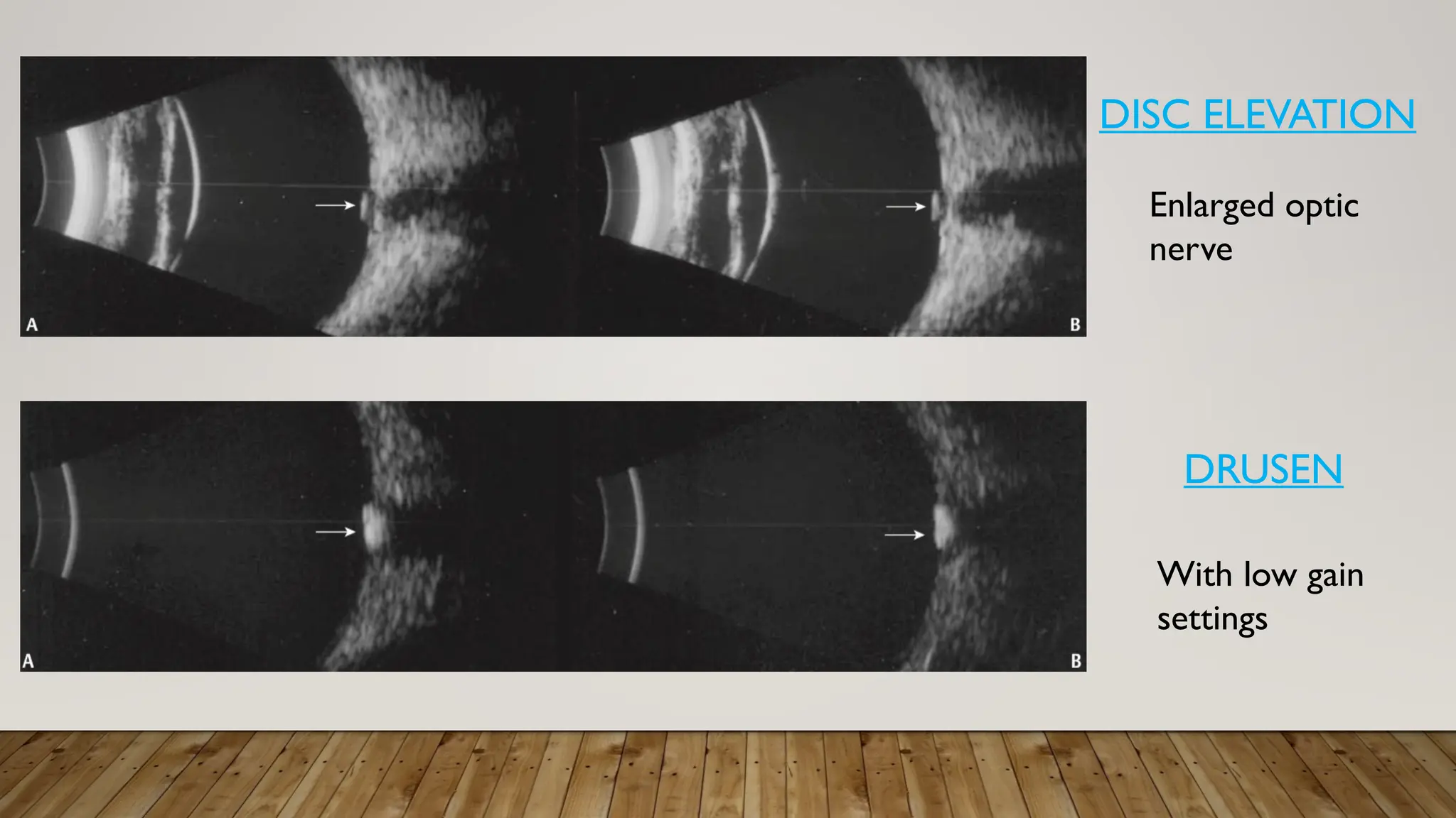
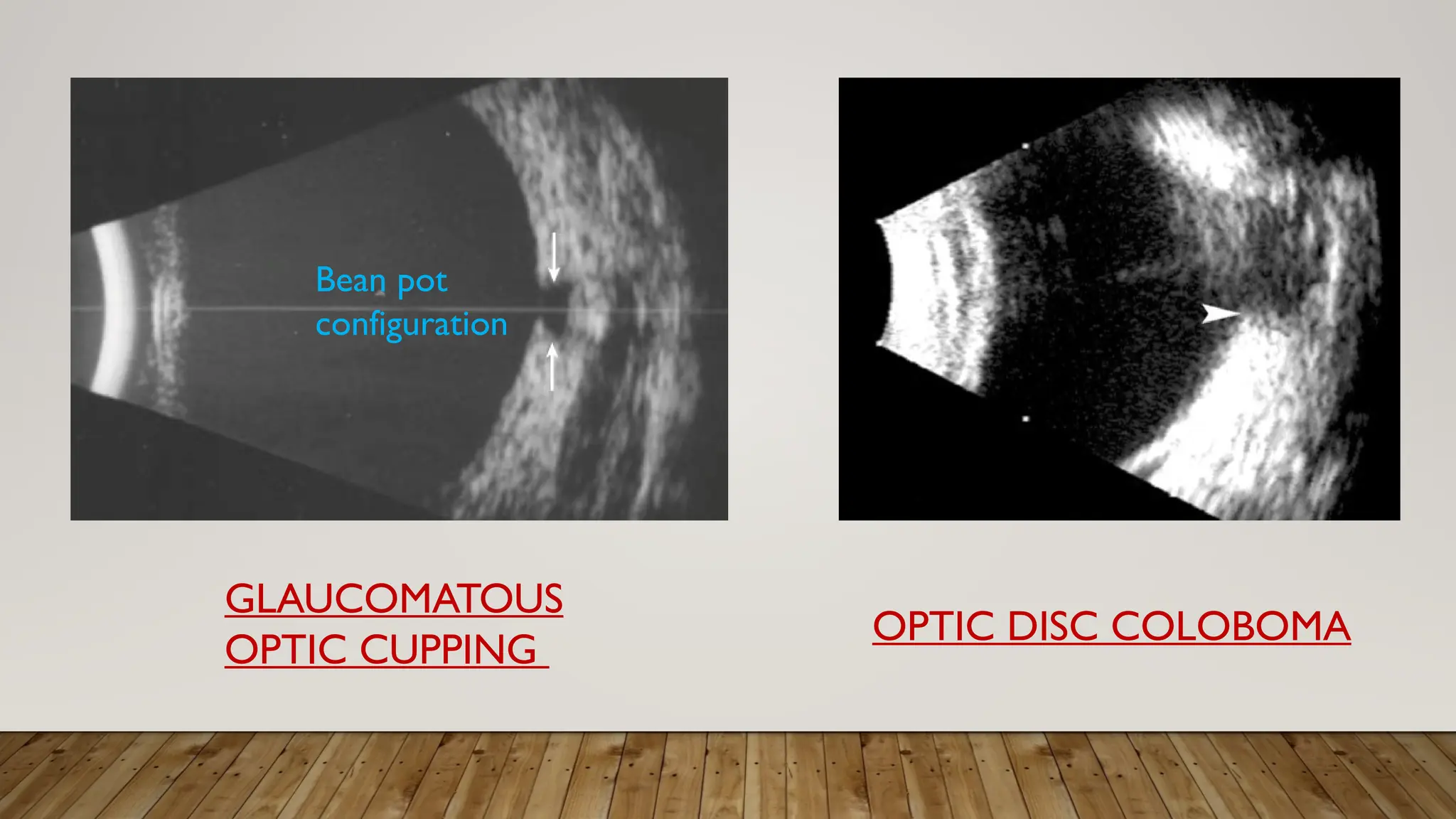
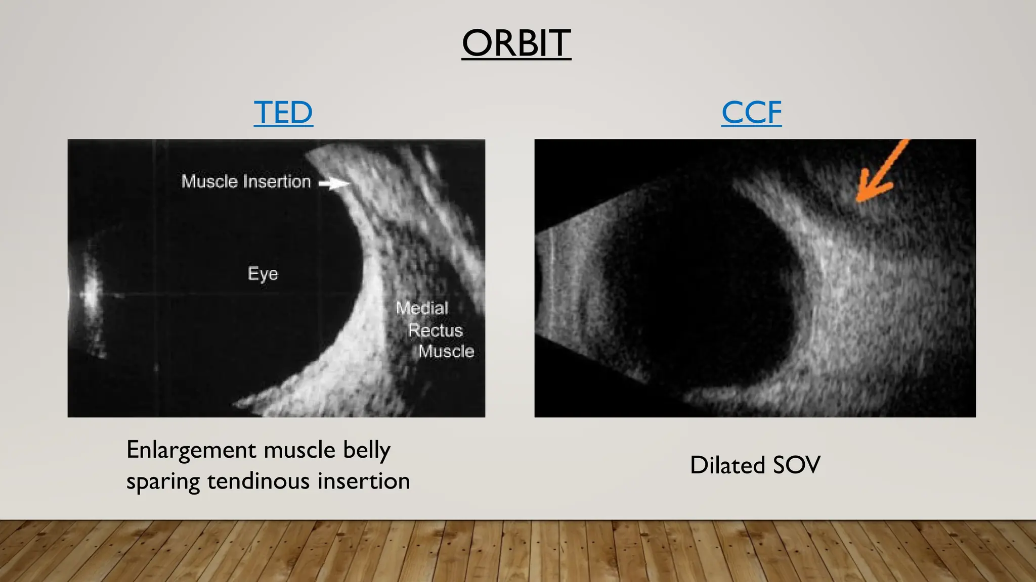
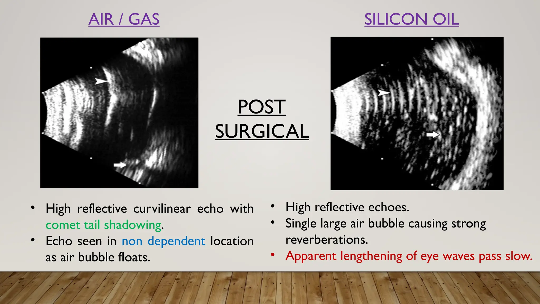
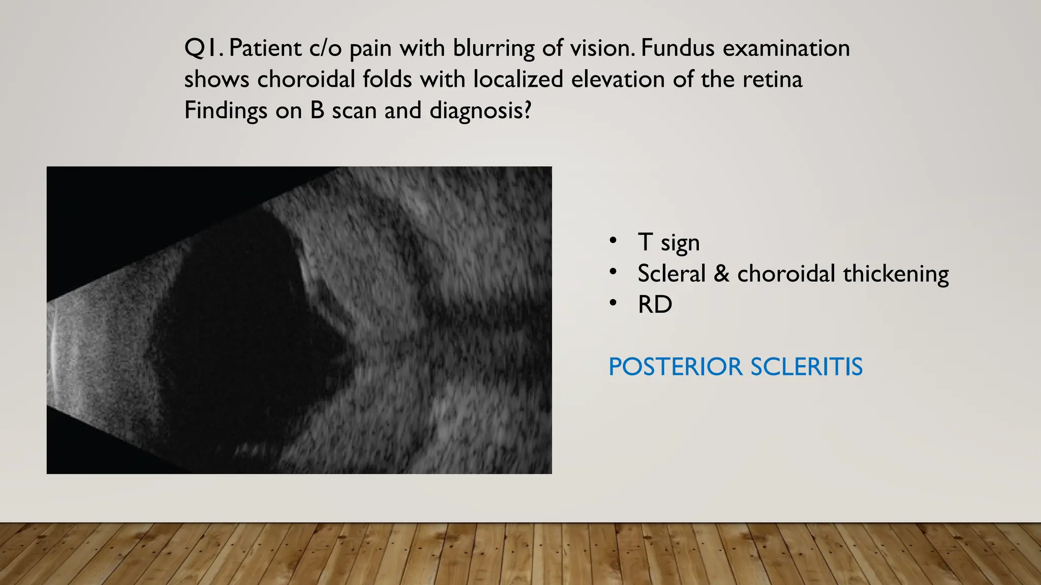
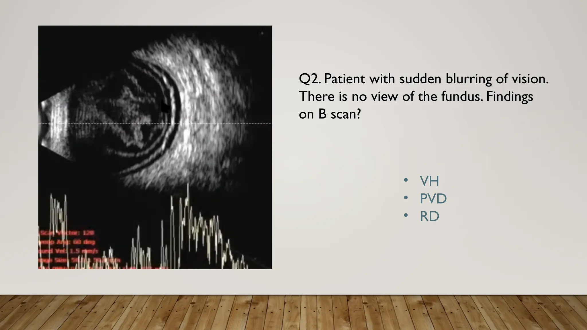
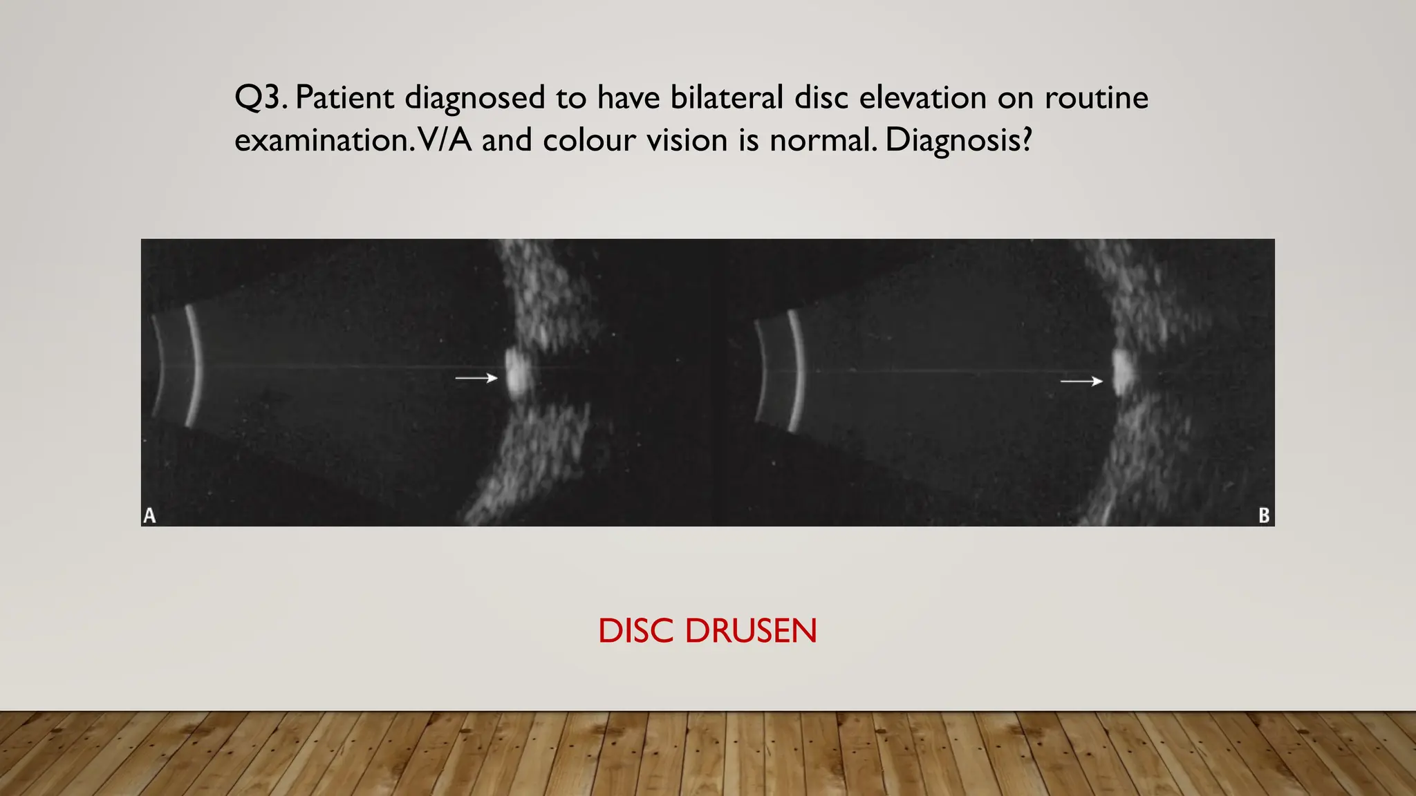
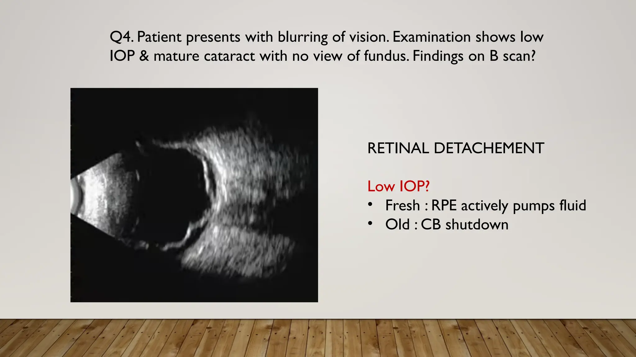
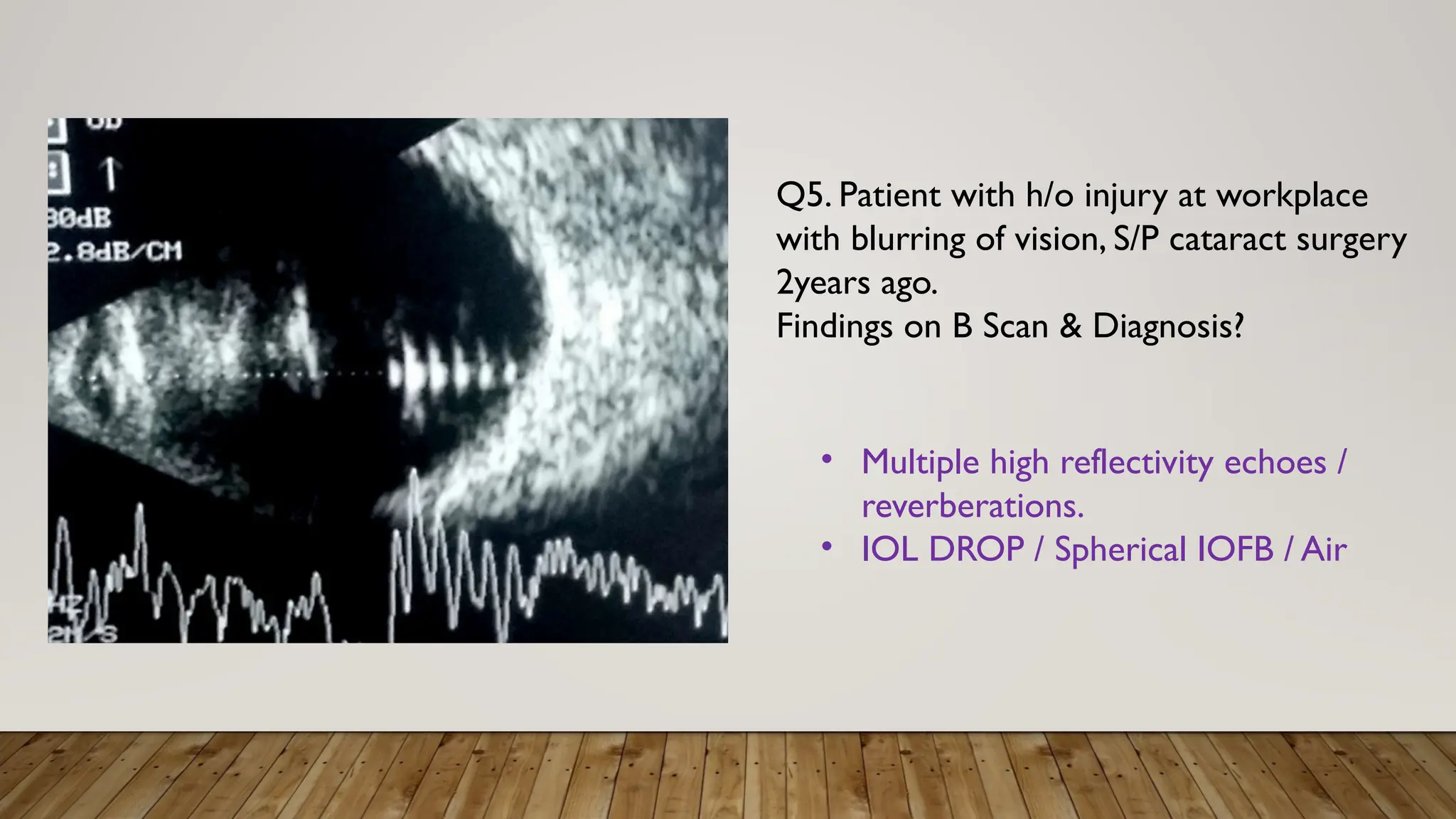
B-SCAN
Moderator : Dr Gladys R
Rodrigues
Presenter : Dr Sriraj Alapati
INTRODUCTION
 Brightness scan
 Complements clinical examination but doesn’t replace it.
 High frequency : good RESOLUTION : UBM (20-50 MHz)
Low frequency : good TISSUE PENETRATION : B Scan (8- 10 MHz)
 Acoustic Impedance – reflectivity (returning echoes)
 Gain (40-100 Db) – amplitude of echoes
 Direct globe / via lid / immersion
INDICATIONS
A. OPAQUE OCULAR MEDIA
Anterior segment :
• Corneal opacification
• Hyphemia / Hypopyon
• Miosis
• Dense Cataract
• Pupillary or retrolental
membrane
Posterior segment :
(Point like in B Scan)
• Vitreous hemorrhage
• Asteroid hyalosis
• Vitreous inflammation
(endophthalmitis)
B. CLEAR OCULAR MEDIA
• Membrane like in B Scan : PVD, Choroidal detachment, RD,
Retinoschisis, PED, ROP, Vitreous cyst, PHPV.
• Infections / Inflammations: VKH, SO, posterior scleritis,
endophthalmitis, neurocysticercosis.
• Trauma : IOFB, IOL / nucleus subluxation / drop, globe / choroidal
rupture, retrobulbar hemorrhage, giant retinal tear.
• Tumors : RB, cavernous hemangioma, choroidal melanoma,
choroidal osteoma.
• Disc abnormalities : Disc oedema, drusen, coloboma.
• Orbit : thyroid, CCF.
• Post surgical : air / gas, silicone oil
PROBE ORIENTATION
Centre of cornea
Perpendicular to cornea
Position & relation of lesion
to lens & ONH
Axial Longitudinal Transverse
Perpendicular to limbus
AP extent of lesion
Parallel to limbus
Lateral extent of lesion
4 transverse scans: 94%
Upper part of screen corresponds to portion of globe
where probe marker is directed.
DESCRIPTION OF A LESION
1.Topographic : size, shape, borders, location (clock, disc)
2. Quantitative :
A. STRUCTURE
Regular homogenous – choroidal melanoma
Regular heterogenous – choroidal hemangioma
Irregular – RB, mets
3. Kinetic : after movements / mobility (move with eye)
Present – PVD
Absent – old RD/CD
B. REFLECTIVITY (A Scan)
High – asteroid, RD, CD
Medium – oldVH
Low – vit cells
POINT LIKE IN B SCAN
VITREOUS CELLS
• Point like on high gain settings
+/- membranes
• Endophthalmitis / uveitis
• Low reflectivity
• After movements present
VITREOUS HAEMORRHAGE
Fresh – Fine granular/ pin point echoes
with high gain settings
PVD / FVP / RD
Medium reflectivity
After movements present
Old – Blood clots and membranes (dot like
echoes organize) of varying reflectivity.
After movements absent
PVD
FVP
ASTEROID HYALOSIS
• Calcium salts in vitreous cavity
• Point like larger, more echogenic than
RBCs.
• Echolucent gap between vitreous and
posterior globe wall.
• High reflectivity
• After movements present
Point like Vitreous cells Vitreous hemorrhage Asteroid hyalosis
Topographic Point like with high gain
Endophthalmitis / uveitis
Point like with high gain
PVD / FVP – PDR / RD
Point like larger, more
echogenic than RBCs.
Echolucent gap
Reflectivity Low Medium (fresh)
low (old)
High
After movement Present Present (fresh) / absent (old) Present
MEMBRANE LIKE IN B SCAN
PVD
• Separation between posterior
vitreous cortex & NSR
• Myopes, old age
• Smooth, thin, mobile membrane,
open funnel +/- disc insertion, inserts
at ORA / CB
• Variable amplitude
• Low reflectivity
• After movements present
Vitreous attached to retina : vitreous base is strongest (ora serrata), optic disc margin,
macula, main retinal vessels, lattice degeneration.
RETINAL DETACHEMENT
• Bright, continuous
• Smooth or folded
• Open or closed funnel
• Disc and ORA insertion
• Steeply rising peak
• 80-100% reflectivity
• After movements present in fresh
(acute) and absent in old RD.
• Rhegmatogenous
Tractional
Exudative
Combined
Long standing RD with intra retinal cyst,
fixed retinal folds, thin retina.
Total RD : triangular / funnel (open/closed) shape
with insertion at optic disc and ORA serrata.
RD RHEGMATOGENOUS TRACTIONAL EXUDATIVE
Topographic Funnel / double membrane
Folds+, retinal break+
Attached to disc
Concave, tent like traction
+/- FVP /VH (PDR)
Convex, bullous
Shifting fluid
Scleral thickening
Mass lesions, Sys causes
Reflectivity High High High
After movement Present (fresh) / absent (old) Absent Absent
CHOROIDAL DETACHEMENT
• Smooth, dome shaped
• Multiconvex / multilobed
• Immobile
• Kissing choroids
• No disc insertion
• Inserts at ORA / CB
• Don’t extend to posterior
pole (vortex veins)
• Steeply rising double peak
(retina, choroid)
• 90-100% reflectivity
• After movements absent
Hypotony :
• GDD
• Sulfonamides
Expulsive choroidal Hg #PCA:
• Uncontrolled HTN
• Sudden decompression of
high pre op IOP
2. HEMORRHAGIC
1. SEROUS
Membrane Like PVD RD CD
Shape Concave Double membrane / concave / convex Multi convex (dome)
Location Variable Variable Periphery (vortex veins)
ONH Attachments Variable Yes No
Types - Rhegmatogenous / Tractional /
Exudative / Combined
Serous / hemorrhagic
Cause Myopia / age Break / FVP / Sys cause Hypotony / expulsive
choroidal hg
Other findings Prominently seen
inferiorly
Folds / FVP / shifting fluid Kissing choroids
Reflectivity (A scan) 40-90% 80-100% 90-100%
Spike Single Single Double (retina, choroid)
Mobility Marked Moderate Absent
Aftermovement + + (fresh Rheg) / - -
Dense anterior membrane
with narrow closed funnel RD
Elevated smooth
thin dome shaped
membrane, often
bilateral
RETINOSCHISIS
ROP
VITREOUS CYST
Dome shaped serous
lesion
PED
PHPV
Tent like retinal dragging
Fluid filled structure with
reflective walls
INFECTIONS / INFLAMMATIONS
CYSTICERCOSIS
• Pork tape worm
• S/R cyst with RD (reverse diamond ring sign)
• High reflectivity spikes
SCOLEX
ENDOPHTHALMITIS
Cobweb appearance
(highly reflective
membranes)
SYMPATHETIC OPHTHALMOPLEGIA
• 2 weeks After penetrating trauma
• Choroidal thickening with exudative RD
• B/L disc oedema
• Choroidal thickening &
areas of bullous retinal
elevation at posterior pole
VKH
1. T sign (tenons fluid + optic nerve hypo intensity)
2. Choroidal thickening
3. Scleral thickening
4. ERD
POSTERIOR SCLERITIS
SUBLUXATED LENS
PCR
IOL DROP
(Multiple high reflectivity echoes)
TRAUMA
NUCLEAS DROP
(Biconvex with shadow)
GLOBE RUPTURE
Hemorrhagic vitreous track
CHOROIDAL RUPTURE
Focal area of fundus thickening
RETROBULBAR
HEMORRHAGE
GIANT RETINALTEAR
• Posterior shadowing
• Persistence at lower gain
• High reflectivity spikes >100%
• Overestimation of size
• Vitreous / retinal / subretinal / orbital
• Metal & stone : higher reflective echoes
• Wood & vegetable matter : intermediate echoes
• Glass is picked only when sound beam strikes
perpendicular along smooth surface of glass.
• Spherical IOFBs produce strong reverberations due
to regular structure.
• Air bubble may mimic foreign body
IOFB
TUMORS
• Calcification produce high
reflectivity and shadowing
• Endophytic – vitreous echoes
• Exophytic – ERD with
subretinal echoes
RETINOBLASTOMA
CHOROIDAL MELANOMA
Collar-button / mushroom
/ dome shape :
Knapp roone tumor
break in bruchs membrabe
Exudative RD
Choroidal excavation
Steep angle
kappa
Acoustic
hollowing
Vascularity on
color doppler
CAVERNOUS HEMANGIOMA
High reflectivity with shadowing at
posterior pole, double optic nerve
CHOROIDAL OSTEOMA
• Intraconal
• Heterogenous structure
• Varying reflectivity
• No color flow on doppler
DISC ABNORMALITIES
Patient to fixate in primary gaze.
Probe placed longitudinal, temporally
with medium gain setting.
Normal ONH seen as round echo
lucent lesion adjacent to globe with
shadowing.
Disc oedema : crescent or doughnut sign
DISC ELEVATION
DRUSEN
Enlarged optic
nerve
With low gain
settings
GLAUCOMATOUS
OPTIC CUPPING
OPTIC DISC COLOBOMA
Bean pot
configuration
ORBIT
Enlargement muscle belly
sparing tendinous insertion
TED
Dilated SOV
CCF
• High reflective curvilinear echo with
comet tail shadowing.
• Echo seen in non dependent location
as air bubble floats.
• High reflective echoes.
• Single large air bubble causing strong
reverberations.
• Apparent lengthening of eye waves pass slow.
AIR / GAS SILICON OIL
POST
SURGICAL
THANKYOU
Q1. Patient c/o pain with blurring of vision. Fundus examination
shows choroidal folds with localized elevation of the retina
Findings on B scan and diagnosis?
• T sign
• Scleral & choroidal thickening
• RD
POSTERIOR SCLERITIS
Q2. Patient with sudden blurring of vision.
There is no view of the fundus. Findings
on B scan?
• VH
• PVD
• RD
Q3. Patient diagnosed to have bilateral disc elevation on routine
examination.V/A and colour vision is normal. Diagnosis?
DISC DRUSEN
Q4. Patient presents with blurring of vision. Examination shows low
IOP & mature cataract with no view of fundus. Findings on B scan?
RETINAL DETACHEMENT
Low IOP?
• Fresh : RPE actively pumps fluid
• Old : CB shutdown
Q5. Patient with h/o injury at workplace
with blurring of vision, S/P cataract surgery
2years ago.
Findings on B Scan & Diagnosis?
• Multiple high reflectivity echoes /
reverberations.
• IOL DROP / Spherical IOFB / Air

B-Scan procedure basics in ophthalmology.pptx

  • 1.
    B-SCAN Moderator : DrGladys R Rodrigues Presenter : Dr Sriraj Alapati
  • 2.
    INTRODUCTION  Brightness scan Complements clinical examination but doesn’t replace it.  High frequency : good RESOLUTION : UBM (20-50 MHz) Low frequency : good TISSUE PENETRATION : B Scan (8- 10 MHz)  Acoustic Impedance – reflectivity (returning echoes)  Gain (40-100 Db) – amplitude of echoes  Direct globe / via lid / immersion
  • 3.
    INDICATIONS A. OPAQUE OCULARMEDIA Anterior segment : • Corneal opacification • Hyphemia / Hypopyon • Miosis • Dense Cataract • Pupillary or retrolental membrane Posterior segment : (Point like in B Scan) • Vitreous hemorrhage • Asteroid hyalosis • Vitreous inflammation (endophthalmitis)
  • 4.
    B. CLEAR OCULARMEDIA • Membrane like in B Scan : PVD, Choroidal detachment, RD, Retinoschisis, PED, ROP, Vitreous cyst, PHPV. • Infections / Inflammations: VKH, SO, posterior scleritis, endophthalmitis, neurocysticercosis. • Trauma : IOFB, IOL / nucleus subluxation / drop, globe / choroidal rupture, retrobulbar hemorrhage, giant retinal tear. • Tumors : RB, cavernous hemangioma, choroidal melanoma, choroidal osteoma. • Disc abnormalities : Disc oedema, drusen, coloboma. • Orbit : thyroid, CCF. • Post surgical : air / gas, silicone oil
  • 5.
    PROBE ORIENTATION Centre ofcornea Perpendicular to cornea Position & relation of lesion to lens & ONH Axial Longitudinal Transverse Perpendicular to limbus AP extent of lesion Parallel to limbus Lateral extent of lesion 4 transverse scans: 94%
  • 6.
    Upper part ofscreen corresponds to portion of globe where probe marker is directed.
  • 7.
    DESCRIPTION OF ALESION 1.Topographic : size, shape, borders, location (clock, disc) 2. Quantitative : A. STRUCTURE Regular homogenous – choroidal melanoma Regular heterogenous – choroidal hemangioma Irregular – RB, mets 3. Kinetic : after movements / mobility (move with eye) Present – PVD Absent – old RD/CD B. REFLECTIVITY (A Scan) High – asteroid, RD, CD Medium – oldVH Low – vit cells
  • 8.
    POINT LIKE INB SCAN VITREOUS CELLS • Point like on high gain settings +/- membranes • Endophthalmitis / uveitis • Low reflectivity • After movements present
  • 9.
    VITREOUS HAEMORRHAGE Fresh –Fine granular/ pin point echoes with high gain settings PVD / FVP / RD Medium reflectivity After movements present Old – Blood clots and membranes (dot like echoes organize) of varying reflectivity. After movements absent PVD FVP
  • 10.
    ASTEROID HYALOSIS • Calciumsalts in vitreous cavity • Point like larger, more echogenic than RBCs. • Echolucent gap between vitreous and posterior globe wall. • High reflectivity • After movements present
  • 11.
    Point like Vitreouscells Vitreous hemorrhage Asteroid hyalosis Topographic Point like with high gain Endophthalmitis / uveitis Point like with high gain PVD / FVP – PDR / RD Point like larger, more echogenic than RBCs. Echolucent gap Reflectivity Low Medium (fresh) low (old) High After movement Present Present (fresh) / absent (old) Present
  • 12.
    MEMBRANE LIKE INB SCAN PVD • Separation between posterior vitreous cortex & NSR • Myopes, old age • Smooth, thin, mobile membrane, open funnel +/- disc insertion, inserts at ORA / CB • Variable amplitude • Low reflectivity • After movements present Vitreous attached to retina : vitreous base is strongest (ora serrata), optic disc margin, macula, main retinal vessels, lattice degeneration.
  • 13.
    RETINAL DETACHEMENT • Bright,continuous • Smooth or folded • Open or closed funnel • Disc and ORA insertion • Steeply rising peak • 80-100% reflectivity • After movements present in fresh (acute) and absent in old RD. • Rhegmatogenous Tractional Exudative Combined
  • 14.
    Long standing RDwith intra retinal cyst, fixed retinal folds, thin retina. Total RD : triangular / funnel (open/closed) shape with insertion at optic disc and ORA serrata.
  • 15.
    RD RHEGMATOGENOUS TRACTIONALEXUDATIVE Topographic Funnel / double membrane Folds+, retinal break+ Attached to disc Concave, tent like traction +/- FVP /VH (PDR) Convex, bullous Shifting fluid Scleral thickening Mass lesions, Sys causes Reflectivity High High High After movement Present (fresh) / absent (old) Absent Absent
  • 16.
    CHOROIDAL DETACHEMENT • Smooth,dome shaped • Multiconvex / multilobed • Immobile • Kissing choroids • No disc insertion • Inserts at ORA / CB • Don’t extend to posterior pole (vortex veins) • Steeply rising double peak (retina, choroid) • 90-100% reflectivity • After movements absent Hypotony : • GDD • Sulfonamides Expulsive choroidal Hg #PCA: • Uncontrolled HTN • Sudden decompression of high pre op IOP 2. HEMORRHAGIC 1. SEROUS
  • 17.
    Membrane Like PVDRD CD Shape Concave Double membrane / concave / convex Multi convex (dome) Location Variable Variable Periphery (vortex veins) ONH Attachments Variable Yes No Types - Rhegmatogenous / Tractional / Exudative / Combined Serous / hemorrhagic Cause Myopia / age Break / FVP / Sys cause Hypotony / expulsive choroidal hg Other findings Prominently seen inferiorly Folds / FVP / shifting fluid Kissing choroids Reflectivity (A scan) 40-90% 80-100% 90-100% Spike Single Single Double (retina, choroid) Mobility Marked Moderate Absent Aftermovement + + (fresh Rheg) / - -
  • 18.
    Dense anterior membrane withnarrow closed funnel RD Elevated smooth thin dome shaped membrane, often bilateral RETINOSCHISIS ROP
  • 19.
    VITREOUS CYST Dome shapedserous lesion PED PHPV Tent like retinal dragging Fluid filled structure with reflective walls
  • 20.
    INFECTIONS / INFLAMMATIONS CYSTICERCOSIS •Pork tape worm • S/R cyst with RD (reverse diamond ring sign) • High reflectivity spikes SCOLEX
  • 21.
    ENDOPHTHALMITIS Cobweb appearance (highly reflective membranes) SYMPATHETICOPHTHALMOPLEGIA • 2 weeks After penetrating trauma • Choroidal thickening with exudative RD
  • 22.
    • B/L discoedema • Choroidal thickening & areas of bullous retinal elevation at posterior pole VKH 1. T sign (tenons fluid + optic nerve hypo intensity) 2. Choroidal thickening 3. Scleral thickening 4. ERD POSTERIOR SCLERITIS
  • 23.
    SUBLUXATED LENS PCR IOL DROP (Multiplehigh reflectivity echoes) TRAUMA NUCLEAS DROP (Biconvex with shadow)
  • 24.
    GLOBE RUPTURE Hemorrhagic vitreoustrack CHOROIDAL RUPTURE Focal area of fundus thickening
  • 25.
  • 26.
    • Posterior shadowing •Persistence at lower gain • High reflectivity spikes >100% • Overestimation of size • Vitreous / retinal / subretinal / orbital • Metal & stone : higher reflective echoes • Wood & vegetable matter : intermediate echoes • Glass is picked only when sound beam strikes perpendicular along smooth surface of glass. • Spherical IOFBs produce strong reverberations due to regular structure. • Air bubble may mimic foreign body IOFB
  • 27.
    TUMORS • Calcification producehigh reflectivity and shadowing • Endophytic – vitreous echoes • Exophytic – ERD with subretinal echoes RETINOBLASTOMA
  • 28.
    CHOROIDAL MELANOMA Collar-button /mushroom / dome shape : Knapp roone tumor break in bruchs membrabe Exudative RD Choroidal excavation Steep angle kappa Acoustic hollowing Vascularity on color doppler
  • 29.
    CAVERNOUS HEMANGIOMA High reflectivitywith shadowing at posterior pole, double optic nerve CHOROIDAL OSTEOMA • Intraconal • Heterogenous structure • Varying reflectivity • No color flow on doppler
  • 30.
    DISC ABNORMALITIES Patient tofixate in primary gaze. Probe placed longitudinal, temporally with medium gain setting. Normal ONH seen as round echo lucent lesion adjacent to globe with shadowing. Disc oedema : crescent or doughnut sign
  • 31.
  • 32.
    GLAUCOMATOUS OPTIC CUPPING OPTIC DISCCOLOBOMA Bean pot configuration
  • 33.
    ORBIT Enlargement muscle belly sparingtendinous insertion TED Dilated SOV CCF
  • 34.
    • High reflectivecurvilinear echo with comet tail shadowing. • Echo seen in non dependent location as air bubble floats. • High reflective echoes. • Single large air bubble causing strong reverberations. • Apparent lengthening of eye waves pass slow. AIR / GAS SILICON OIL POST SURGICAL
  • 35.
  • 36.
    Q1. Patient c/opain with blurring of vision. Fundus examination shows choroidal folds with localized elevation of the retina Findings on B scan and diagnosis? • T sign • Scleral & choroidal thickening • RD POSTERIOR SCLERITIS
  • 37.
    Q2. Patient withsudden blurring of vision. There is no view of the fundus. Findings on B scan? • VH • PVD • RD
  • 38.
    Q3. Patient diagnosedto have bilateral disc elevation on routine examination.V/A and colour vision is normal. Diagnosis? DISC DRUSEN
  • 39.
    Q4. Patient presentswith blurring of vision. Examination shows low IOP & mature cataract with no view of fundus. Findings on B scan? RETINAL DETACHEMENT Low IOP? • Fresh : RPE actively pumps fluid • Old : CB shutdown
  • 40.
    Q5. Patient withh/o injury at workplace with blurring of vision, S/P cataract surgery 2years ago. Findings on B Scan & Diagnosis? • Multiple high reflectivity echoes / reverberations. • IOL DROP / Spherical IOFB / Air