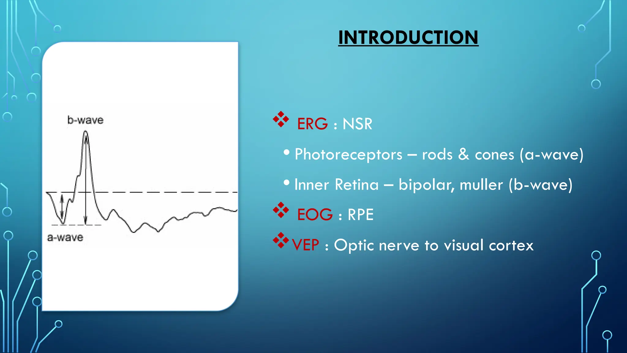
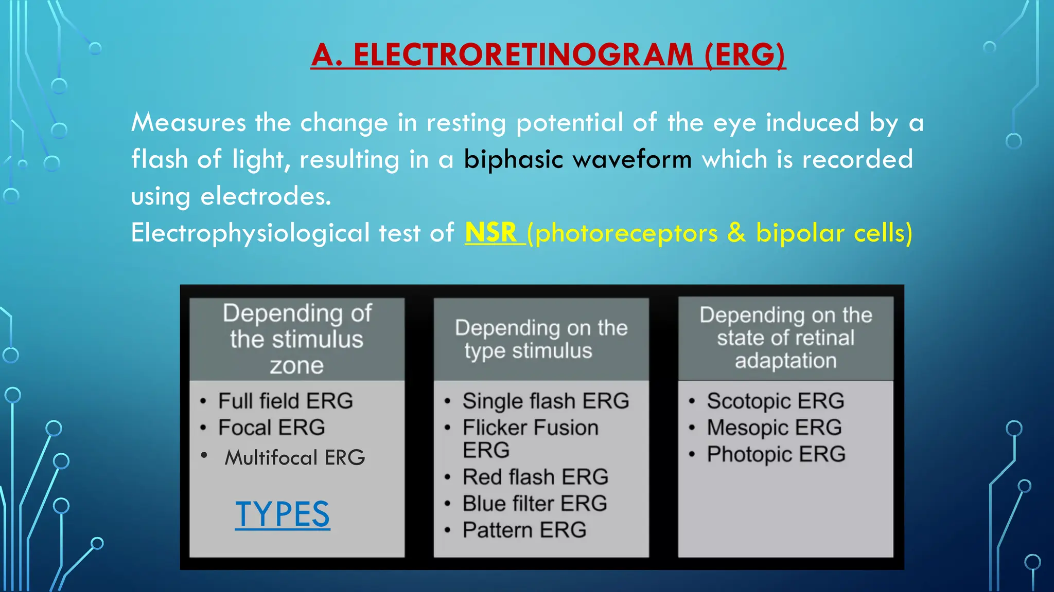
ELECTRO
PHYSIOLOGICAL
TESTS
Moderator : Dr Rajesh R Nayak
Presenter : Dr Sriraj Alapati
INTRODUCTION
 ERG : NSR
• Photoreceptors – rods & cones (a-wave)
• Inner Retina – bipolar, muller (b-wave)
 EOG : RPE
VEP : Optic nerve to visual cortex
Measures the change in resting potential of the eye induced by a
flash of light, resulting in a biphasic waveform which is recorded
using electrodes.
Electrophysiological test of NSR (photoreceptors & bipolar cells)
A. ELECTRORETINOGRAM (ERG)
TYPES
• Multifocal ERG
1ST
order : Bipolar cells
2nd
order : Ganglion cells
3rd
order : LGB
PHYSIOLOGY
Description RODS CONES
Number 120 M 6 M
Location Mid periphery (bony spicules in RP) Fovea
Types 1 3 (L, M; S) : 2nd
CN#; retina, macula#
Pigment Rhodopsin Phorphyropsin (proton): red (XLR) 555nm
Iodopsin (deutron): green (XLR) 530nm
Cyanopsin (triton): blue (Chr 7) 426nm
LGB (3rd
order) Ventral Magnocellular (WHERE) 1
Affected in glaucoma
Dorsal Parvocellular (WHAT) 8
Light conditions Dim light (Night vision) : scotopic Bright light (Day vision) : photopic
Type of vision Black and white (Achromatic) Color vision (chromatic)
Functions Peripheral vision
Gross vision, stereopsis
Contrast & motion sense
Central vision
Fine vision, stereopsis
Abnormality Nyctalopia Dyschromatopsia, Hemeralopia
Electrodes:
• Ground - forehead
• Reference – outer canthus
• Active - cornea contact, conjunctival
HOW TO PERFORM?
WAVE FORMS
A wave B wave
Negative (hyperpolarization) Positive (depolarization)
Outer retina (Photoreceptors) – rods & cones Inner retina (INL) – bipolar ON, muller
Choroidal pathology ? (20 short PCA) Retinal pathology ? (CRA)
Rod a wave (scotopic) – broad
Cone a wave (photopic) – sharp
B1 – rod cone (scotopic) : slow rising, broad peak
B2 – cone (photopic) : rapid rising, sharp peak
C wave:
Small positive
RPE
D wave:
Positive, photopic
Bipolar OFF
OP’s (oscillatory potentials):
Amacrine
Flicker response:
Cone driven
Scotopic/DA 0.01 - rods (b wave)
Scotopic/DA 3.0 - mixed
Scotopic/DA 10.0 – mixed, strong
Scotopic/DA 3.0 OP’s - amacrine
Photopic/LA 3.0 - mixed
Photopic/LA 3.0 flicker - cones
1. Full Field (Ff ERG)
Indications
NEGATIVE ERG
Reduced b wave or b/a ratio
Negative b, a waves
• RP
• CRVO, CRAO
• CSNB
• Cone dystrophy
• HCQ
EXTINGUISHED ERG
Absent b, a waves
• Advanced RP
• OIS
• Leber's congenital amaurosis
• Retinal aplasia
• RD
• Choroideremia
Reduced cone responses in
cone dystrophy
Reduced scotopic rod &
combined responses in early RP
Retina has dual Blood Supply
1. Outer 4 Retinal layers (Photoreceptors) : PCA
'a' wave lost in choroidal #
2. Inner 6 Retinal layers (INL) : CRA
'b' wave lost in retinal #
INNER RETINAL ISCHAEMIA
I. b/a ratio or b wave amplitude reduced
II. OP’s absent (DA 3.0 OP – amacrine)
III. Flicker implicit time prolonged (LA 3.0 flicker – cones)
IV. Interocular difference
ERG In Vascular Retinopathies
USES :
1. Non ischemic Vs
Ischemic CRVO
2. CRAO Vs OIS
3. NPDR Vs PDR
Non ischemic Vs Ischemic CRVO
ERG > FFA ? in CRVO:
1.Non-Invasive
2.Detects Inner retinal ischaemia
3.Not limited by the wide field
4.Not limited by media opacity
5.Not limited by Retinal hg’s / Oedema
6.Not limited by Systemic co-morbidities
• RAPD
• ERG
• RAPD + ERG (most reliable 97%)
• HFA (VF)
• FFA (RETINAL CAPILLARIES obliterated - non perfusion)
• Fundus could mislead (splashed tomato sauce appearance)
• Ophthalmic artery occlusion
• Mid periphery retinal hg’s, MA,
dilated veins, NVE, NVG
• Suspect in U/L DR changes
• Do carotid artery doppler
• Extinguished ERG (a, b absent)
OIS
CRAO
• Retinal whitening, Cherry red spot,
attenuated arteries, dilated veins,
cattle truckling of veins.
• Do carotid artery doppler
• Negative ERG (a, b negative)
NPDR Vs PDR
2. Multifocal (Mf ERG)
Only cones (LA)
Mathematical extraction
Topographic mapping
Pupillary dilation
Refractive correction
103 hexagonal units
Kernel responses
Central fixation dots & circles
Mf ERG don’t replace Ff ERG
USES : HCQ, desferoxamine toxicity
CSCR, RP, stargardts
> 5 mg/kg/day
Cv abnormal
Fundus : bulls eye maculopathy
HFA 10-2 : paracentral scotomas
OCT : disruption of parafoveal EZ
FAF : parafoveal hyperAF
Abnormal Mf.ERG
HCQ
3. Pattern (P ERG)
P50 - macular photoreceptors
N95 - GCL
Physiological -
• Age : newborns have immature ERG
• Gender : females have higher amplitude b waves
• Refraction : myopes have lesser amplitude b waves
• Diurnal Variation : daylight have lesser amplitude rod b waves
• Pupillary size : small pupil have lesser amplitude waves
• Media clarity : VH have unrecordable ERG
• Uveal pigment : albinism have higher amplitude waves
• Dark adaptation
• Medications : alcohol, GA, steroids
Technical - electrodes, stimulus parameters, filters, amplifiers
Variations In ERG
B. ELECTRO-OCULOGRAM (EOG)
Standing potential of eye : Difference in electrical
potential between anterior & posterior part of the eye
Electrophysiological test of RPE
• Dilated pupils
• Electrodes
• Ganzfeld dome
1. Saccades 10s of each min
2. Wave form
3. Dt (dark trough) : 15 mins
4. Lp (light peak) : 15 mins
5. Ardens ratio Lp/Dt
Components of EOG
 Normal > 1.80
 Subnormal 1.65 to 1.80
 Abnormal < 1.65
Ardens ratio :
light peak (Lp)/dark trough (Dt)
1. Good saccades
2. Sine wave
3. No dark trough
4. No light peak
5. Abn Arden ratio
V/A - Normal
Fundus ERG EOG
AD Best vitelliform (V/A – N) Macula Normal Abnormal
AR Bestrophinopathy Vascular arcades Abnormal Abnormal
AD vitreoretinochoroidopathy Vitreous Abnormal Abnormal
C. VISUAL EVOKED POTENTIAL (VEP)
Electrical signal generated at visual cortex in response
to visual stimulation.
Electrophysiological test from Optic nerve to visual
Cortex (Central VF).
Electrodes :
• Occipital lobe (Oz, active/positive)
• Forehead (Fz, reference/negative)
• Earlobe/vertex/mastoid
(ground/neutral)
Types
Waveforms
Optic neuritis - Multiple sclerosis
AION
Optic atrophy
Toxic neuropathy
TON
Pattern Reversal VEP
(optic neuropathy)
Visual function of babies
Non-responsive subjects
Unexplained visual loss
Flash VEP
Amblyopia
Malingering
Nystagmus
Pattern on/off VEP
Indications
VEP Amplitude Latency/Implicit time
AION Reduced Normal
Optic neuritis Reduced Increased/Delayed
TON Reduced Increased/Delayed
Optic atrophy Absent No signal
Toxic neuropathy Normal Increased P100
Pattern reversal
VEP
THANK YOU

Electrophysiological Tests - ERG, EOG, VEP in ophthalmology.pptx

  • 1.
    ELECTRO PHYSIOLOGICAL TESTS Moderator : DrRajesh R Nayak Presenter : Dr Sriraj Alapati
  • 2.
    INTRODUCTION  ERG :NSR • Photoreceptors – rods & cones (a-wave) • Inner Retina – bipolar, muller (b-wave)  EOG : RPE VEP : Optic nerve to visual cortex
  • 3.
    Measures the changein resting potential of the eye induced by a flash of light, resulting in a biphasic waveform which is recorded using electrodes. Electrophysiological test of NSR (photoreceptors & bipolar cells) A. ELECTRORETINOGRAM (ERG) TYPES • Multifocal ERG
  • 4.
    1ST order : Bipolarcells 2nd order : Ganglion cells 3rd order : LGB PHYSIOLOGY
  • 5.
    Description RODS CONES Number120 M 6 M Location Mid periphery (bony spicules in RP) Fovea Types 1 3 (L, M; S) : 2nd CN#; retina, macula# Pigment Rhodopsin Phorphyropsin (proton): red (XLR) 555nm Iodopsin (deutron): green (XLR) 530nm Cyanopsin (triton): blue (Chr 7) 426nm LGB (3rd order) Ventral Magnocellular (WHERE) 1 Affected in glaucoma Dorsal Parvocellular (WHAT) 8 Light conditions Dim light (Night vision) : scotopic Bright light (Day vision) : photopic Type of vision Black and white (Achromatic) Color vision (chromatic) Functions Peripheral vision Gross vision, stereopsis Contrast & motion sense Central vision Fine vision, stereopsis Abnormality Nyctalopia Dyschromatopsia, Hemeralopia
  • 7.
    Electrodes: • Ground -forehead • Reference – outer canthus • Active - cornea contact, conjunctival HOW TO PERFORM?
  • 8.
  • 9.
    A wave Bwave Negative (hyperpolarization) Positive (depolarization) Outer retina (Photoreceptors) – rods & cones Inner retina (INL) – bipolar ON, muller Choroidal pathology ? (20 short PCA) Retinal pathology ? (CRA) Rod a wave (scotopic) – broad Cone a wave (photopic) – sharp B1 – rod cone (scotopic) : slow rising, broad peak B2 – cone (photopic) : rapid rising, sharp peak
  • 10.
    C wave: Small positive RPE Dwave: Positive, photopic Bipolar OFF OP’s (oscillatory potentials): Amacrine Flicker response: Cone driven
  • 11.
    Scotopic/DA 0.01 -rods (b wave) Scotopic/DA 3.0 - mixed Scotopic/DA 10.0 – mixed, strong Scotopic/DA 3.0 OP’s - amacrine Photopic/LA 3.0 - mixed Photopic/LA 3.0 flicker - cones 1. Full Field (Ff ERG)
  • 13.
  • 14.
    NEGATIVE ERG Reduced bwave or b/a ratio Negative b, a waves • RP • CRVO, CRAO • CSNB • Cone dystrophy • HCQ EXTINGUISHED ERG Absent b, a waves • Advanced RP • OIS • Leber's congenital amaurosis • Retinal aplasia • RD • Choroideremia
  • 15.
    Reduced cone responsesin cone dystrophy Reduced scotopic rod & combined responses in early RP
  • 16.
    Retina has dualBlood Supply 1. Outer 4 Retinal layers (Photoreceptors) : PCA 'a' wave lost in choroidal # 2. Inner 6 Retinal layers (INL) : CRA 'b' wave lost in retinal # INNER RETINAL ISCHAEMIA I. b/a ratio or b wave amplitude reduced II. OP’s absent (DA 3.0 OP – amacrine) III. Flicker implicit time prolonged (LA 3.0 flicker – cones) IV. Interocular difference ERG In Vascular Retinopathies USES : 1. Non ischemic Vs Ischemic CRVO 2. CRAO Vs OIS 3. NPDR Vs PDR
  • 17.
    Non ischemic VsIschemic CRVO
  • 18.
    ERG > FFA? in CRVO: 1.Non-Invasive 2.Detects Inner retinal ischaemia 3.Not limited by the wide field 4.Not limited by media opacity 5.Not limited by Retinal hg’s / Oedema 6.Not limited by Systemic co-morbidities • RAPD • ERG • RAPD + ERG (most reliable 97%) • HFA (VF) • FFA (RETINAL CAPILLARIES obliterated - non perfusion) • Fundus could mislead (splashed tomato sauce appearance)
  • 19.
    • Ophthalmic arteryocclusion • Mid periphery retinal hg’s, MA, dilated veins, NVE, NVG • Suspect in U/L DR changes • Do carotid artery doppler • Extinguished ERG (a, b absent) OIS
  • 20.
    CRAO • Retinal whitening,Cherry red spot, attenuated arteries, dilated veins, cattle truckling of veins. • Do carotid artery doppler • Negative ERG (a, b negative)
  • 21.
  • 22.
    2. Multifocal (MfERG) Only cones (LA) Mathematical extraction Topographic mapping Pupillary dilation Refractive correction 103 hexagonal units Kernel responses Central fixation dots & circles Mf ERG don’t replace Ff ERG USES : HCQ, desferoxamine toxicity CSCR, RP, stargardts
  • 24.
    > 5 mg/kg/day Cvabnormal Fundus : bulls eye maculopathy HFA 10-2 : paracentral scotomas OCT : disruption of parafoveal EZ FAF : parafoveal hyperAF Abnormal Mf.ERG HCQ
  • 25.
    3. Pattern (PERG) P50 - macular photoreceptors N95 - GCL
  • 26.
    Physiological - • Age: newborns have immature ERG • Gender : females have higher amplitude b waves • Refraction : myopes have lesser amplitude b waves • Diurnal Variation : daylight have lesser amplitude rod b waves • Pupillary size : small pupil have lesser amplitude waves • Media clarity : VH have unrecordable ERG • Uveal pigment : albinism have higher amplitude waves • Dark adaptation • Medications : alcohol, GA, steroids Technical - electrodes, stimulus parameters, filters, amplifiers Variations In ERG
  • 27.
    B. ELECTRO-OCULOGRAM (EOG) Standingpotential of eye : Difference in electrical potential between anterior & posterior part of the eye Electrophysiological test of RPE • Dilated pupils • Electrodes • Ganzfeld dome
  • 28.
    1. Saccades 10sof each min 2. Wave form 3. Dt (dark trough) : 15 mins 4. Lp (light peak) : 15 mins 5. Ardens ratio Lp/Dt Components of EOG
  • 29.
     Normal >1.80  Subnormal 1.65 to 1.80  Abnormal < 1.65 Ardens ratio : light peak (Lp)/dark trough (Dt)
  • 30.
    1. Good saccades 2.Sine wave 3. No dark trough 4. No light peak 5. Abn Arden ratio
  • 31.
  • 32.
    Fundus ERG EOG ADBest vitelliform (V/A – N) Macula Normal Abnormal AR Bestrophinopathy Vascular arcades Abnormal Abnormal AD vitreoretinochoroidopathy Vitreous Abnormal Abnormal
  • 33.
    C. VISUAL EVOKEDPOTENTIAL (VEP) Electrical signal generated at visual cortex in response to visual stimulation. Electrophysiological test from Optic nerve to visual Cortex (Central VF). Electrodes : • Occipital lobe (Oz, active/positive) • Forehead (Fz, reference/negative) • Earlobe/vertex/mastoid (ground/neutral)
  • 34.
  • 35.
    Optic neuritis -Multiple sclerosis AION Optic atrophy Toxic neuropathy TON Pattern Reversal VEP (optic neuropathy) Visual function of babies Non-responsive subjects Unexplained visual loss Flash VEP Amblyopia Malingering Nystagmus Pattern on/off VEP Indications
  • 36.
    VEP Amplitude Latency/Implicittime AION Reduced Normal Optic neuritis Reduced Increased/Delayed TON Reduced Increased/Delayed Optic atrophy Absent No signal Toxic neuropathy Normal Increased P100 Pattern reversal VEP
  • 37.