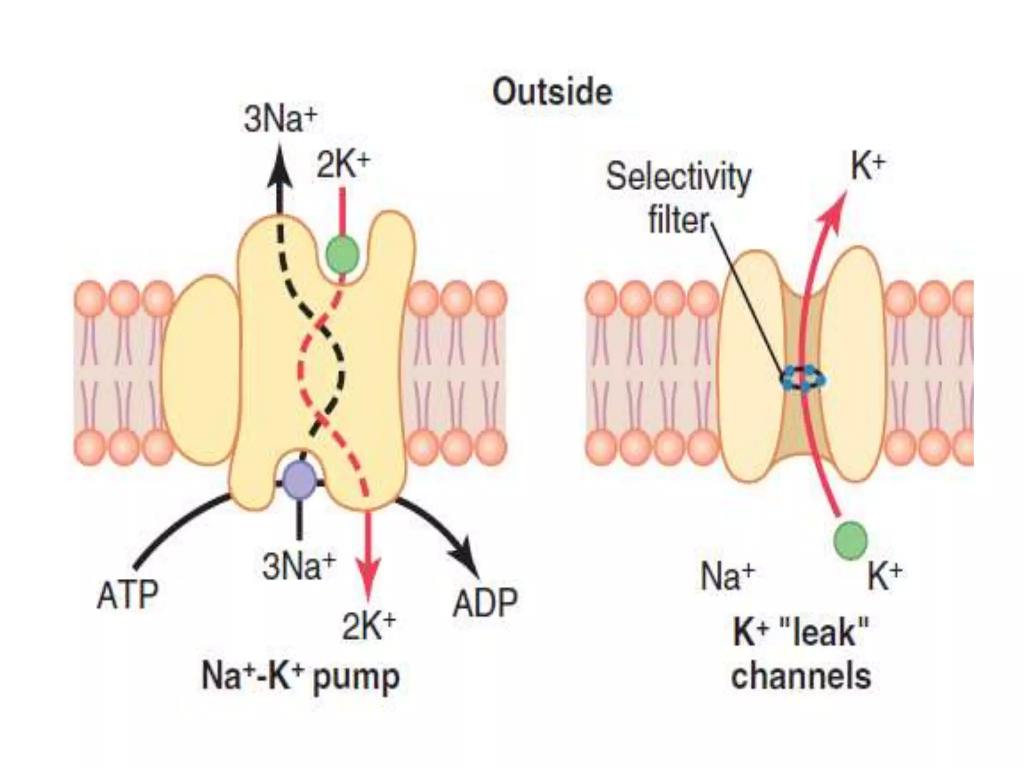
Action potential By Dr. Mrs. Padmaja R Desai Physiology Dept
To study the Concept of Action Potential and describe the stages of action potential.
Ionic basis of Action Potential & its Propogation.
Properties of Action Potential.
Types action Potential
Diffusion potential. Large Nerve. Na -K ATPase. Guyton and Hall. Medical Physiology. Dr. Nusrat Tariq. Professor Of Physiology. M.I.M.D.C. GOLDMAN HODGKIN KATZ EQUATION
Homeostasis I Negative and Positive Feedback Mechanism I Feedforward Mechanis...HM Learnings
Homeostasis I Negative and Positive Feedback Mechanism I Feedforward Mechanism I General Physiology I
The slide will be about :
1. Definition of homeostasis
2. What is internal environment ?
3. Why ECF is considered as an internal environment for cell ?
4. Homeostatic mechanism
5. Components of homeostatic mechanism
6. Feedback mechanism
7. Negative feedback mechanism
8. Positive feedback mechanism
9. Feedforward mechanism
You can also watch the same topic on HM Learnings Youtube channel.
You can also follow HM Learnings on facebook, instagram and twitter for daily updates
It is over 60 years since Hodgkin and
Huxley1 made the first direct recording of
the electrical changes across the neuronal
membrane that mediate the action
potential. Using an electrode placed inside a
squid giant axon they were able to measure a
transmembrane potential of around 260 mV
inside relative to outside, under resting
conditions (this is called the resting membrane
potential). The action potential is a
transient (,1 millisecond) reversal in the
polarity of this transmembrane potential
which then moves from its point of initiation,
down the axon, to the axon terminals. In a
subsequent series of elegant experiments
Hodgkin and Huxley, along with Bernard
Katz, discovered that the action potential
results from transient changes in the permeability
of the axon membrane to sodium (Na+)
and potassium (K+) ions. Importantly, Na+ and
K+ cross the membrane through independent
pathways that open in response to a change
in membrane potential.
As testimony to their pioneering work, the
fundamental mechanisms described by
Hodgkin, Huxley and Katz remain applicable
to all excitable cells today. Indeed, the
predictions they made about the molecular
mechanisms that might underlie the changes
in membrane permeability showed remarkable
foresight. The molecular basis of the action
potential lies in the presence of proteins
called ion channels that form the permeation
pathways across the neuronal membrane.
Although the first electrophysiological
recordings from individual ion channels were
not made until the mid 1970s,2 Hodgkin and
Huxley predicted many of the properties now
known to be key components of their
function: ion selectivity, the electrical basis
of voltage-sensitivity and, importantly, a
mechanism for quickly closing down the
permeability pathways to ensure that the
action potential only moves along the axon in
one direction.
Action potential (the guyton and hall physiology)Maryam Fida
ACTION POTENTIAL
Action potential is abrupt pulse like change in the membrane potential lasting for a fraction of second
During action potential there is reversal of membrane potential i.e. inside becomes positive and outside becomes Negative.
We can see the action potential on cathode ray oscilloscope
Abrupt or sudden in onset
2. Have limited magnitude or amplitude i.e. Inside, the potential will go to + 35 or + 45 mV and not beyond that.
3. It is of short duration. Duration is in milli seconds. Duration of spike potential Is 1 -2 milli second. Action potential with plateau has longer duration i.e. may be up to 300 m sec
4. It obeys All or None law i.e. if stimulus is sub threshold it is not produced and when the stimulus is threshold or supra threshold it will be produced with maximum amplitude.
5. It is self propagated i.e. once produced in a membrane it is automatically propagated in both directions.
6. It is not decremented with distance i.e. it will travel with same amplitude through all the distance.
7. It has refractory period. The period during which the tissue will not respond to second stimulus after the application of first stimulus. It could be Absolute and Refractory.
Absolute no response of tissue what so ever may be the strength of stimulus example closure of inactivation gate of sodium channels.
Relative response with higher stimulus than threshold stimulus
DEPOLARIZATION: Sudden loss of Negativity inside the membrane is depolarization.
REPOLARIZATION: return of negativity inside the membrane is Repolarization.
HYPERPOLARIZATION: More Negativity inside
Resting Membrane Potential
Understanding of
Channels Involved
Voltage gated Sodium Channels
Voltage gated Potassium Channels
Sodium Potassium ATPase Pump
Movements of ions
Concentrations of Sodium and Potassium in ECF and ICF
Direction of movement
Plateau is known as Sustained depolarization.
In some instances, the excited membrane does not repolarize immediately after depolarization.
Duration of depolarization of cardiac muscle is 300 milli sec.
Plateau phase has got advantages:
1. It prolongs the duration of depolarization, AP and Contraction. It prolongs the refractory period. Cardiac muscle cannot be tetanized because of this.
2. There is influx of calcium into the sarcoplasm from the ECF which is used for muscle contraction.
How membrane potential of a cell is reached, we discuss about nernst equation. goldman equation and pumps that are involved in the working of cell membrane. Concentration and working of ions across cell membrane
Glomerular Filtration rate and its determinants.pptxDr.Nusrat Tariq
The first step in urine formation is filtration of large
amounts of fluid through the glomerular capillaries into
Bowman’s capsule—almost 180 L/day. Most of this filtrate is reabsorbed, leaving only about 1 liter of fluid to be
excreted each day, although the renal fluid excretion rate
is highly variable, depending on fluid intake. The high rate
of glomerular filtration depends on a high rate of kidney
blood flow, as well as the special properties of the glomerular capillary membranes.
Glomerular Filtration and determinants of glomerular filtration .pptxDr.Nusrat Tariq
The first step in urine formation is filtration of large
amounts of fluid through the glomerular capillaries into
Bowman’s capsule—almost 180 L/day. Most of this filtrate is reabsorbed, leaving only about 1 liter of fluid to be
excreted each day, although the renal fluid excretion rate
is highly variable, depending on fluid intake. The high rate
of glomerular filtration depends on a high rate of kidney
blood flow, as well as the special properties of the glomerular capillary membranes.
this lecture gives detailed account of functions of liver as an organ, secretion, regulation and functions of biliary secretion. exocrine and endocrine functions of pancreas. composition of pancreatic secretions
These simplified slides by Dr. Sidra Arshad present an overview of the non-respiratory functions of the respiratory tract.
Learning objectives:
1. Enlist the non-respiratory functions of the respiratory tract
2. Briefly explain how these functions are carried out
3. Discuss the significance of dead space
4. Differentiate between minute ventilation and alveolar ventilation
5. Describe the cough and sneeze reflexes
Study Resources:
1. Chapter 39, Guyton and Hall Textbook of Medical Physiology, 14th edition
2. Chapter 34, Ganong’s Review of Medical Physiology, 26th edition
3. Chapter 17, Human Physiology by Lauralee Sherwood, 9th edition
4. Non-respiratory functions of the lungs https://academic.oup.com/bjaed/article/13/3/98/278874
Muktapishti is a traditional Ayurvedic preparation made from Shoditha Mukta (Purified Pearl), is believed to help regulate thyroid function and reduce symptoms of hyperthyroidism due to its cooling and balancing properties. Clinical evidence on its efficacy remains limited, necessitating further research to validate its therapeutic benefits.
Basavarajeeyam is a Sreshta Sangraha grantha (Compiled book ), written by Neelkanta kotturu Basavaraja Virachita. It contains 25 Prakaranas, First 24 Chapters related to Rogas& 25th to Rasadravyas.
ABDOMINAL TRAUMA in pediatrics part one.drhasanrajab
Abdominal trauma in pediatrics refers to injuries or damage to the abdominal organs in children. It can occur due to various causes such as falls, motor vehicle accidents, sports-related injuries, and physical abuse. Children are more vulnerable to abdominal trauma due to their unique anatomical and physiological characteristics. Signs and symptoms include abdominal pain, tenderness, distension, vomiting, and signs of shock. Diagnosis involves physical examination, imaging studies, and laboratory tests. Management depends on the severity and may involve conservative treatment or surgical intervention. Prevention is crucial in reducing the incidence of abdominal trauma in children.
Tom Selleck Health: A Comprehensive Look at the Iconic Actor’s Wellness Journeygreendigital
Tom Selleck, an enduring figure in Hollywood. has captivated audiences for decades with his rugged charm, iconic moustache. and memorable roles in television and film. From his breakout role as Thomas Magnum in Magnum P.I. to his current portrayal of Frank Reagan in Blue Bloods. Selleck's career has spanned over 50 years. But beyond his professional achievements. fans have often been curious about Tom Selleck Health. especially as he has aged in the public eye.
Follow us on: Pinterest
Introduction
Many have been interested in Tom Selleck health. not only because of his enduring presence on screen but also because of the challenges. and lifestyle choices he has faced and made over the years. This article delves into the various aspects of Tom Selleck health. exploring his fitness regimen, diet, mental health. and the challenges he has encountered as he ages. We'll look at how he maintains his well-being. the health issues he has faced, and his approach to ageing .
Early Life and Career
Childhood and Athletic Beginnings
Tom Selleck was born on January 29, 1945, in Detroit, Michigan, and grew up in Sherman Oaks, California. From an early age, he was involved in sports, particularly basketball. which played a significant role in his physical development. His athletic pursuits continued into college. where he attended the University of Southern California (USC) on a basketball scholarship. This early involvement in sports laid a strong foundation for his physical health and disciplined lifestyle.
Transition to Acting
Selleck's transition from an athlete to an actor came with its physical demands. His first significant role in "Magnum P.I." required him to perform various stunts and maintain a fit appearance. This role, which he played from 1980 to 1988. necessitated a rigorous fitness routine to meet the show's demands. setting the stage for his long-term commitment to health and wellness.
Fitness Regimen
Workout Routine
Tom Selleck health and fitness regimen has evolved. adapting to his changing roles and age. During his "Magnum, P.I." days. Selleck's workouts were intense and focused on building and maintaining muscle mass. His routine included weightlifting, cardiovascular exercises. and specific training for the stunts he performed on the show.
Selleck adjusted his fitness routine as he aged to suit his body's needs. Today, his workouts focus on maintaining flexibility, strength, and cardiovascular health. He incorporates low-impact exercises such as swimming, walking, and light weightlifting. This balanced approach helps him stay fit without putting undue strain on his joints and muscles.
Importance of Flexibility and Mobility
In recent years, Selleck has emphasized the importance of flexibility and mobility in his fitness regimen. Understanding the natural decline in muscle mass and joint flexibility with age. he includes stretching and yoga in his routine. These practices help prevent injuries, improve posture, and maintain mobilit
Local Advanced Lung Cancer: Artificial Intelligence, Synergetics, Complex Sys...Oleg Kshivets
Overall life span (LS) was 1671.7±1721.6 days and cumulative 5YS reached 62.4%, 10 years – 50.4%, 20 years – 44.6%. 94 LCP lived more than 5 years without cancer (LS=2958.6±1723.6 days), 22 – more than 10 years (LS=5571±1841.8 days). 67 LCP died because of LC (LS=471.9±344 days). AT significantly improved 5YS (68% vs. 53.7%) (P=0.028 by log-rank test). Cox modeling displayed that 5YS of LCP significantly depended on: N0-N12, T3-4, blood cell circuit, cell ratio factors (ratio between cancer cells-CC and blood cells subpopulations), LC cell dynamics, recalcification time, heparin tolerance, prothrombin index, protein, AT, procedure type (P=0.000-0.031). Neural networks, genetic algorithm selection and bootstrap simulation revealed relationships between 5YS and N0-12 (rank=1), thrombocytes/CC (rank=2), segmented neutrophils/CC (3), eosinophils/CC (4), erythrocytes/CC (5), healthy cells/CC (6), lymphocytes/CC (7), stick neutrophils/CC (8), leucocytes/CC (9), monocytes/CC (10). Correct prediction of 5YS was 100% by neural networks computing (error=0.000; area under ROC curve=1.0).
Title: Sense of Smell
Presenter: Dr. Faiza, Assistant Professor of Physiology
Qualifications:
MBBS (Best Graduate, AIMC Lahore)
FCPS Physiology
ICMT, CHPE, DHPE (STMU)
MPH (GC University, Faisalabad)
MBA (Virtual University of Pakistan)
Learning Objectives:
Describe the primary categories of smells and the concept of odor blindness.
Explain the structure and location of the olfactory membrane and mucosa, including the types and roles of cells involved in olfaction.
Describe the pathway and mechanisms of olfactory signal transmission from the olfactory receptors to the brain.
Illustrate the biochemical cascade triggered by odorant binding to olfactory receptors, including the role of G-proteins and second messengers in generating an action potential.
Identify different types of olfactory disorders such as anosmia, hyposmia, hyperosmia, and dysosmia, including their potential causes.
Key Topics:
Olfactory Genes:
3% of the human genome accounts for olfactory genes.
400 genes for odorant receptors.
Olfactory Membrane:
Located in the superior part of the nasal cavity.
Medially: Folds downward along the superior septum.
Laterally: Folds over the superior turbinate and upper surface of the middle turbinate.
Total surface area: 5-10 square centimeters.
Olfactory Mucosa:
Olfactory Cells: Bipolar nerve cells derived from the CNS (100 million), with 4-25 olfactory cilia per cell.
Sustentacular Cells: Produce mucus and maintain ionic and molecular environment.
Basal Cells: Replace worn-out olfactory cells with an average lifespan of 1-2 months.
Bowman’s Gland: Secretes mucus.
Stimulation of Olfactory Cells:
Odorant dissolves in mucus and attaches to receptors on olfactory cilia.
Involves a cascade effect through G-proteins and second messengers, leading to depolarization and action potential generation in the olfactory nerve.
Quality of a Good Odorant:
Small (3-20 Carbon atoms), volatile, water-soluble, and lipid-soluble.
Facilitated by odorant-binding proteins in mucus.
Membrane Potential and Action Potential:
Resting membrane potential: -55mV.
Action potential frequency in the olfactory nerve increases with odorant strength.
Adaptation Towards the Sense of Smell:
Rapid adaptation within the first second, with further slow adaptation.
Psychological adaptation greater than receptor adaptation, involving feedback inhibition from the central nervous system.
Primary Sensations of Smell:
Camphoraceous, Musky, Floral, Pepperminty, Ethereal, Pungent, Putrid.
Odor Detection Threshold:
Examples: Hydrogen sulfide (0.0005 ppm), Methyl-mercaptan (0.002 ppm).
Some toxic substances are odorless at lethal concentrations.
Characteristics of Smell:
Odor blindness for single substances due to lack of appropriate receptor protein.
Behavioral and emotional influences of smell.
Transmission of Olfactory Signals:
From olfactory cells to glomeruli in the olfactory bulb, involving lateral inhibition.
Primitive, less old, and new olfactory systems with different path
Knee anatomy and clinical tests 2024.pdfvimalpl1234
This includes all relevant anatomy and clinical tests compiled from standard textbooks, Campbell,netter etc..It is comprehensive and best suited for orthopaedicians and orthopaedic residents.
2. LEARNING OBJECTIVES
By the end of this lecture you should be able to:
• Describe the transport properties of the Resting
Nerve Membrane.
• Describe the Origin of Normal Resting Membrane
Potential
• Describe the factors that determine the level of
Resting Membrane Potential.
3. RESTING MEMBRANE POTENTIAL
The resting membrane potential of large nerve
fibers when they are not transmitting nerve
signals is about −90 millivolts.
The potential inside the fiber is 90 millivolts
more negative than the potential in the ECF on
the outside of the fiber.
4. ORIGIN OF THE NORMAL RESTING
MEMBRANE POTENTIAL
• Contribution of the Potassium Diffusion
Potential.
• Contribution of Sodium Diffusion Through
the Nerve Membrane.
• Contribution of the Na + -K+ Pump
5.
6.
7. Contribution of the Potassium And
Sodium Diffusion Potential
The resting membrane potential is established
by diffusion potentials that result from
concentration differences of permeant ions.
8. Each permeable ion attempts to drive the
membrane potential toward its equilibrium
potential.
• Ions with the highest permeabilities, or
conductances, will make the greatest
contributions to the resting membrane potential.
• those with the lowest permeabilities will make
little or no contribution.
9. EXAMPLE
The resting membrane potential of nerve is –90 mV,
which is close to the calculated K+ equilibrium
potential of –94 mV, but far from the calculated Na+
equilibrium potential of +65 mV.
At rest, the nerve membrane is far more
permeable to K+ than to Na+.
10. Contribution of the Na + -K+ Pump
Active transport of Sodium and
Potassium ions through the
membrane takes place by the sodium-
potassium (Na+-K+) Pump.
11.
12. The Na+-K+ pump also causes large concentration
gradients for sodium and potassium across the resting
nerve
membrane. These gradients are as follows:
Na+ (outside):142mEq/L
Na+ (inside):14mEq/L
K+ (outside): 4mEq/L
K+ (inside):140mEq/L
The ratios of these two respective ions from the inside to
the outside are:
Na inside /Na outside + + = 0.1
K inside /K outside + + = 35.0
13. The Na+–K+ pump contributes only
indirectly
• The Na+–K+ pump contributes only indirectly
to the resting membrane potential by
maintaining, across the cell membrane, the
Na+ and K+ concentration gradients that then
produce diffusion potentials.
• The direct electrogenic contribution of the
pump (3 Na+ pumped out of the cell for every
2 K+ pumped into the cell) is small.