Downloaded 131 times


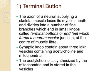


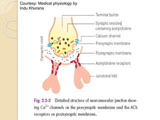


The document provides an overview of the neuromuscular junction, detailing its structure, function, and the processes involved in muscle contraction such as the release and action of acetylcholine. It discusses the mechanisms by which various drugs can affect neuromuscular transmission, including neuromuscular blockers and stimulators. Additionally, the document describes clinical conditions like myasthenia gravis and Lambert-Eaton syndrome, which impair normal neuromuscular function.


