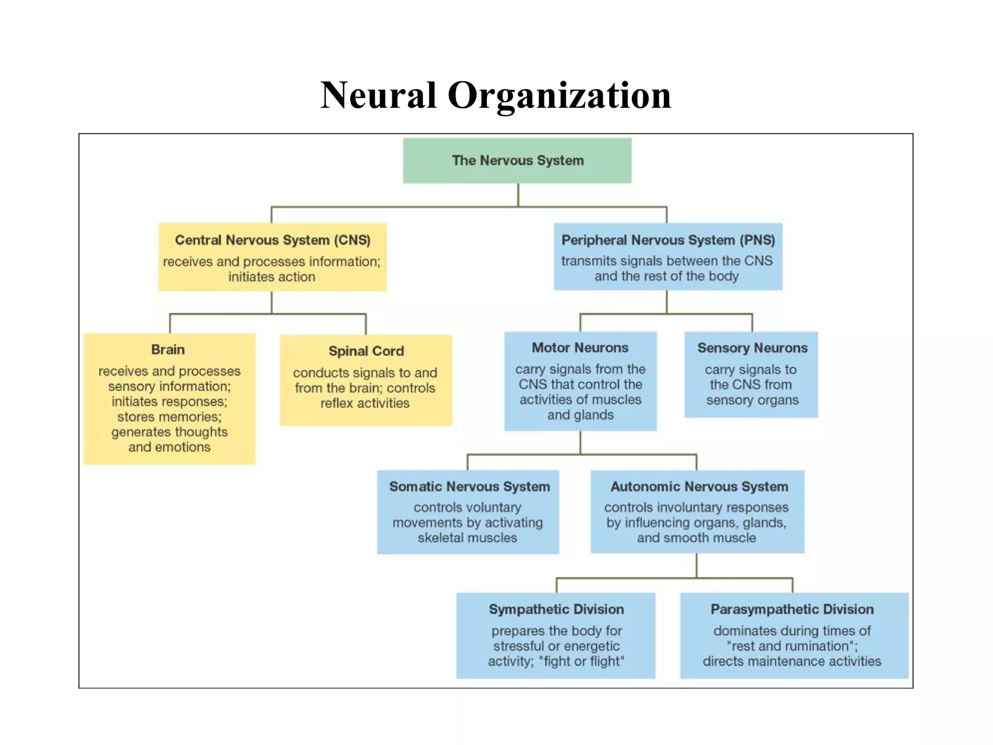
The document summarizes the organization and function of the nervous system in three parts:
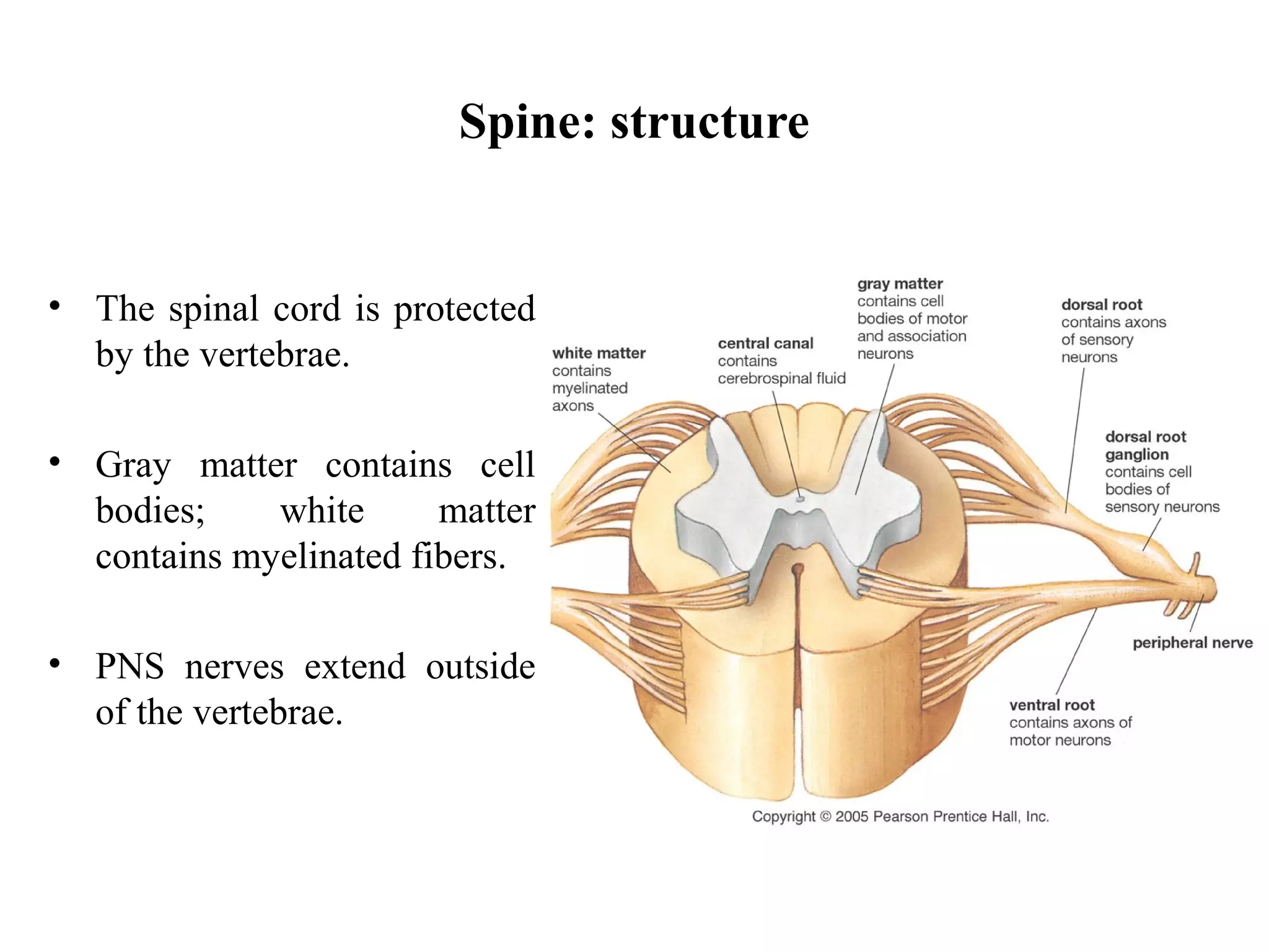
1) The central nervous system (CNS) consists of the brain and spinal cord. It receives sensory signals, determines responses, stores memory, and enables thought.
2) The peripheral nervous system (PNS) is outside the CNS and sends signals to the CNS, receives and transmits motor signals from the CNS, and stimulates effectors.
3) Within the PNS, the somatic nervous system controls voluntary movement via skeletal muscles, while the autonomic nervous system controls involuntary responses like digestion via organs and glands.


![Apporach to lung biopsy [Auto-saved].pptx latest](https://cdn.slidesharecdn.com/ss_thumbnails/apporachtolungbiopsyauto-saved-251211225655-93258539-thumbnail.jpg?width=640&height=640&fit=bounds)


