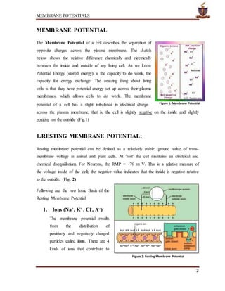
SlideShare a Scribd company logo
1 of 21
MEMBRANE POTENTIALS
1
Topic Content Table
MEMBRANE POTENTIAL
1. RESTING MEMBRANE POTENTIAL:
1.1. Equilibrium Potentials, the Na+/K+ pump and the RMP
1.2. Approximation of RMP by Nernst Equation:
1.3. Different Cells have Different RMP’s Values:
2. GRADED POTENTIAL AND ACTIONS POTENTIALS
2.1 GRADED POTENTIAL
2.1.1. Excitatory Postsynaptic Potentials (EPSPs)
2.1.2. Inhibitory Post Synaptic Potentials (IPSPs)
2.1.3. The Integration of Postsynaptic Potentials and the Generation of
Action Potentials
2.2. ACTION POTENTIAL
2.2.1. Voltage-Gated Sodium and Potassium Channels
2.2.2. Voltage-Gated Potassium Channel and Its Activation
2.2.3. Roles of Other Ions during the Action Potential
2.2.4. The General Sequence Events of an Action Potential
2.2.5. Initiation of Action Potentials
2.2.6. Summation
3. COMPARISON OF GRADED AND ACTION POTENTIALS
REFERENCES
MEMBRANE POTENTIALS
2
MEMBRANE POTENTIAL
The Membrane Potential of a cell describes the separation of
opposite charges across the plasma membrane. The sketch
below shows the relative difference chemically and electrically
between the inside and outside of any living cell. As we know
Potential Energy (stored energy) is the capacity to do work, the
capacity for energy exchange. The amazing thing about living
cells is that they have potential energy set up across their plasma
membranes, which allows cells to do work. The membrane
potential of a cell has a slight imbalance in electrical charge
across the plasma membrane, that is, the cell is slightly negative on the inside and slightly
positive on the outside (Fig.1)
1.RESTING MEMBRANE POTENTIAL:
Resting membrane potential can be defined as a relatively stable, ground value of trans-
membrane voltage in animal and plant cells. At 'rest' the cell maintains an electrical and
chemical disequilibrium. For Neurons, the RMP = -70 m V. This is a relative measure of
the voltage inside of the cell; the negative value indicates that the inside is negative relative
to the outside. (Fig. 2)
Following are the two Ionic Basis of the
Resting Membrane Potential
1. Ions (Na+
, K+
, Cl-
, A--
)
The membrane potential results
from the distribution of
positively and negatively charged
particles called ions. There are 4
kinds of ions that contribute to
Figure 1: Membrane Potential
Figure 2: Resting Membrane Potential
MEMBRANE POTENTIALS
3
the resting membrane potential: Sodium (Na+), potassium (K+), chloride (Cl-), and
negatively charged protein ions sometimes called Anions (A--).
The concentration of Na+ and Cl- ions are greatest outside of the resting cell,
whereas the concentrations of K+ is greatest inside the cell and negatively charged
protein ions which are synthesized inside the neuron are trapped there. These ions
concentrations from one side of the cell membrane to the other are different and
cause chemical gradient or disequilibrium.
2. Differential permeability
The neuronal membrane is porous (i.e., contains ion channels) and allows certain
ions to pass in and out of the cell more readily than others. This passive property of
the cell membrane is called differential permeability and contributes to the
polarized resting potential. For example, both K+ and Cl- ions readily diffuse
through the neural membrane; Na+ ions diffuse with more difficulty and anions
cannot diffuse at all. The electrical charge they contribute from one side of the cell
membrane to the other also differs. This is referred as electrical gradient or
disequilibrium.
Table. 1: A comparison of the permeabilities of ions responsible for creating the
membrane potential.
Ion ECF
Concentration
(mM)
ICF
Concentration
(mM)
Permeability
Na+ 150 15 1
K+ 5 150 50-75
Pro- 0 65 0
As Table 1 above shows, K+ is the most permeable of the ions. In this way, K+ is the most
influential ion in establishing the RMP.
MEMBRANE POTENTIALS
4
1.1. Equilibrium Potentials, the Na+
/K+
pump and the RMP
If we examine the equilibrium potential of the important ions
Na+ and K+ it nicely illustrates how the differences in permeabilities of these
ions contribute to the value of the RMP. To understand the equilibrium
potentials for Na+ and K+ ions, we must examine a hypothetical cell and assume
in each case (separately) that the Na+ and K+ ions are freely permeable, thus can
cross the cell membrane freely.(Fig. 3)
1.1.1. The Movement of Na+ ions
alone: If it is assumed that Na+ ions
are freely permeable, with no
restrictions to its movement, then Na+
ions will move back and forth across
the membrane until the
Electrochemical Gradient has
Equilibrated. The value of the voltage
across the membrane for the Equilibrium Potential of Na+ = +60 mV (ENa+ =
+60mV)
1.1.2. The movement of K+ ions alone: If it is assumed that K+ ions are freely
permeable, with no restrictions to its movement, then K+ ions will move back
and forth across the membrane until the Electrochemical Gradient has
Equilibrated. The value of the voltage across the membrane for the Equilibrium
Potential of K+ = -90 mV (EK+ = -90m V) If these ions were both equally
permeable, then the RMP would be somewhere in between these two values (in
between -90 and +60 mV). However, K+ ions are 50 to 75 times more
permeable than Na+ and therefore the RMP is much closer to the EK+ than the
ENa+. The value of -70 mV is much closer to -90mV than to +60 mV.
1.1.3. The Na+/ K+ Pump (also called the Na+/K+ ATPase): A transport
membrane spanning protein embedded in the plasma membrane that 'pumps'
Na+ and K+ ions across the membrane against their concentration gradients. To
do this, it requires ATP directly, and so it is a primary active transport
mechanism. It pumps out or ejects 3 Na+ ions from the inside of the cell and
Figure 3: Different Ions Contribution in RMP)
MEMBRANE POTENTIALS
5
pumps in or imports 2K+ into the cell from the outside at the cost of 1 ATP for
one cycle of the Na+/K+ pumps. The pump is a protein that has catalytic ability
(is an enzyme as well) and
hydrolyzes ATP to ADP + Pi
and heat. (Fig. 4)
Both Na+ and K+ ions
continuously "leak" across the
cell membrane down their
concentration gradients
(through open protein channels
or ‘pores’ in the membrane).
Because of this, the Na+/ K+ pump must be active all the time in order to
constantly bailout the leaky ship and maintain the RMP. In summary, it is these
three issues that contribute to the maintenance of the RMP.
1.2. Approximation of RMP by Nernst Equation:
RMP Approximation for the equilibrium potential of a given ion only needs the
concentrations on either side of the membrane and the temperature. It can be calculated
using the Nernst equation:
Where
 Eeq,K
+ is the equilibrium potential for potassium, measured in volts
 R is the universal gas constant, equal to 8.314 joules·K−1·mol−1
 T is the absolute temperature, measured in kelvins (= K = degrees Celsius + 273.15)
 z is the number of elementary charges of the ion in question involved in the reaction
 F is the Faraday constant, equal to 96,485 coulombs·mol−1 or J·V−1·mol−1
Figure 4
MEMBRANE POTENTIALS
6
 [K+]o is the extracellular concentration of potassium, measured in mol·m−3 or
mmol·l−1
 [K+]i is likewise the intracellular concentration of potassium
Potassium equilibrium potentials of around −80 millivolts (inside negative) are common.
Differences are observed in different species, different tissues within the same animal, and
the same tissues under different environmental conditions. Applying the Nernst Equation
above, one may account for these differences by changes in relative K+ concentration or
differences in temperature.
Common usage of the Nernst equation is often given in a simplified form by assuming
typical human body temperature (37 °C), reducing the constants and switching to Log base
10. (The units used for concentration are unimportant as they will cancel out into a ratio).
For Potassium at normal body temperature one may calculate the equilibrium potential in
millivolts as:
Likewise the equilibrium potential for sodium (Na+) at normal human body temperature is
calculated using the same simplified constant. You can calculate E assuming an outside
concentration, [K+]o, of 10mM and an inside concentration, [K+]i, of 100mM. For chloride
ions (Cl−) the sign of the constant must be reversed (−61.54 mV). If calculating the
equilibrium potential for calcium (Ca2+) the 2+ charge halves the simplified constant to
30.77 mV. If working at room temperature, about 21 °C, the calculated constants are
approximately 58 mV for K+ and Na+, −58 mV for Cl− and 29 mV for Ca2+. At
physiological temperature, about 29.5 °C, and physiological concentrations (which vary for
each ion), the calculated potentials are approximately 67 mV for Na+, −90 mV for K+,
−86 mV for Cl− and 123 mV for Ca2+.
MEMBRANE POTENTIALS
7
1.3. Different Cells have Different RMP’s Values:
Mainly 4 types of primary tissues are found in the human body that mainly contributes
toward the overall functionality of the body:
1. Epithelium Tissue
2. Connective Tissue
3. Muscle Tissue*
4. Nervous Tissue*
*indicating tissue excitable tissue which respond to the excitement.
The excitable tissues have various RMP's, for example; neurons have a RMP of -70mV
whereas most cardiac muscle cells have a RMP of -90mV. Excitable means that they are
capable of producing electrical signals when excited. As we know the flow of charged
particles is an electrical current, and these currents are used to send signals or do work.
Table.2: Resting potential values in different types of cells
Cell types Resting potential
Skeletal muscle cells −95 mV
Smooth muscle cells –60 mV
Astroglia –80 to –90 mV
Neurons –60 to –70 mV
Erythrocytes –9 mV
Photoreceptor cells –40 mV
MEMBRANE POTENTIALS
8
2. GRADED POTENTIAL AND ACTIONS POTENTIALS
Neurons are the basic cell of communication in the Nervous System, There are two ways
that a neuron can undergo rapid changes in RMP and this really means that there are two
ways that neurons can be electrically communicated. These ways include following main
types of membrane potentials:
1. Graded Potentials
2. Action Potentials.
2.1. GRADED POTENTIAL
Graded potentials are Local change in membrane potential with variable
degrees of magnitude and die out within 1 to 2 mm of their site of origin. They are
usually produced by some specific change in the cell’s environment acting on a
specialized region of the membrane, and they are called “graded potentials” simply
because the magnitude of the potential change can vary (is graded). These are means for
short distance communication. We encounter a number of graded potentials, which are
given various names related to the location of the potential or to the function it performs:
receptor potential, synaptic potential, and pacemaker potential. (Fig. 5)
Let us have brief Introduction of each type of these potentials.
1. Synaptic potential: A graded potential change produced in the postsynaptic
neuron in response to release of a neurotransmitter by a presynaptic terminal; it
may be depolarizing (an excitatory postsynaptic potential or EPSP) or
hyperpolarizing (an inhibitory postsynaptic potential or IPSP).
2. Receptor potential: A graded potential produced at the peripheral endings of
afferent neurons (or in separate receptor cells) in response to a stimulus.
3. Pacemaker potential: A spontaneously occurring graded potential change that
occurs in certain specialized cells.
MEMBRANE POTENTIALS
9
These potentials arise from the summation of the individual actions of ligand-gated ion
channel proteins, and decrease over time and space. They do not typically involve voltage-
gated sodium and potassium channels. These impulses are incremental and may be
excitatory or inhibitory.
Figure 5: Graded Potential
They particularly occur at the postsynaptic dendrite as a result of presynaptic neuron firing
and release of neurotransmitter, or may occur in skeletal, smooth, or cardiac muscle in
response to nerve input. The magnitude of a graded potential is determined by the strength
of the stimulus.When neurotransmitter molecules bind to postsynaptic receptors, they
have one of two effects: depolarization or hyperpolarization.
1. Depolarizations are called Excitatory postsynaptic potentials (or EPSPs) because
they increase the likelihood that the neuron will fire;
2. Hyperpolarizations are called Inhibitory postsynaptic potentials (IPSPs) and
decrease the likelihood that the neuron will fire.
Both events are graded potentials because the strength of their effects are proportional to
the intensity of the signal.EPSPs and IPSPs travel passively through the neuron like an
MEMBRANE POTENTIALS
10
electrical signal travels through a cable. This result in rapid transmission that is
decremental i.e., the signal gets weaker (decreases in amplitude) the farther it travels. (Fig.
6a & 6b)
2.1.1. Excitatory Postsynaptic Potentials (EPSPs)
Graded potentials
that make the
membrane
potential less
negative or more
positive, thus
making the
postsynaptic cell
more likely to have an action potential, are called excitatory postsynaptic
potentials (EPSPs). Depolarizing local potentials sum together, and if the
voltage reaches the threshold potential, an action potential occurs in that cell.
EPSPs are caused by the influx of Na+ or Ca+2 from the extracellular space into
the neuron or muscle cell. When the presynaptic neuron has an action potential,
Ca+2 enters the axon terminal via voltage-dependent calcium channels and
causes exocytosis of synaptic vesicles, causing neurotransmitter to be released.
The transmitter diffuses across the synaptic cleft and activates ligand-gated ion
channels that mediate the EPSP. The amplitude of the EPSP is directly
proportional to the number of synaptic vesicles that were released.
If the EPSP is not large enough to trigger an action potential, the membrane
subsequently repolarizes to its resting membrane potential. This shows the
temporary and reversible nature of graded potentials.
2.1.2. Inhibitory Post Synaptic Potentials (IPSPs)
Graded potentials that make the membrane potential more negative,
and make the postsynaptic cell less likely to have an action potential, are called
inhibitory post synaptic potentials (IPSPs). Hyperpolarization of membranes is
caused by influx of Cl− or efflux of K+. As with EPSPs, the amplitude of the
Figure 6a Figure 6b
MEMBRANE POTENTIALS
11
IPSP is directly proportional to the number of synaptic vesicles that were
released.
2.1.3. The Integration of Postsynaptic Potentials and the Generationof
Action Potentials
Each neuron receives thousands of synaptic contacts which produce
graded potentials. Whether or not a neuron fires depends on the summation of
the signals that reach the axon hillock. The integration of graded potentials
summates in two ways: temporally and spatially (Fig. 7).
1. Temporal summation refers to the combining of signals from a single
synapse across time. The potentials summate because there is a greater
number of open ions channels and, therefore a greater flow of positive
ions into the cell.
2. Spatial summation refers to the combination of signals from different
synapses that are located in close proximity to each other.
3. If the combined stimulation results in a sufficient depolarization at the
hillock then the neuron will generate an action potential; the threshold
of excitation is about -65mV for many neurons.
In graded potentials because the electric signal decreases with distance, they
can function as signals only over very short distances (a few millimeters).
Nevertheless, graded potentials are the only means of communication used by
some neurons and, as we shall see, play very important roles in the initiation
and integration of the long-distance signals by neurons and some other cells.
Figure 7
MEMBRANE POTENTIALS
12
Stronger the triggering event, the stronger the graded potential.
What is a trigger? Here are some examples of what can trigger a graded
potential:
1. A Specific Stimulus - a change in temperature, pH, light intensity, etc.
2. A Surface Receptor on plasma membrane - binding of the receptor by a
ligand.
3. Spontaneous change in membrane potential - may be caused by 'leaky'
channels, etc.
2.2. ACTION POTENTIAL
Action Potential = a brief reversal of resting membrane potential
by a rapid change in plasma membrane permeability. 'Reversal' => from -70mV to +30mV
back to -90mV. Nerve and muscle cells as well as some endocrine, immune, and
reproductive cells have plasma membranes capable of producing action potentials. These
membranes are called excitable membranes, and their ability to generate action potentials is
known as excitability. Whereas all cells are capable of conducting graded potentials, only
excitable membranes can conduct action potentials. The propagation of action potentials is
the mechanism used by the nervous system to communicate over long distances.
The spread of an action potential is non-decremental, that is, the strength of the signal does
not diminish over distance, and it is maintained from the site of origin to destination. An
action potential can be described as an All or None event. During an action potential,
significant changes occur in membrane permeability for Na+ and K+. This causes rapid
fluxes of theses ions down their electrochemical gradients.
There are 4 main phases of an action potential:
1. Threshold
2. Depolarization phase
3. Repolarization phase
4. Hyperpolarization phase
MEMBRANE POTENTIALS
13
For an action potential to occur, threshold must be reached. The threshold value in neurons
is -55 mV. When the RMP is altered and it reaches threshold, this change in the voltage of
the membrane causes voltage gated Na+ channels to open, and this triggers the onset of an
action potential (Fig. 8)
Resting Stage. This is the resting membrane potential before the action potential begins.
The membrane is said to be “polarized” during this stage because of the –90 millivolts
negative membrane potential that is present.
Depolarization Phase. At this time, the
membrane suddenly becomes very permeable to
sodium ions, allowing tremendous numbers of
positively charged sodium ions to diffuse to the
interior of the axon. The normal “polarized”
state of –90 millivolts is immediately
neutralized by the inflowing positively charged
sodium ions, with the potential rising rapidly in the positive direction. This is called
depolarization. In large nerve fibers, the great excess of positive sodium ions moving to the
inside causes the membrane potential to actually “overshoot” beyond the zero level and to
become somewhat positive. In some smaller fibers, as well as in many central nervous
system neurons, the potential merely approaches the zero level and does not overshoot to
the positive state.
Repolarization Phase. Within a few 10,000ths of a second after the membrane
becomes highly permeable to sodium ions, the sodium channels begin to close and the
potassium channels open more than normal. Then, rapid diffusion of potassium ions to
the exterior re-establishes the normal negative resting membrane potential. This is called
repolarization of the membrane.
To explain more fully the factors that cause both depolarization and repolarization, we need
to describe the special characteristics of other types of transport channels through the nerve
membrane.
Figure 8: Different Stages of Action Potential
MEMBRANE POTENTIALS
14
2.2.1. Voltage-Gated Sodium and Potassium Channels
The necessary in causing both depolarization and repolarization of the nerve
membrane during the action potential is the voltage-gated sodium channel. A voltage-
gated potassium channel also plays an impor- tant role in increasing the rapidity of
repolarization of the membrane. These two voltage-gated channels are in addition to the
- p and t - (Fig. 9).
Voltage-Gated Sodium Channel—Activation and Inactivation of
the Channel
This channel has two gates—one near the outside of the channel
called the activation gate, and another near the inside called the inactivation gate.
In the normal resting membrane when the membrane potential is –90 millivolts,
the activation gate is
closed, which prevents
any entry of sodium ions
to the interior of the fiber
through these sodium
channels.
Activation of the
Sodium Channel.
When the membrane
potential becomes less
negative than during
the resting state, rising
from –90 millivolts
toward zero, it finally
reaches a voltage—
usually somewhere between –70 and –50 millivolts—that causes a sudden
conformational change in the activation gate, flipping it all the way to the open
position. This is called the activated state; during this state, sodium ions can pour
Figure 9
MEMBRANE POTENTIALS
15
inward through the channel, increasing the sodium permeability of the membrane
as much as 500- to 5000-fold.
Inactivation of the Sodium Channel. The same increase in voltage that
opens the activation gate also closes the inactivation gate. The inactivation gate,
however, closes a few 10,000ths of a second after the activation gate opens. That
is, the conformational change that flips the inactivation gate to the closed state is a
slower process than the conformational change that opens the activation gate.
Therefore, after the sodium channel has remained open for a few 10,000ths of
a second, the inactivation gate closes, and sodium ions no longer can pour to the
inside of the membrane. At this point, the membrane potential begins to recover
back toward the resting membrane state, which is the repolarization process.
Another important characteristic of the sodium channel inactivation process is that
the inactivation gate will not reopen until the membrane potential returns to or
near the original resting membrane potential level. Therefore, it usually is not
possible for the sodium channels to open again without the nerve fibers first
repolarizing.
2.2.2. Voltage-Gated Potassium Channel and Its Activation
During the resting state, the gate of the potassium channel is closed, and potassium ions are
prevented from passing through this channel to the exterior. When the membrane potential
rises from –90 millivolts toward zero, this voltage change causes a conformational opening
of the gate and allows increased potassium diffusion outward through the channel.
However, because of the slight delay in opening of the potassium channels, for the most
part, they open just at the same time that the sodium channels are beginning to close
because of inactivation. Thus, the decrease in sodium entry to the cell and the simultaneous
increase in potassium exit from the cell combine to speed the repolarization process,
leading to full recovery of the resting membrane potential within another few 10,000ths of
a second.
MEMBRANE POTENTIALS
16
2.2.3. Roles of Other Ions during the Action Potential
Two other types of ions must be considered: negative anions and calcium
ions for the action potentials of the membranes.
Negatively Charged Ions inside the Nerve Axon. Inside the axon there are many
negatively charged ions that cannot pass through the membrane channels. They include the
anions of protein molecules and of many organic phosphate compounds, sulfate
compounds, and so forth. Because these ions cannot leave the interior of the axon, any
deficit of positive ions inside the membrane leaves an excess of these impermeant negative
anions. Therefore, these impermeant negative ions are responsible for the negative charge
inside the fiber when there is a net deficit of positively charged potassium ions and other
positive ions.
Calcium Ions. The membranes of almost all cells of the body have a calcium pump
similar to the sodium pump, and calcium serves along with sodium in some cells to cause
most of the action potential. Like the sodium pump, the calcium pump pumps calcium ions
from the interior to the exterior of the cell membrane (or into the endoplasmic reticulum of
the cell), creating a calcium ion gradient of about 10,000-fold. This leaves an internal cell
concentration of calcium ions of about 10–7 molar, in contrast to an external concentration
of about 10–3 molar.
In addition, there are voltage-gated calcium channels. These channels are slightly
permeable to sodium ions as well as to calcium ions; when they open, both calcium and
sodium ions flow to the interior of the fiber. Therefore, these channels are also called
- um channels are slow to become activated, requiring
10 to 20 times as long for activation as the sodium channels. Therefore, they are called
slow channels, in contrast to the sodium channels, which are called fast channels. Calcium
channels are numerous in both cardiac muscle and smooth muscle. In fact, in some types of
smooth muscle, the fast sodium channels are hardly present, so that the action potentials are
caused almost entirely by activation of slow calcium channels
MEMBRANE POTENTIALS
17
2.2.4. The General Sequence Events of an Action Potential
The result of the opening of voltage gated Na+ channels when
threshold is reached (and the positive feedback loop that ensues) is that Na+ floods into cell
and the inside of the cell becomes more positive very quickly, going from -55 mV (resting)
towards a positive value of +30 mV. Recall that the ENa+ = +60 mV, therefore the
membrane is getting closer to this value. At the 'Peak' of the action potential (+30mV), the
Na+ channels close (become deactivated) and remain closed and inactive until RMP is
restored.
All the while, the slow to open K+ channels continue to open and at the peak of the action
potential K+ rush out of the cell, down their concentration gradient. This outward
movement of K+ starts to restore membrane potential back toward RMP (the membrane
voltage is decreasing now but the potential is increasing). This is the repolarization phase;
the cell is becoming more negative inside as the positively charged K+ leaves the cell.
These K+ channels are also slow to close and continue to allow the positively charged K+ to
leave the cell. This leads to a more negatively charged cell inside and represents the
Hyperpolarization phase of the action potential. As the slow closing K+ finally close, the
resting permeability of the cell is restored, RMP is restored and the action potential is over.
An Action Potentials has 2 Refractory Periods
1. Absolute Refractory Period: During this period, the cell is unresponsive to any further
stimuli. No other action potential can be fired at this point, regardless of the strength of the
stimuli. The role of the Absolute refractory period is to ensure one-way propagation of
action potentials.
2. Relative Refractory Period: During this period, another action potential can be
produced but the strength of the stimuli must be greater than normal to trigger an action
potential. The role of the Relative refractory period: helps to limit the frequency of action
potentials.
MEMBRANE POTENTIALS
18
2.2.5. Initiation of Action Potentials
Stimuli” is the initiators of action potentials. Various types of neurons generate action
potentials which can be summarized as fellow (Fig. 10)
In afferent neurons, the initial
depolarization to threshold is achieved
by a graded potential—here called a
receptor potential, which is generated
in the sensory receptors at the
peripheral ends of the neurons. These
are the ends farthest from the central
nervous system, and where the
nervous system functionally
encounters the outside world. In all
other neurons, the depolarization to
threshold is due either to a graded
potential generated by synaptic input
to the neuron or to a spontaneous
change in the neuron’s membrane
potential, known as a pacemaker
potential. Spontaneous generation of
pacemaker potentials occurs in the absence of any identifiable external stimulus and is an
inherent property of certain neurons (and other excitable cells, including certain smooth-
muscle and cardiac-muscle cells). In these cells, the activity of different types of ion
channels in the plasma membrane causes a graded depolarization of the membrane—the
pacemaker potential. If threshold is reached, an action potential occurs; the membrane then
depolarizes and again begins to depolarize. There is no stable, resting membrane potential
in such cells because of the continuous change in membrane permeability. The rate at
which the membrane depolarizes to threshold determines the action-potential frequency.
Pacemaker potentials are implicated in many rhythmical behaviors, such as breathing, the
heartbeat, and movements within the walls of the stomach and intestines.
Figure 10
MEMBRANE POTENTIALS
19
2.2.6. Summation:
Summation is when the magnitude of graded potentials can be added together, to have a
combined effect on the postsynaptic membrane. Summation of graded potentials can occur
in two ways: Temporal Summation and Spatial Summation.
Temporal Summation occurs from the summation of graded potentials overlapping in
time. In other words (using the example in class), as the frequency of signals (action
potentials) from neuron A to another neuron, (neuron X) increases, the graded potentials
(from A) can summate.
Spatial Summation occurs from the summation of several graded potentials from several
converging neurons simultaneously. In other words (again using the example in class),
when several different neurons in space (e.g., A and B) send a signal simultaneously to
neuron X, these graded potentials that are sent at the same time are summated by neuron X.
Speed of the Conduction of the Signal
Although the magnitude of an action potential is always the same, the speed of the
propagation of an action potential down an axon can vary.
1. Diameter of Axon
Compare the cross sectional diameter of axons A and B.
Which of these axons will conduct a signal faster and why?
A B
The larger axon will conduct a signal faster than a smaller axon. This is because there is
less friction between the moving charged particles (Na+ and K+) and the sides of the axon
in the larger axon. Axons in the human body do vary in their diameter, but there is a limit
to how large the diameter of an axon can be within the confines of the entire human body.
MEMBRANE POTENTIALS
20
2. Temperature
When the surrounding temperature increases, chemical reactions speed up. Thus, if axon
temperatures increase, the rate of conduction of the impulse down the axon will increase.
Conversely, if temperatures decrease, the rate of conduction of the impulse down the axon
will also decrease. Normally, body temperature remains very constant but can change
dramatically in some situations. Typically a dramatic drop in Tb will significantly slow
down neuronal transmission. For example, if a person falls into the very cold water of a
frozen over lake, all of their nervous responses will be significantly slowed.
3. Myelination of Axon
The myelin sheath that covers some axon is made from the cytoplasm of glial cells
(Schwann cells in the PNS and oligodendrocytes in the CNS). The myelin sheath is mostly
composed of lipids and therefore is a good insulator, which is the same as saying it is a
poor conductor of electrical charge. In this way, it reduces the electrical 'leakiness' along
the axon and helps to conduct the signal more quickly.
Little gaps in the myelin sheath, called 'Nodes of Ranvier', allow the action potential to
move faster along the axon. The electrical signal is said to jump from node to node, thus it
is called Saltatory Conduction. This is not what actually happens at the Nodes of Ranvier,
but at this stage it is convenient to think of the signal 'jumping' down the myelinated axon
significantly faster than a non-myelinated axon.
Of these three factors that can affect the speed of an action potential traveling down an
axon, (diameter, temperature and myelination), it is axon myelination that is the most
significant. This is mainly because axon diameter and body temperature are kept fairly
constant.
The degenerative disease multiple sclerosis is due to the destruction of the myelin sheath
on somatic motor neurons that control skeletal muscle movement. Initially it causes a
slowing of the signal and eventually it can stop motor signals to skeletal muscle all
together. The sensory neurons that are bringing in sensory information are not affected by
multiple sclerosis. So, you could feel your legs normally but would have problems sending
signals out for muscle control.
MEMBRANE POTENTIALS
21
3.COMPARISON OF GRADED AND ACTION POTENTIALS
Below is a side-by-side comparison of graded and action potentials.
Graded Potentials Action Potentials
1) Magnitude varies 1) No variation - All or None
2) Decremental (passive spread) 2) Non-decremental (self-regenerating)
3) No Refractory Periods 3) Two Refractory Periods (absolute
and relative)
4) Summation is possible 4) No Summation possible
5) Trigger: NT's, hormones, etc. 5) Trigger: Threshold reached
6) Occurs at cell body (direction can vary) 6) Occurs at axon hillock (one way
direction)
REFERENCES
1. Hall, John E., and Arthur C. Guyton., (2006). The Textbook of Medical Physiology.11e.
Philadelphia, PA: Saunders Elsevier. Pp-58-67.
2. Hille, B., (2001). Ion Channels of Excitable Membranes (3rd Ed.). Sunderland,
Massachusetts: Sinauer. pp. 169–200.
3. Vander, A. J., Luciano, D., Sherman, J., (2001). Human Physiology: The Mechanisms of
Body Function, 8th Ed. McGraw-Hill Higher Education, Boston, MA, pp. 188-190.
4. Wright, S. H.,(2004) “Generation of resting membrane potential “ Advances in Physiology
Education, 28(1–4): 139–142,
5. Barnett, M. W., Larkman, P.M., Larkman, (2007). "The action potential". Pract Neurol 7
(3): 192–197.
6. Stevens, C.F., (1966). Neurophysiology: A Primer. New York: John Wiley and Sons. pp.
127-128.
7. Bullock, T. H., Orkand, R., Grinnell, A., (1977). Introduction to Nervous Systems. A series
of books in biology. San Francisco: W. H. Freeman. pp. 160-164.

More Related Content

What's hot

Action potential (niraj)
Action potential (niraj)Action potential (niraj)
Action potential (niraj)Niraj Kumar
 
Muscle contraction
Muscle contractionMuscle contraction
Muscle contractionvajira54
 
Action potential
Action potentialAction potential
Action potentialIlyas Raza
 
General Physiology - Action potential
General Physiology -  Action potentialGeneral Physiology -  Action potential
General Physiology - Action potentialHamzeh AlBattikhi
 
Resting Membrane potential
Resting Membrane potentialResting Membrane potential
Resting Membrane potentialNathan Peter
 
ACTION POTENTIAL - IONIC BASIS AND RECORDING
ACTION POTENTIAL - IONIC BASIS AND RECORDINGACTION POTENTIAL - IONIC BASIS AND RECORDING
ACTION POTENTIAL - IONIC BASIS AND RECORDINGAnu Priya
 
Nerve and muscle physiology
Nerve and muscle physiologyNerve and muscle physiology
Nerve and muscle physiologySamrah Jawed
 
Tubular-functions-of-kidney
Tubular-functions-of-kidneyTubular-functions-of-kidney
Tubular-functions-of-kidneyRaghu Veer
 
Membrane Potentials
Membrane PotentialsMembrane Potentials
Membrane Potentialsvajira54
 
Physiology of muscle contraction.pptx
Physiology of muscle contraction.pptxPhysiology of muscle contraction.pptx
Physiology of muscle contraction.pptxvinay nandimalla
 
Action potential (the guyton and hall physiology)
Action potential (the guyton and hall physiology)Action potential (the guyton and hall physiology)
Action potential (the guyton and hall physiology)Maryam Fida
 
The Gibbs- Donnan equilibrium.
The Gibbs- Donnan equilibrium.The Gibbs- Donnan equilibrium.
The Gibbs- Donnan equilibrium.Arindam Sain
 
Action potential
Action potential Action potential
Action potential Jinny Shaw
 
Structure, function and classification of neuron
Structure, function and classification of neuronStructure, function and classification of neuron
Structure, function and classification of neuronDr Sara Sadiq
 

What's hot (20)

Action potential (niraj)
Action potential (niraj)Action potential (niraj)
Action potential (niraj)
 
Synapse
SynapseSynapse
Synapse
 
Muscle contraction
Muscle contractionMuscle contraction
Muscle contraction
 
RESTING MEMBRANE POTENTIAL & ACTION POTENTIAL
RESTING MEMBRANE POTENTIAL & ACTION POTENTIALRESTING MEMBRANE POTENTIAL & ACTION POTENTIAL
RESTING MEMBRANE POTENTIAL & ACTION POTENTIAL
 
Resting membrane potential
Resting membrane potentialResting membrane potential
Resting membrane potential
 
Action potential
Action potentialAction potential
Action potential
 
Ans
AnsAns
Ans
 
General Physiology - Action potential
General Physiology -  Action potentialGeneral Physiology -  Action potential
General Physiology - Action potential
 
Resting Membrane potential
Resting Membrane potentialResting Membrane potential
Resting Membrane potential
 
ACTION POTENTIAL - IONIC BASIS AND RECORDING
ACTION POTENTIAL - IONIC BASIS AND RECORDINGACTION POTENTIAL - IONIC BASIS AND RECORDING
ACTION POTENTIAL - IONIC BASIS AND RECORDING
 
SYNAPSE
SYNAPSESYNAPSE
SYNAPSE
 
Nerve and muscle physiology
Nerve and muscle physiologyNerve and muscle physiology
Nerve and muscle physiology
 
MEMBRANE POTENTIALS
MEMBRANE POTENTIALS MEMBRANE POTENTIALS
MEMBRANE POTENTIALS
 
Tubular-functions-of-kidney
Tubular-functions-of-kidneyTubular-functions-of-kidney
Tubular-functions-of-kidney
 
Membrane Potentials
Membrane PotentialsMembrane Potentials
Membrane Potentials
 
Physiology of muscle contraction.pptx
Physiology of muscle contraction.pptxPhysiology of muscle contraction.pptx
Physiology of muscle contraction.pptx
 
Action potential (the guyton and hall physiology)
Action potential (the guyton and hall physiology)Action potential (the guyton and hall physiology)
Action potential (the guyton and hall physiology)
 
The Gibbs- Donnan equilibrium.
The Gibbs- Donnan equilibrium.The Gibbs- Donnan equilibrium.
The Gibbs- Donnan equilibrium.
 
Action potential
Action potential Action potential
Action potential
 
Structure, function and classification of neuron
Structure, function and classification of neuronStructure, function and classification of neuron
Structure, function and classification of neuron
 

Similar to Membrane potentials

Membrane and Resting Membrane potential .ppt
Membrane  and Resting Membrane potential .pptMembrane  and Resting Membrane potential .ppt
Membrane and Resting Membrane potential .pptJamakala Obaiah
 
RESTING MEMBRANE POTENTIAL BY DR QAZI IMTIAZ RASOOL
RESTING MEMBRANE POTENTIAL BY DR QAZI IMTIAZ RASOOLRESTING MEMBRANE POTENTIAL BY DR QAZI IMTIAZ RASOOL
RESTING MEMBRANE POTENTIAL BY DR QAZI IMTIAZ RASOOLDr Qazi Imtiaz RASOOL
 
Active transport, biological signals
Active transport, biological signalsActive transport, biological signals
Active transport, biological signalsJuliana Knocikova
 
Resting membrane potential by DR. IRUM
Resting membrane potential by DR. IRUMResting membrane potential by DR. IRUM
Resting membrane potential by DR. IRUMSMS_2015
 
RESTING MEMBRANE POTENTIAL.ppt
RESTING MEMBRANE POTENTIAL.pptRESTING MEMBRANE POTENTIAL.ppt
RESTING MEMBRANE POTENTIAL.pptShivamJindal71
 
Muscloskeletal system.pdf
Muscloskeletal system.pdfMuscloskeletal system.pdf
Muscloskeletal system.pdfssuser7d3c1b
 
Resting membrane potential
Resting membrane potentialResting membrane potential
Resting membrane potentialarijita02
 
Nerve and Muscle 1.pdf
Nerve and Muscle 1.pdfNerve and Muscle 1.pdf
Nerve and Muscle 1.pdfssuser00317c1
 
Commentary: The number of Electrons in the Nernst Equation: Energetic Conside...
Commentary: The number of Electrons in the Nernst Equation: Energetic Conside...Commentary: The number of Electrons in the Nernst Equation: Energetic Conside...
Commentary: The number of Electrons in the Nernst Equation: Energetic Conside...crimsonpublishersOJCHD
 
1 Sem Chap-3 Neuromuscular phys..pptx
1 Sem Chap-3 Neuromuscular phys..pptx1 Sem Chap-3 Neuromuscular phys..pptx
1 Sem Chap-3 Neuromuscular phys..pptxMegersa4
 
Membranes new
Membranes newMembranes new
Membranes newMUBOSScz
 
Membrane potential + action potential
Membrane  potential + action potentialMembrane  potential + action potential
Membrane potential + action potentialMubashir Iqbal
 
Bioelectric potential
Bioelectric potentialBioelectric potential
Bioelectric potentialanju jha
 
Neural Transmission
Neural TransmissionNeural Transmission
Neural Transmissionvacagodx
 
2. BIOPHYSICS – Membrane Potentials and Action Potential.pptx
2. BIOPHYSICS – Membrane Potentials and Action Potential.pptx2. BIOPHYSICS – Membrane Potentials and Action Potential.pptx
2. BIOPHYSICS – Membrane Potentials and Action Potential.pptxDr Gaurav Saxena
 

Similar to Membrane potentials (20)

RMP.pptx
RMP.pptxRMP.pptx
RMP.pptx
 
Membrane and Resting Membrane potential .ppt
Membrane  and Resting Membrane potential .pptMembrane  and Resting Membrane potential .ppt
Membrane and Resting Membrane potential .ppt
 
RESTING MEMBRANE POTENTIAL BY DR QAZI IMTIAZ RASOOL
RESTING MEMBRANE POTENTIAL BY DR QAZI IMTIAZ RASOOLRESTING MEMBRANE POTENTIAL BY DR QAZI IMTIAZ RASOOL
RESTING MEMBRANE POTENTIAL BY DR QAZI IMTIAZ RASOOL
 
Active transport, biological signals
Active transport, biological signalsActive transport, biological signals
Active transport, biological signals
 
Resting membrane potential by DR. IRUM
Resting membrane potential by DR. IRUMResting membrane potential by DR. IRUM
Resting membrane potential by DR. IRUM
 
Na katp
Na katpNa katp
Na katp
 
Na k pump
Na k pumpNa k pump
Na k pump
 
RESTING MEMBRANE POTENTIAL.ppt
RESTING MEMBRANE POTENTIAL.pptRESTING MEMBRANE POTENTIAL.ppt
RESTING MEMBRANE POTENTIAL.ppt
 
Lecture 40 resting membrane potential
Lecture  40 resting membrane potentialLecture  40 resting membrane potential
Lecture 40 resting membrane potential
 
Muscloskeletal system.pdf
Muscloskeletal system.pdfMuscloskeletal system.pdf
Muscloskeletal system.pdf
 
Resting membrane potential
Resting membrane potentialResting membrane potential
Resting membrane potential
 
Nerve and Muscle 1.pdf
Nerve and Muscle 1.pdfNerve and Muscle 1.pdf
Nerve and Muscle 1.pdf
 
Commentary: The number of Electrons in the Nernst Equation: Energetic Conside...
Commentary: The number of Electrons in the Nernst Equation: Energetic Conside...Commentary: The number of Electrons in the Nernst Equation: Energetic Conside...
Commentary: The number of Electrons in the Nernst Equation: Energetic Conside...
 
1 Sem Chap-3 Neuromuscular phys..pptx
1 Sem Chap-3 Neuromuscular phys..pptx1 Sem Chap-3 Neuromuscular phys..pptx
1 Sem Chap-3 Neuromuscular phys..pptx
 
Membranes new
Membranes newMembranes new
Membranes new
 
Membrane potential + action potential
Membrane  potential + action potentialMembrane  potential + action potential
Membrane potential + action potential
 
Bioelectric potential
Bioelectric potentialBioelectric potential
Bioelectric potential
 
Neural Transmission
Neural TransmissionNeural Transmission
Neural Transmission
 
2. BIOPHYSICS – Membrane Potentials and Action Potential.pptx
2. BIOPHYSICS – Membrane Potentials and Action Potential.pptx2. BIOPHYSICS – Membrane Potentials and Action Potential.pptx
2. BIOPHYSICS – Membrane Potentials and Action Potential.pptx
 
RMP.pptx
RMP.pptxRMP.pptx
RMP.pptx
 

Recently uploaded

Call Girls Kochi Just Call 9907093804 Top Class Call Girl Service Available
Call Girls Kochi Just Call 9907093804 Top Class Call Girl Service AvailableCall Girls Kochi Just Call 9907093804 Top Class Call Girl Service Available
Call Girls Kochi Just Call 9907093804 Top Class Call Girl Service AvailableDipal Arora
 
Call Girls Faridabad Just Call 9907093804 Top Class Call Girl Service Available
Call Girls Faridabad Just Call 9907093804 Top Class Call Girl Service AvailableCall Girls Faridabad Just Call 9907093804 Top Class Call Girl Service Available
Call Girls Faridabad Just Call 9907093804 Top Class Call Girl Service AvailableDipal Arora
 
Top Rated Bangalore Call Girls Ramamurthy Nagar ⟟ 9332606886 ⟟ Call Me For G...
Top Rated Bangalore Call Girls Ramamurthy Nagar ⟟  9332606886 ⟟ Call Me For G...Top Rated Bangalore Call Girls Ramamurthy Nagar ⟟  9332606886 ⟟ Call Me For G...
Top Rated Bangalore Call Girls Ramamurthy Nagar ⟟ 9332606886 ⟟ Call Me For G...narwatsonia7
 
(👑VVIP ISHAAN ) Russian Call Girls Service Navi Mumbai🖕9920874524🖕Independent...
(👑VVIP ISHAAN ) Russian Call Girls Service Navi Mumbai🖕9920874524🖕Independent...(👑VVIP ISHAAN ) Russian Call Girls Service Navi Mumbai🖕9920874524🖕Independent...
(👑VVIP ISHAAN ) Russian Call Girls Service Navi Mumbai🖕9920874524🖕Independent...Taniya Sharma
 
Call Girls Bangalore Just Call 9907093804 Top Class Call Girl Service Available
Call Girls Bangalore Just Call 9907093804 Top Class Call Girl Service AvailableCall Girls Bangalore Just Call 9907093804 Top Class Call Girl Service Available
Call Girls Bangalore Just Call 9907093804 Top Class Call Girl Service AvailableDipal Arora
 
Call Girls Aurangabad Just Call 9907093804 Top Class Call Girl Service Available
Call Girls Aurangabad Just Call 9907093804 Top Class Call Girl Service AvailableCall Girls Aurangabad Just Call 9907093804 Top Class Call Girl Service Available
Call Girls Aurangabad Just Call 9907093804 Top Class Call Girl Service AvailableDipal Arora
 
Call Girls Coimbatore Just Call 9907093804 Top Class Call Girl Service Available
Call Girls Coimbatore Just Call 9907093804 Top Class Call Girl Service AvailableCall Girls Coimbatore Just Call 9907093804 Top Class Call Girl Service Available
Call Girls Coimbatore Just Call 9907093804 Top Class Call Girl Service AvailableDipal Arora
 
Book Paid Powai Call Girls Mumbai 𖠋 9930245274 𖠋Low Budget Full Independent H...
Book Paid Powai Call Girls Mumbai 𖠋 9930245274 𖠋Low Budget Full Independent H...Book Paid Powai Call Girls Mumbai 𖠋 9930245274 𖠋Low Budget Full Independent H...
Book Paid Powai Call Girls Mumbai 𖠋 9930245274 𖠋Low Budget Full Independent H...Call Girls in Nagpur High Profile
 
♛VVIP Hyderabad Call Girls Chintalkunta🖕7001035870🖕Riya Kappor Top Call Girl ...
♛VVIP Hyderabad Call Girls Chintalkunta🖕7001035870🖕Riya Kappor Top Call Girl ...♛VVIP Hyderabad Call Girls Chintalkunta🖕7001035870🖕Riya Kappor Top Call Girl ...
♛VVIP Hyderabad Call Girls Chintalkunta🖕7001035870🖕Riya Kappor Top Call Girl ...astropune
 
Call Girls Bareilly Just Call 9907093804 Top Class Call Girl Service Available
Call Girls Bareilly Just Call 9907093804 Top Class Call Girl Service AvailableCall Girls Bareilly Just Call 9907093804 Top Class Call Girl Service Available
Call Girls Bareilly Just Call 9907093804 Top Class Call Girl Service AvailableDipal Arora
 
Call Girls Cuttack Just Call 9907093804 Top Class Call Girl Service Available
Call Girls Cuttack Just Call 9907093804 Top Class Call Girl Service AvailableCall Girls Cuttack Just Call 9907093804 Top Class Call Girl Service Available
Call Girls Cuttack Just Call 9907093804 Top Class Call Girl Service AvailableDipal Arora
 
Call Girls Gwalior Just Call 8617370543 Top Class Call Girl Service Available
Call Girls Gwalior Just Call 8617370543 Top Class Call Girl Service AvailableCall Girls Gwalior Just Call 8617370543 Top Class Call Girl Service Available
Call Girls Gwalior Just Call 8617370543 Top Class Call Girl Service AvailableDipal Arora
 
Call Girls Varanasi Just Call 9907093804 Top Class Call Girl Service Available
Call Girls Varanasi Just Call 9907093804 Top Class Call Girl Service AvailableCall Girls Varanasi Just Call 9907093804 Top Class Call Girl Service Available
Call Girls Varanasi Just Call 9907093804 Top Class Call Girl Service AvailableDipal Arora
 
Premium Call Girls Cottonpet Whatsapp 7001035870 Independent Escort Service
Premium Call Girls Cottonpet Whatsapp 7001035870 Independent Escort ServicePremium Call Girls Cottonpet Whatsapp 7001035870 Independent Escort Service
Premium Call Girls Cottonpet Whatsapp 7001035870 Independent Escort Servicevidya singh
 
Call Girls Ooty Just Call 9907093804 Top Class Call Girl Service Available
Call Girls Ooty Just Call 9907093804 Top Class Call Girl Service AvailableCall Girls Ooty Just Call 9907093804 Top Class Call Girl Service Available
Call Girls Ooty Just Call 9907093804 Top Class Call Girl Service AvailableDipal Arora
 
VIP Hyderabad Call Girls Bahadurpally 7877925207 ₹5000 To 25K With AC Room 💚😋
VIP Hyderabad Call Girls Bahadurpally 7877925207 ₹5000 To 25K With AC Room 💚😋VIP Hyderabad Call Girls Bahadurpally 7877925207 ₹5000 To 25K With AC Room 💚😋
VIP Hyderabad Call Girls Bahadurpally 7877925207 ₹5000 To 25K With AC Room 💚😋TANUJA PANDEY
 
Top Rated Bangalore Call Girls Richmond Circle ⟟ 9332606886 ⟟ Call Me For Ge...
Top Rated Bangalore Call Girls Richmond Circle ⟟  9332606886 ⟟ Call Me For Ge...Top Rated Bangalore Call Girls Richmond Circle ⟟  9332606886 ⟟ Call Me For Ge...
Top Rated Bangalore Call Girls Richmond Circle ⟟ 9332606886 ⟟ Call Me For Ge...narwatsonia7
 
VIP Call Girls Indore Kirti 💚😋 9256729539 🚀 Indore Escorts
VIP Call Girls Indore Kirti 💚😋  9256729539 🚀 Indore EscortsVIP Call Girls Indore Kirti 💚😋  9256729539 🚀 Indore Escorts
VIP Call Girls Indore Kirti 💚😋 9256729539 🚀 Indore Escortsaditipandeya
 
College Call Girls in Haridwar 9667172968 Short 4000 Night 10000 Best call gi...
College Call Girls in Haridwar 9667172968 Short 4000 Night 10000 Best call gi...College Call Girls in Haridwar 9667172968 Short 4000 Night 10000 Best call gi...
College Call Girls in Haridwar 9667172968 Short 4000 Night 10000 Best call gi...perfect solution
 
Call Girls Horamavu WhatsApp Number 7001035870 Meeting With Bangalore Escorts
Call Girls Horamavu WhatsApp Number 7001035870 Meeting With Bangalore EscortsCall Girls Horamavu WhatsApp Number 7001035870 Meeting With Bangalore Escorts
Call Girls Horamavu WhatsApp Number 7001035870 Meeting With Bangalore Escortsvidya singh
 

Recently uploaded (20)

Call Girls Kochi Just Call 9907093804 Top Class Call Girl Service Available
Call Girls Kochi Just Call 9907093804 Top Class Call Girl Service AvailableCall Girls Kochi Just Call 9907093804 Top Class Call Girl Service Available
Call Girls Kochi Just Call 9907093804 Top Class Call Girl Service Available
 
Call Girls Faridabad Just Call 9907093804 Top Class Call Girl Service Available
Call Girls Faridabad Just Call 9907093804 Top Class Call Girl Service AvailableCall Girls Faridabad Just Call 9907093804 Top Class Call Girl Service Available
Call Girls Faridabad Just Call 9907093804 Top Class Call Girl Service Available
 
Top Rated Bangalore Call Girls Ramamurthy Nagar ⟟ 9332606886 ⟟ Call Me For G...
Top Rated Bangalore Call Girls Ramamurthy Nagar ⟟  9332606886 ⟟ Call Me For G...Top Rated Bangalore Call Girls Ramamurthy Nagar ⟟  9332606886 ⟟ Call Me For G...
Top Rated Bangalore Call Girls Ramamurthy Nagar ⟟ 9332606886 ⟟ Call Me For G...
 
(👑VVIP ISHAAN ) Russian Call Girls Service Navi Mumbai🖕9920874524🖕Independent...
(👑VVIP ISHAAN ) Russian Call Girls Service Navi Mumbai🖕9920874524🖕Independent...(👑VVIP ISHAAN ) Russian Call Girls Service Navi Mumbai🖕9920874524🖕Independent...
(👑VVIP ISHAAN ) Russian Call Girls Service Navi Mumbai🖕9920874524🖕Independent...
 
Call Girls Bangalore Just Call 9907093804 Top Class Call Girl Service Available
Call Girls Bangalore Just Call 9907093804 Top Class Call Girl Service AvailableCall Girls Bangalore Just Call 9907093804 Top Class Call Girl Service Available
Call Girls Bangalore Just Call 9907093804 Top Class Call Girl Service Available
 
Call Girls Aurangabad Just Call 9907093804 Top Class Call Girl Service Available
Call Girls Aurangabad Just Call 9907093804 Top Class Call Girl Service AvailableCall Girls Aurangabad Just Call 9907093804 Top Class Call Girl Service Available
Call Girls Aurangabad Just Call 9907093804 Top Class Call Girl Service Available
 
Call Girls Coimbatore Just Call 9907093804 Top Class Call Girl Service Available
Call Girls Coimbatore Just Call 9907093804 Top Class Call Girl Service AvailableCall Girls Coimbatore Just Call 9907093804 Top Class Call Girl Service Available
Call Girls Coimbatore Just Call 9907093804 Top Class Call Girl Service Available
 
Book Paid Powai Call Girls Mumbai 𖠋 9930245274 𖠋Low Budget Full Independent H...
Book Paid Powai Call Girls Mumbai 𖠋 9930245274 𖠋Low Budget Full Independent H...Book Paid Powai Call Girls Mumbai 𖠋 9930245274 𖠋Low Budget Full Independent H...
Book Paid Powai Call Girls Mumbai 𖠋 9930245274 𖠋Low Budget Full Independent H...
 
♛VVIP Hyderabad Call Girls Chintalkunta🖕7001035870🖕Riya Kappor Top Call Girl ...
♛VVIP Hyderabad Call Girls Chintalkunta🖕7001035870🖕Riya Kappor Top Call Girl ...♛VVIP Hyderabad Call Girls Chintalkunta🖕7001035870🖕Riya Kappor Top Call Girl ...
♛VVIP Hyderabad Call Girls Chintalkunta🖕7001035870🖕Riya Kappor Top Call Girl ...
 
Call Girls Bareilly Just Call 9907093804 Top Class Call Girl Service Available
Call Girls Bareilly Just Call 9907093804 Top Class Call Girl Service AvailableCall Girls Bareilly Just Call 9907093804 Top Class Call Girl Service Available
Call Girls Bareilly Just Call 9907093804 Top Class Call Girl Service Available
 
Call Girls Cuttack Just Call 9907093804 Top Class Call Girl Service Available
Call Girls Cuttack Just Call 9907093804 Top Class Call Girl Service AvailableCall Girls Cuttack Just Call 9907093804 Top Class Call Girl Service Available
Call Girls Cuttack Just Call 9907093804 Top Class Call Girl Service Available
 
Call Girls Gwalior Just Call 8617370543 Top Class Call Girl Service Available
Call Girls Gwalior Just Call 8617370543 Top Class Call Girl Service AvailableCall Girls Gwalior Just Call 8617370543 Top Class Call Girl Service Available
Call Girls Gwalior Just Call 8617370543 Top Class Call Girl Service Available
 
Call Girls Varanasi Just Call 9907093804 Top Class Call Girl Service Available
Call Girls Varanasi Just Call 9907093804 Top Class Call Girl Service AvailableCall Girls Varanasi Just Call 9907093804 Top Class Call Girl Service Available
Call Girls Varanasi Just Call 9907093804 Top Class Call Girl Service Available
 
Premium Call Girls Cottonpet Whatsapp 7001035870 Independent Escort Service
Premium Call Girls Cottonpet Whatsapp 7001035870 Independent Escort ServicePremium Call Girls Cottonpet Whatsapp 7001035870 Independent Escort Service
Premium Call Girls Cottonpet Whatsapp 7001035870 Independent Escort Service
 
Call Girls Ooty Just Call 9907093804 Top Class Call Girl Service Available
Call Girls Ooty Just Call 9907093804 Top Class Call Girl Service AvailableCall Girls Ooty Just Call 9907093804 Top Class Call Girl Service Available
Call Girls Ooty Just Call 9907093804 Top Class Call Girl Service Available
 
VIP Hyderabad Call Girls Bahadurpally 7877925207 ₹5000 To 25K With AC Room 💚😋
VIP Hyderabad Call Girls Bahadurpally 7877925207 ₹5000 To 25K With AC Room 💚😋VIP Hyderabad Call Girls Bahadurpally 7877925207 ₹5000 To 25K With AC Room 💚😋
VIP Hyderabad Call Girls Bahadurpally 7877925207 ₹5000 To 25K With AC Room 💚😋
 
Top Rated Bangalore Call Girls Richmond Circle ⟟ 9332606886 ⟟ Call Me For Ge...
Top Rated Bangalore Call Girls Richmond Circle ⟟  9332606886 ⟟ Call Me For Ge...Top Rated Bangalore Call Girls Richmond Circle ⟟  9332606886 ⟟ Call Me For Ge...
Top Rated Bangalore Call Girls Richmond Circle ⟟ 9332606886 ⟟ Call Me For Ge...
 
VIP Call Girls Indore Kirti 💚😋 9256729539 🚀 Indore Escorts
VIP Call Girls Indore Kirti 💚😋  9256729539 🚀 Indore EscortsVIP Call Girls Indore Kirti 💚😋  9256729539 🚀 Indore Escorts
VIP Call Girls Indore Kirti 💚😋 9256729539 🚀 Indore Escorts
 
College Call Girls in Haridwar 9667172968 Short 4000 Night 10000 Best call gi...
College Call Girls in Haridwar 9667172968 Short 4000 Night 10000 Best call gi...College Call Girls in Haridwar 9667172968 Short 4000 Night 10000 Best call gi...
College Call Girls in Haridwar 9667172968 Short 4000 Night 10000 Best call gi...
 
Call Girls Horamavu WhatsApp Number 7001035870 Meeting With Bangalore Escorts
Call Girls Horamavu WhatsApp Number 7001035870 Meeting With Bangalore EscortsCall Girls Horamavu WhatsApp Number 7001035870 Meeting With Bangalore Escorts
Call Girls Horamavu WhatsApp Number 7001035870 Meeting With Bangalore Escorts
 

Membrane potentials

  • 1. MEMBRANE POTENTIALS 1 Topic Content Table MEMBRANE POTENTIAL 1. RESTING MEMBRANE POTENTIAL: 1.1. Equilibrium Potentials, the Na+/K+ pump and the RMP 1.2. Approximation of RMP by Nernst Equation: 1.3. Different Cells have Different RMP’s Values: 2. GRADED POTENTIAL AND ACTIONS POTENTIALS 2.1 GRADED POTENTIAL 2.1.1. Excitatory Postsynaptic Potentials (EPSPs) 2.1.2. Inhibitory Post Synaptic Potentials (IPSPs) 2.1.3. The Integration of Postsynaptic Potentials and the Generation of Action Potentials 2.2. ACTION POTENTIAL 2.2.1. Voltage-Gated Sodium and Potassium Channels 2.2.2. Voltage-Gated Potassium Channel and Its Activation 2.2.3. Roles of Other Ions during the Action Potential 2.2.4. The General Sequence Events of an Action Potential 2.2.5. Initiation of Action Potentials 2.2.6. Summation 3. COMPARISON OF GRADED AND ACTION POTENTIALS REFERENCES
  • 2. MEMBRANE POTENTIALS 2 MEMBRANE POTENTIAL The Membrane Potential of a cell describes the separation of opposite charges across the plasma membrane. The sketch below shows the relative difference chemically and electrically between the inside and outside of any living cell. As we know Potential Energy (stored energy) is the capacity to do work, the capacity for energy exchange. The amazing thing about living cells is that they have potential energy set up across their plasma membranes, which allows cells to do work. The membrane potential of a cell has a slight imbalance in electrical charge across the plasma membrane, that is, the cell is slightly negative on the inside and slightly positive on the outside (Fig.1) 1.RESTING MEMBRANE POTENTIAL: Resting membrane potential can be defined as a relatively stable, ground value of trans- membrane voltage in animal and plant cells. At 'rest' the cell maintains an electrical and chemical disequilibrium. For Neurons, the RMP = -70 m V. This is a relative measure of the voltage inside of the cell; the negative value indicates that the inside is negative relative to the outside. (Fig. 2) Following are the two Ionic Basis of the Resting Membrane Potential 1. Ions (Na+ , K+ , Cl- , A-- ) The membrane potential results from the distribution of positively and negatively charged particles called ions. There are 4 kinds of ions that contribute to Figure 1: Membrane Potential Figure 2: Resting Membrane Potential
  • 3. MEMBRANE POTENTIALS 3 the resting membrane potential: Sodium (Na+), potassium (K+), chloride (Cl-), and negatively charged protein ions sometimes called Anions (A--). The concentration of Na+ and Cl- ions are greatest outside of the resting cell, whereas the concentrations of K+ is greatest inside the cell and negatively charged protein ions which are synthesized inside the neuron are trapped there. These ions concentrations from one side of the cell membrane to the other are different and cause chemical gradient or disequilibrium. 2. Differential permeability The neuronal membrane is porous (i.e., contains ion channels) and allows certain ions to pass in and out of the cell more readily than others. This passive property of the cell membrane is called differential permeability and contributes to the polarized resting potential. For example, both K+ and Cl- ions readily diffuse through the neural membrane; Na+ ions diffuse with more difficulty and anions cannot diffuse at all. The electrical charge they contribute from one side of the cell membrane to the other also differs. This is referred as electrical gradient or disequilibrium. Table. 1: A comparison of the permeabilities of ions responsible for creating the membrane potential. Ion ECF Concentration (mM) ICF Concentration (mM) Permeability Na+ 150 15 1 K+ 5 150 50-75 Pro- 0 65 0 As Table 1 above shows, K+ is the most permeable of the ions. In this way, K+ is the most influential ion in establishing the RMP.
  • 4. MEMBRANE POTENTIALS 4 1.1. Equilibrium Potentials, the Na+ /K+ pump and the RMP If we examine the equilibrium potential of the important ions Na+ and K+ it nicely illustrates how the differences in permeabilities of these ions contribute to the value of the RMP. To understand the equilibrium potentials for Na+ and K+ ions, we must examine a hypothetical cell and assume in each case (separately) that the Na+ and K+ ions are freely permeable, thus can cross the cell membrane freely.(Fig. 3) 1.1.1. The Movement of Na+ ions alone: If it is assumed that Na+ ions are freely permeable, with no restrictions to its movement, then Na+ ions will move back and forth across the membrane until the Electrochemical Gradient has Equilibrated. The value of the voltage across the membrane for the Equilibrium Potential of Na+ = +60 mV (ENa+ = +60mV) 1.1.2. The movement of K+ ions alone: If it is assumed that K+ ions are freely permeable, with no restrictions to its movement, then K+ ions will move back and forth across the membrane until the Electrochemical Gradient has Equilibrated. The value of the voltage across the membrane for the Equilibrium Potential of K+ = -90 mV (EK+ = -90m V) If these ions were both equally permeable, then the RMP would be somewhere in between these two values (in between -90 and +60 mV). However, K+ ions are 50 to 75 times more permeable than Na+ and therefore the RMP is much closer to the EK+ than the ENa+. The value of -70 mV is much closer to -90mV than to +60 mV. 1.1.3. The Na+/ K+ Pump (also called the Na+/K+ ATPase): A transport membrane spanning protein embedded in the plasma membrane that 'pumps' Na+ and K+ ions across the membrane against their concentration gradients. To do this, it requires ATP directly, and so it is a primary active transport mechanism. It pumps out or ejects 3 Na+ ions from the inside of the cell and Figure 3: Different Ions Contribution in RMP)
  • 5. MEMBRANE POTENTIALS 5 pumps in or imports 2K+ into the cell from the outside at the cost of 1 ATP for one cycle of the Na+/K+ pumps. The pump is a protein that has catalytic ability (is an enzyme as well) and hydrolyzes ATP to ADP + Pi and heat. (Fig. 4) Both Na+ and K+ ions continuously "leak" across the cell membrane down their concentration gradients (through open protein channels or ‘pores’ in the membrane). Because of this, the Na+/ K+ pump must be active all the time in order to constantly bailout the leaky ship and maintain the RMP. In summary, it is these three issues that contribute to the maintenance of the RMP. 1.2. Approximation of RMP by Nernst Equation: RMP Approximation for the equilibrium potential of a given ion only needs the concentrations on either side of the membrane and the temperature. It can be calculated using the Nernst equation: Where  Eeq,K + is the equilibrium potential for potassium, measured in volts  R is the universal gas constant, equal to 8.314 joules·K−1·mol−1  T is the absolute temperature, measured in kelvins (= K = degrees Celsius + 273.15)  z is the number of elementary charges of the ion in question involved in the reaction  F is the Faraday constant, equal to 96,485 coulombs·mol−1 or J·V−1·mol−1 Figure 4
  • 6. MEMBRANE POTENTIALS 6  [K+]o is the extracellular concentration of potassium, measured in mol·m−3 or mmol·l−1  [K+]i is likewise the intracellular concentration of potassium Potassium equilibrium potentials of around −80 millivolts (inside negative) are common. Differences are observed in different species, different tissues within the same animal, and the same tissues under different environmental conditions. Applying the Nernst Equation above, one may account for these differences by changes in relative K+ concentration or differences in temperature. Common usage of the Nernst equation is often given in a simplified form by assuming typical human body temperature (37 °C), reducing the constants and switching to Log base 10. (The units used for concentration are unimportant as they will cancel out into a ratio). For Potassium at normal body temperature one may calculate the equilibrium potential in millivolts as: Likewise the equilibrium potential for sodium (Na+) at normal human body temperature is calculated using the same simplified constant. You can calculate E assuming an outside concentration, [K+]o, of 10mM and an inside concentration, [K+]i, of 100mM. For chloride ions (Cl−) the sign of the constant must be reversed (−61.54 mV). If calculating the equilibrium potential for calcium (Ca2+) the 2+ charge halves the simplified constant to 30.77 mV. If working at room temperature, about 21 °C, the calculated constants are approximately 58 mV for K+ and Na+, −58 mV for Cl− and 29 mV for Ca2+. At physiological temperature, about 29.5 °C, and physiological concentrations (which vary for each ion), the calculated potentials are approximately 67 mV for Na+, −90 mV for K+, −86 mV for Cl− and 123 mV for Ca2+.
  • 7. MEMBRANE POTENTIALS 7 1.3. Different Cells have Different RMP’s Values: Mainly 4 types of primary tissues are found in the human body that mainly contributes toward the overall functionality of the body: 1. Epithelium Tissue 2. Connective Tissue 3. Muscle Tissue* 4. Nervous Tissue* *indicating tissue excitable tissue which respond to the excitement. The excitable tissues have various RMP's, for example; neurons have a RMP of -70mV whereas most cardiac muscle cells have a RMP of -90mV. Excitable means that they are capable of producing electrical signals when excited. As we know the flow of charged particles is an electrical current, and these currents are used to send signals or do work. Table.2: Resting potential values in different types of cells Cell types Resting potential Skeletal muscle cells −95 mV Smooth muscle cells –60 mV Astroglia –80 to –90 mV Neurons –60 to –70 mV Erythrocytes –9 mV Photoreceptor cells –40 mV
  • 8. MEMBRANE POTENTIALS 8 2. GRADED POTENTIAL AND ACTIONS POTENTIALS Neurons are the basic cell of communication in the Nervous System, There are two ways that a neuron can undergo rapid changes in RMP and this really means that there are two ways that neurons can be electrically communicated. These ways include following main types of membrane potentials: 1. Graded Potentials 2. Action Potentials. 2.1. GRADED POTENTIAL Graded potentials are Local change in membrane potential with variable degrees of magnitude and die out within 1 to 2 mm of their site of origin. They are usually produced by some specific change in the cell’s environment acting on a specialized region of the membrane, and they are called “graded potentials” simply because the magnitude of the potential change can vary (is graded). These are means for short distance communication. We encounter a number of graded potentials, which are given various names related to the location of the potential or to the function it performs: receptor potential, synaptic potential, and pacemaker potential. (Fig. 5) Let us have brief Introduction of each type of these potentials. 1. Synaptic potential: A graded potential change produced in the postsynaptic neuron in response to release of a neurotransmitter by a presynaptic terminal; it may be depolarizing (an excitatory postsynaptic potential or EPSP) or hyperpolarizing (an inhibitory postsynaptic potential or IPSP). 2. Receptor potential: A graded potential produced at the peripheral endings of afferent neurons (or in separate receptor cells) in response to a stimulus. 3. Pacemaker potential: A spontaneously occurring graded potential change that occurs in certain specialized cells.
  • 9. MEMBRANE POTENTIALS 9 These potentials arise from the summation of the individual actions of ligand-gated ion channel proteins, and decrease over time and space. They do not typically involve voltage- gated sodium and potassium channels. These impulses are incremental and may be excitatory or inhibitory. Figure 5: Graded Potential They particularly occur at the postsynaptic dendrite as a result of presynaptic neuron firing and release of neurotransmitter, or may occur in skeletal, smooth, or cardiac muscle in response to nerve input. The magnitude of a graded potential is determined by the strength of the stimulus.When neurotransmitter molecules bind to postsynaptic receptors, they have one of two effects: depolarization or hyperpolarization. 1. Depolarizations are called Excitatory postsynaptic potentials (or EPSPs) because they increase the likelihood that the neuron will fire; 2. Hyperpolarizations are called Inhibitory postsynaptic potentials (IPSPs) and decrease the likelihood that the neuron will fire. Both events are graded potentials because the strength of their effects are proportional to the intensity of the signal.EPSPs and IPSPs travel passively through the neuron like an
  • 10. MEMBRANE POTENTIALS 10 electrical signal travels through a cable. This result in rapid transmission that is decremental i.e., the signal gets weaker (decreases in amplitude) the farther it travels. (Fig. 6a & 6b) 2.1.1. Excitatory Postsynaptic Potentials (EPSPs) Graded potentials that make the membrane potential less negative or more positive, thus making the postsynaptic cell more likely to have an action potential, are called excitatory postsynaptic potentials (EPSPs). Depolarizing local potentials sum together, and if the voltage reaches the threshold potential, an action potential occurs in that cell. EPSPs are caused by the influx of Na+ or Ca+2 from the extracellular space into the neuron or muscle cell. When the presynaptic neuron has an action potential, Ca+2 enters the axon terminal via voltage-dependent calcium channels and causes exocytosis of synaptic vesicles, causing neurotransmitter to be released. The transmitter diffuses across the synaptic cleft and activates ligand-gated ion channels that mediate the EPSP. The amplitude of the EPSP is directly proportional to the number of synaptic vesicles that were released. If the EPSP is not large enough to trigger an action potential, the membrane subsequently repolarizes to its resting membrane potential. This shows the temporary and reversible nature of graded potentials. 2.1.2. Inhibitory Post Synaptic Potentials (IPSPs) Graded potentials that make the membrane potential more negative, and make the postsynaptic cell less likely to have an action potential, are called inhibitory post synaptic potentials (IPSPs). Hyperpolarization of membranes is caused by influx of Cl− or efflux of K+. As with EPSPs, the amplitude of the Figure 6a Figure 6b
  • 11. MEMBRANE POTENTIALS 11 IPSP is directly proportional to the number of synaptic vesicles that were released. 2.1.3. The Integration of Postsynaptic Potentials and the Generationof Action Potentials Each neuron receives thousands of synaptic contacts which produce graded potentials. Whether or not a neuron fires depends on the summation of the signals that reach the axon hillock. The integration of graded potentials summates in two ways: temporally and spatially (Fig. 7). 1. Temporal summation refers to the combining of signals from a single synapse across time. The potentials summate because there is a greater number of open ions channels and, therefore a greater flow of positive ions into the cell. 2. Spatial summation refers to the combination of signals from different synapses that are located in close proximity to each other. 3. If the combined stimulation results in a sufficient depolarization at the hillock then the neuron will generate an action potential; the threshold of excitation is about -65mV for many neurons. In graded potentials because the electric signal decreases with distance, they can function as signals only over very short distances (a few millimeters). Nevertheless, graded potentials are the only means of communication used by some neurons and, as we shall see, play very important roles in the initiation and integration of the long-distance signals by neurons and some other cells. Figure 7
  • 12. MEMBRANE POTENTIALS 12 Stronger the triggering event, the stronger the graded potential. What is a trigger? Here are some examples of what can trigger a graded potential: 1. A Specific Stimulus - a change in temperature, pH, light intensity, etc. 2. A Surface Receptor on plasma membrane - binding of the receptor by a ligand. 3. Spontaneous change in membrane potential - may be caused by 'leaky' channels, etc. 2.2. ACTION POTENTIAL Action Potential = a brief reversal of resting membrane potential by a rapid change in plasma membrane permeability. 'Reversal' => from -70mV to +30mV back to -90mV. Nerve and muscle cells as well as some endocrine, immune, and reproductive cells have plasma membranes capable of producing action potentials. These membranes are called excitable membranes, and their ability to generate action potentials is known as excitability. Whereas all cells are capable of conducting graded potentials, only excitable membranes can conduct action potentials. The propagation of action potentials is the mechanism used by the nervous system to communicate over long distances. The spread of an action potential is non-decremental, that is, the strength of the signal does not diminish over distance, and it is maintained from the site of origin to destination. An action potential can be described as an All or None event. During an action potential, significant changes occur in membrane permeability for Na+ and K+. This causes rapid fluxes of theses ions down their electrochemical gradients. There are 4 main phases of an action potential: 1. Threshold 2. Depolarization phase 3. Repolarization phase 4. Hyperpolarization phase
  • 13. MEMBRANE POTENTIALS 13 For an action potential to occur, threshold must be reached. The threshold value in neurons is -55 mV. When the RMP is altered and it reaches threshold, this change in the voltage of the membrane causes voltage gated Na+ channels to open, and this triggers the onset of an action potential (Fig. 8) Resting Stage. This is the resting membrane potential before the action potential begins. The membrane is said to be “polarized” during this stage because of the –90 millivolts negative membrane potential that is present. Depolarization Phase. At this time, the membrane suddenly becomes very permeable to sodium ions, allowing tremendous numbers of positively charged sodium ions to diffuse to the interior of the axon. The normal “polarized” state of –90 millivolts is immediately neutralized by the inflowing positively charged sodium ions, with the potential rising rapidly in the positive direction. This is called depolarization. In large nerve fibers, the great excess of positive sodium ions moving to the inside causes the membrane potential to actually “overshoot” beyond the zero level and to become somewhat positive. In some smaller fibers, as well as in many central nervous system neurons, the potential merely approaches the zero level and does not overshoot to the positive state. Repolarization Phase. Within a few 10,000ths of a second after the membrane becomes highly permeable to sodium ions, the sodium channels begin to close and the potassium channels open more than normal. Then, rapid diffusion of potassium ions to the exterior re-establishes the normal negative resting membrane potential. This is called repolarization of the membrane. To explain more fully the factors that cause both depolarization and repolarization, we need to describe the special characteristics of other types of transport channels through the nerve membrane. Figure 8: Different Stages of Action Potential
  • 14. MEMBRANE POTENTIALS 14 2.2.1. Voltage-Gated Sodium and Potassium Channels The necessary in causing both depolarization and repolarization of the nerve membrane during the action potential is the voltage-gated sodium channel. A voltage- gated potassium channel also plays an impor- tant role in increasing the rapidity of repolarization of the membrane. These two voltage-gated channels are in addition to the - p and t - (Fig. 9). Voltage-Gated Sodium Channel—Activation and Inactivation of the Channel This channel has two gates—one near the outside of the channel called the activation gate, and another near the inside called the inactivation gate. In the normal resting membrane when the membrane potential is –90 millivolts, the activation gate is closed, which prevents any entry of sodium ions to the interior of the fiber through these sodium channels. Activation of the Sodium Channel. When the membrane potential becomes less negative than during the resting state, rising from –90 millivolts toward zero, it finally reaches a voltage— usually somewhere between –70 and –50 millivolts—that causes a sudden conformational change in the activation gate, flipping it all the way to the open position. This is called the activated state; during this state, sodium ions can pour Figure 9
  • 15. MEMBRANE POTENTIALS 15 inward through the channel, increasing the sodium permeability of the membrane as much as 500- to 5000-fold. Inactivation of the Sodium Channel. The same increase in voltage that opens the activation gate also closes the inactivation gate. The inactivation gate, however, closes a few 10,000ths of a second after the activation gate opens. That is, the conformational change that flips the inactivation gate to the closed state is a slower process than the conformational change that opens the activation gate. Therefore, after the sodium channel has remained open for a few 10,000ths of a second, the inactivation gate closes, and sodium ions no longer can pour to the inside of the membrane. At this point, the membrane potential begins to recover back toward the resting membrane state, which is the repolarization process. Another important characteristic of the sodium channel inactivation process is that the inactivation gate will not reopen until the membrane potential returns to or near the original resting membrane potential level. Therefore, it usually is not possible for the sodium channels to open again without the nerve fibers first repolarizing. 2.2.2. Voltage-Gated Potassium Channel and Its Activation During the resting state, the gate of the potassium channel is closed, and potassium ions are prevented from passing through this channel to the exterior. When the membrane potential rises from –90 millivolts toward zero, this voltage change causes a conformational opening of the gate and allows increased potassium diffusion outward through the channel. However, because of the slight delay in opening of the potassium channels, for the most part, they open just at the same time that the sodium channels are beginning to close because of inactivation. Thus, the decrease in sodium entry to the cell and the simultaneous increase in potassium exit from the cell combine to speed the repolarization process, leading to full recovery of the resting membrane potential within another few 10,000ths of a second.
  • 16. MEMBRANE POTENTIALS 16 2.2.3. Roles of Other Ions during the Action Potential Two other types of ions must be considered: negative anions and calcium ions for the action potentials of the membranes. Negatively Charged Ions inside the Nerve Axon. Inside the axon there are many negatively charged ions that cannot pass through the membrane channels. They include the anions of protein molecules and of many organic phosphate compounds, sulfate compounds, and so forth. Because these ions cannot leave the interior of the axon, any deficit of positive ions inside the membrane leaves an excess of these impermeant negative anions. Therefore, these impermeant negative ions are responsible for the negative charge inside the fiber when there is a net deficit of positively charged potassium ions and other positive ions. Calcium Ions. The membranes of almost all cells of the body have a calcium pump similar to the sodium pump, and calcium serves along with sodium in some cells to cause most of the action potential. Like the sodium pump, the calcium pump pumps calcium ions from the interior to the exterior of the cell membrane (or into the endoplasmic reticulum of the cell), creating a calcium ion gradient of about 10,000-fold. This leaves an internal cell concentration of calcium ions of about 10–7 molar, in contrast to an external concentration of about 10–3 molar. In addition, there are voltage-gated calcium channels. These channels are slightly permeable to sodium ions as well as to calcium ions; when they open, both calcium and sodium ions flow to the interior of the fiber. Therefore, these channels are also called - um channels are slow to become activated, requiring 10 to 20 times as long for activation as the sodium channels. Therefore, they are called slow channels, in contrast to the sodium channels, which are called fast channels. Calcium channels are numerous in both cardiac muscle and smooth muscle. In fact, in some types of smooth muscle, the fast sodium channels are hardly present, so that the action potentials are caused almost entirely by activation of slow calcium channels
  • 17. MEMBRANE POTENTIALS 17 2.2.4. The General Sequence Events of an Action Potential The result of the opening of voltage gated Na+ channels when threshold is reached (and the positive feedback loop that ensues) is that Na+ floods into cell and the inside of the cell becomes more positive very quickly, going from -55 mV (resting) towards a positive value of +30 mV. Recall that the ENa+ = +60 mV, therefore the membrane is getting closer to this value. At the 'Peak' of the action potential (+30mV), the Na+ channels close (become deactivated) and remain closed and inactive until RMP is restored. All the while, the slow to open K+ channels continue to open and at the peak of the action potential K+ rush out of the cell, down their concentration gradient. This outward movement of K+ starts to restore membrane potential back toward RMP (the membrane voltage is decreasing now but the potential is increasing). This is the repolarization phase; the cell is becoming more negative inside as the positively charged K+ leaves the cell. These K+ channels are also slow to close and continue to allow the positively charged K+ to leave the cell. This leads to a more negatively charged cell inside and represents the Hyperpolarization phase of the action potential. As the slow closing K+ finally close, the resting permeability of the cell is restored, RMP is restored and the action potential is over. An Action Potentials has 2 Refractory Periods 1. Absolute Refractory Period: During this period, the cell is unresponsive to any further stimuli. No other action potential can be fired at this point, regardless of the strength of the stimuli. The role of the Absolute refractory period is to ensure one-way propagation of action potentials. 2. Relative Refractory Period: During this period, another action potential can be produced but the strength of the stimuli must be greater than normal to trigger an action potential. The role of the Relative refractory period: helps to limit the frequency of action potentials.
  • 18. MEMBRANE POTENTIALS 18 2.2.5. Initiation of Action Potentials Stimuli” is the initiators of action potentials. Various types of neurons generate action potentials which can be summarized as fellow (Fig. 10) In afferent neurons, the initial depolarization to threshold is achieved by a graded potential—here called a receptor potential, which is generated in the sensory receptors at the peripheral ends of the neurons. These are the ends farthest from the central nervous system, and where the nervous system functionally encounters the outside world. In all other neurons, the depolarization to threshold is due either to a graded potential generated by synaptic input to the neuron or to a spontaneous change in the neuron’s membrane potential, known as a pacemaker potential. Spontaneous generation of pacemaker potentials occurs in the absence of any identifiable external stimulus and is an inherent property of certain neurons (and other excitable cells, including certain smooth- muscle and cardiac-muscle cells). In these cells, the activity of different types of ion channels in the plasma membrane causes a graded depolarization of the membrane—the pacemaker potential. If threshold is reached, an action potential occurs; the membrane then depolarizes and again begins to depolarize. There is no stable, resting membrane potential in such cells because of the continuous change in membrane permeability. The rate at which the membrane depolarizes to threshold determines the action-potential frequency. Pacemaker potentials are implicated in many rhythmical behaviors, such as breathing, the heartbeat, and movements within the walls of the stomach and intestines. Figure 10
  • 19. MEMBRANE POTENTIALS 19 2.2.6. Summation: Summation is when the magnitude of graded potentials can be added together, to have a combined effect on the postsynaptic membrane. Summation of graded potentials can occur in two ways: Temporal Summation and Spatial Summation. Temporal Summation occurs from the summation of graded potentials overlapping in time. In other words (using the example in class), as the frequency of signals (action potentials) from neuron A to another neuron, (neuron X) increases, the graded potentials (from A) can summate. Spatial Summation occurs from the summation of several graded potentials from several converging neurons simultaneously. In other words (again using the example in class), when several different neurons in space (e.g., A and B) send a signal simultaneously to neuron X, these graded potentials that are sent at the same time are summated by neuron X. Speed of the Conduction of the Signal Although the magnitude of an action potential is always the same, the speed of the propagation of an action potential down an axon can vary. 1. Diameter of Axon Compare the cross sectional diameter of axons A and B. Which of these axons will conduct a signal faster and why? A B The larger axon will conduct a signal faster than a smaller axon. This is because there is less friction between the moving charged particles (Na+ and K+) and the sides of the axon in the larger axon. Axons in the human body do vary in their diameter, but there is a limit to how large the diameter of an axon can be within the confines of the entire human body.
  • 20. MEMBRANE POTENTIALS 20 2. Temperature When the surrounding temperature increases, chemical reactions speed up. Thus, if axon temperatures increase, the rate of conduction of the impulse down the axon will increase. Conversely, if temperatures decrease, the rate of conduction of the impulse down the axon will also decrease. Normally, body temperature remains very constant but can change dramatically in some situations. Typically a dramatic drop in Tb will significantly slow down neuronal transmission. For example, if a person falls into the very cold water of a frozen over lake, all of their nervous responses will be significantly slowed. 3. Myelination of Axon The myelin sheath that covers some axon is made from the cytoplasm of glial cells (Schwann cells in the PNS and oligodendrocytes in the CNS). The myelin sheath is mostly composed of lipids and therefore is a good insulator, which is the same as saying it is a poor conductor of electrical charge. In this way, it reduces the electrical 'leakiness' along the axon and helps to conduct the signal more quickly. Little gaps in the myelin sheath, called 'Nodes of Ranvier', allow the action potential to move faster along the axon. The electrical signal is said to jump from node to node, thus it is called Saltatory Conduction. This is not what actually happens at the Nodes of Ranvier, but at this stage it is convenient to think of the signal 'jumping' down the myelinated axon significantly faster than a non-myelinated axon. Of these three factors that can affect the speed of an action potential traveling down an axon, (diameter, temperature and myelination), it is axon myelination that is the most significant. This is mainly because axon diameter and body temperature are kept fairly constant. The degenerative disease multiple sclerosis is due to the destruction of the myelin sheath on somatic motor neurons that control skeletal muscle movement. Initially it causes a slowing of the signal and eventually it can stop motor signals to skeletal muscle all together. The sensory neurons that are bringing in sensory information are not affected by multiple sclerosis. So, you could feel your legs normally but would have problems sending signals out for muscle control.
  • 21. MEMBRANE POTENTIALS 21 3.COMPARISON OF GRADED AND ACTION POTENTIALS Below is a side-by-side comparison of graded and action potentials. Graded Potentials Action Potentials 1) Magnitude varies 1) No variation - All or None 2) Decremental (passive spread) 2) Non-decremental (self-regenerating) 3) No Refractory Periods 3) Two Refractory Periods (absolute and relative) 4) Summation is possible 4) No Summation possible 5) Trigger: NT's, hormones, etc. 5) Trigger: Threshold reached 6) Occurs at cell body (direction can vary) 6) Occurs at axon hillock (one way direction) REFERENCES 1. Hall, John E., and Arthur C. Guyton., (2006). The Textbook of Medical Physiology.11e. Philadelphia, PA: Saunders Elsevier. Pp-58-67. 2. Hille, B., (2001). Ion Channels of Excitable Membranes (3rd Ed.). Sunderland, Massachusetts: Sinauer. pp. 169–200. 3. Vander, A. J., Luciano, D., Sherman, J., (2001). Human Physiology: The Mechanisms of Body Function, 8th Ed. McGraw-Hill Higher Education, Boston, MA, pp. 188-190. 4. Wright, S. H.,(2004) “Generation of resting membrane potential “ Advances in Physiology Education, 28(1–4): 139–142, 5. Barnett, M. W., Larkman, P.M., Larkman, (2007). "The action potential". Pract Neurol 7 (3): 192–197. 6. Stevens, C.F., (1966). Neurophysiology: A Primer. New York: John Wiley and Sons. pp. 127-128. 7. Bullock, T. H., Orkand, R., Grinnell, A., (1977). Introduction to Nervous Systems. A series of books in biology. San Francisco: W. H. Freeman. pp. 160-164.