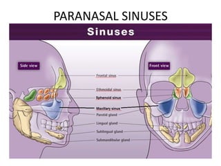
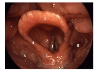
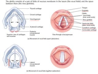
This document provides information on the anatomy and physiology of the respiratory system. It describes the structures of the upper respiratory tract including the nose, pharynx, and larynx. It then discusses the lower respiratory tract including the trachea, bronchi, lungs, and alveoli. Key points include:
- The nose functions to warm, moisten, and filter inhaled air, and contains paranasal sinuses.
- The pharynx is a passageway for both air and food that connects to the larynx and esophagus.
- The larynx contains vocal folds that vibrate to produce voice. Contraction of laryngeal muscles changes the tension of the vocal folds.
- The trache
Respiratory system
a) Anatomy of respiratory organs and functions
b) Mechanism / physiology of respiration and regulation of respiration
c) Transport of respiratory gases
d) Respiratory volumes and capacities, and Definition of: Hypoxia, Asphyxia, Dybarism, Oxygen therapy and resuscitation
Anatomically the respiratory system is divided into
Upper respiratory tract
From the nostril to the vocal cord
Lower respiratory tract
The lower respiratory tract is from bellow the vocal cord upto the alveoli
This was a presentation that was carried out in our research method class by our group. It will be useful for PHD and master students quantitative and qualitative method. It consist sample definition, purpose of sampling, stages in the selection of a sample, types of sampling in quantitative researches, types of sampling in qualitative researches, and ethical Considerations in Data Collection.
Students, digital devices and success - Andreas Schleicher - 27 May 2024..pptxEduSkills OECD
Andreas Schleicher presents at the OECD webinar ‘Digital devices in schools: detrimental distraction or secret to success?’ on 27 May 2024. The presentation was based on findings from PISA 2022 results and the webinar helped launch the PISA in Focus ‘Managing screen time: How to protect and equip students against distraction’ https://www.oecd-ilibrary.org/education/managing-screen-time_7c225af4-en and the OECD Education Policy Perspective ‘Students, digital devices and success’ can be found here - https://oe.cd/il/5yV
How to Create Map Views in the Odoo 17 ERPCeline George
The map views are useful for providing a geographical representation of data. They allow users to visualize and analyze the data in a more intuitive manner.
2024.06.01 Introducing a competency framework for languag learning materials ...Sandy Millin
http://sandymillin.wordpress.com/iateflwebinar2024
Published classroom materials form the basis of syllabuses, drive teacher professional development, and have a potentially huge influence on learners, teachers and education systems. All teachers also create their own materials, whether a few sentences on a blackboard, a highly-structured fully-realised online course, or anything in between. Despite this, the knowledge and skills needed to create effective language learning materials are rarely part of teacher training, and are mostly learnt by trial and error.
Knowledge and skills frameworks, generally called competency frameworks, for ELT teachers, trainers and managers have existed for a few years now. However, until I created one for my MA dissertation, there wasn’t one drawing together what we need to know and do to be able to effectively produce language learning materials.
This webinar will introduce you to my framework, highlighting the key competencies I identified from my research. It will also show how anybody involved in language teaching (any language, not just English!), teacher training, managing schools or developing language learning materials can benefit from using the framework.
Palestine last event orientationfvgnh .pptxRaedMohamed3
An EFL lesson about the current events in Palestine. It is intended to be for intermediate students who wish to increase their listening skills through a short lesson in power point.
This is a presentation by Dada Robert in a Your Skill Boost masterclass organised by the Excellence Foundation for South Sudan (EFSS) on Saturday, the 25th and Sunday, the 26th of May 2024.
He discussed the concept of quality improvement, emphasizing its applicability to various aspects of life, including personal, project, and program improvements. He defined quality as doing the right thing at the right time in the right way to achieve the best possible results and discussed the concept of the "gap" between what we know and what we do, and how this gap represents the areas we need to improve. He explained the scientific approach to quality improvement, which involves systematic performance analysis, testing and learning, and implementing change ideas. He also highlighted the importance of client focus and a team approach to quality improvement.
Unit 8 - Information and Communication Technology (Paper I).pdfThiyagu K
This slides describes the basic concepts of ICT, basics of Email, Emerging Technology and Digital Initiatives in Education. This presentations aligns with the UGC Paper I syllabus.
The Art Pastor's Guide to Sabbath | Steve ThomasonSteve Thomason
What is the purpose of the Sabbath Law in the Torah. It is interesting to compare how the context of the law shifts from Exodus to Deuteronomy. Who gets to rest, and why?
Ethnobotany and Ethnopharmacology:
Ethnobotany in herbal drug evaluation,
Impact of Ethnobotany in traditional medicine,
New development in herbals,
Bio-prospecting tools for drug discovery,
Role of Ethnopharmacology in drug evaluation,
Reverse Pharmacology.
1. Respiratory system
Upper respiratory tract - nose, pharynx and
associated structures.
Lower respiratory tract - larynx, trachea, bronchi
and lungs.
Study Oto-Rhino-Laryng-Ology - ENT
Pulmonologist - lung diseases.
2. 1.Nose
• External & internal nose
• External nose – supportive frame work of
– bone – frontal bone, nasal bone, maxillae and bony
framework of external nose, and
– hyaline cartilage – septal cartilage – anterior portion,
lateral cartilage – inferior and alar cartilage – wall of
nostrils with muscle, skin and mucus membrane.
– external opening – external nares (naris – singular)
nostrils.
Functions :
1.Warming, moistening & filtering.
2.Detecting olfactory stimuli.
3.Modifying speech vibrations.
– Rhino – rhinitis, rhinoplasty.
3.
4. CONT….
• Internal nose – large cavity (nasal cavity) in the
interior aspect of skull lined with muscle and
mucus
– Internal nares or choane – 2 openings for
communication with pharynx (divided into series of
groove like passages – superior, middle and inferior
meatuses)
– Paranasal sinus and nasolacrimal ducts opens into
internal nose
– lined with olfactory epithelium
– Nasal septum – divides nasal cavity into right & left.
8. 2. Pharynx (throat)
• Funnel shaped tube approximately 13 cm long.
• Starts at internal nares ends at larynx’s cricoid cartilage.
• Anterior to cervical vertebrae and posterior to nasal/oral cavities.
• Composed of skeletal muscles.
• Functions
– as passage of air and food.
– resonating chamber for sound.
– house of tonsils in immunological reactions.
Anatomically 3 portions
• 1. Nasopharynx – (helps to adjust pressure b/w pharynx and
middle ear) superior portion with openings to internal nares, ear
and oropharynx, contain pharyngeal tonsil.
• 2. Oropharynx – middle portion, opening called fauces to mouth-
posses respiratory and digestive function, palatine and lingual
tonsils found here.
• 3. Laryngopharynx – opens down to esophagus also has
respiratory and digestive function.
9.
10. 3. Larynx (voice box)
• Connects pharynx and trachea, in the midline of neck anterior
to esophagus
Made of nine piece of cartilage
• 1. Thyroid cartilage – Adam’s apple- 2 fused cartilages in
triangular shape, larger in male.
• 2. Epiglottis (glottis- tongue)large leaf shaped with tapered stem and
broad superior leafy portion.
– Glottis portion consists of pair of folds of mucus membrane called
vocal folds and space b/w them called Rima glottides.
• 3. Cricoid cartilage – ring of hyaline cartilage attaches with
trachea and thyroid cartilage by ligaments.
• 4. Arytenoid cartilage (paired) – triangular attached above
cricoid attached to vocal folds.
• 5. Corniculate cartilages – (paired) at the apex of arytenoid
supporting epiglottis.
• 6. Cuneiform cartilages – (paired) club shaped cartilages above
cuneiform support vocal folds.
11.
12.
13.
14.
15.
16.
17. 4.Trachea (wind pipe)
• Tubular passage for air, approximately 12 cm long &
2.5 cm diameter.
• Anterior to esophagus.
• Starts from larynx to 5th thoracic vertebra and divides
into two primary bronchi.
• 4 layered –
– mucosa & sub mucosa - which provide protection from
dust and produce mucus.
– hyaline cartilage – 16-20 horizontal, incomplete C shaped
rings stalked upon each other, open ends has trachealis
muscle helps to adjust with esophagus expansion.
– adventitia connect trachea to other surrounding tissues.
18. 5.Bronchi (wind pipe)
• At 5th thoracic vertebra trachea divides into 2.
• Right and left primary bronchi – right into right lung
and left into left lung.
• Primary bronchi also has incomplete cartilages.
• Internal ridge where right and left bronchi divides is
called carina – where mucus membrane is most
sensitive.
• Primary bronchi divides to secondary (lobar) and then
to tertiary (segmental) to bronchioles and then to
terminal bronchioles.
• This extensive branching called bronchial tree.
19.
20. • Right pulmonary bronchus – more vertical,
longer, wider, aspired air object more into right
lung.
• Right bronchi – 3 lobes.
• Bronchi to bronchioles – pseudostratified
cuboidal. columnar becomes – non ciliated
simple cuboidal.
• No Goblet cells.
• Incomplete rings to cartilages and cartilage
decreases replaced by more and more smooth
muscle - muscle spasm can occur.
21. Lungs – light weight
• Paired, cone shaped, in the thoracic cavity.
• Heart lies in between.
• Lobes and fissures.
• Oblique fissure – on both lungs anteriorly and
posteriorly - to superior and inferior lobes.
• Horizontal fissure – right lung - middle and inferior
lobes.
22. • Pleural membrane – 2 layers of serous membrane -
enclose and protect lung.
– Parietal pleura – superficial to thoracic wall.
– Visceral pleura – cover the lungs.
– Pleural cavity – b/w pleura – lubricates, reduce friction
and helps in breathing.
• Pneumothorax – if air inside, haemothorax if blood,
atelectasis – collapse of lung.
• A hemothorax (or haemothorax/Haemorrhagic
pleural effusion) is a condition that results from
blood accumulating in the pleural cavity. Atelectasis
(from Greek: "incomplete" + "extension") is defined
as the collapse or closure of the lung resulting in
reduced or absent gas exchange.
23.
24. • Position
– Above diaphragm, just inferior to clavicles.
– Base broad and concave.
– Apex – narrow superior.
– Coastal surface – surface against ribs.
– Hilum - where bronchi, pulmonary vessels, lymph, blood
vessels enters in to the lungs.
– Root - above structures, pleura, connective tissue held
together.
– Cardiac notch – concave cavity of left lung where the heart is
situated.
• Right lung - shorter broader, thicker, due to liver
accommodation.
• Left lung - 10 % shorter than right.
25.
26. • 10 bronchi – divides into 20 bronchi – right lung 3 lobes
and left lung 2 lobes.
• 20 to 30 bronchi – one 20 to 10 – 30 30 bronchi.
• Bronchopulmonary segment - each 30 and segment of
lung tissue.
• Lobules – small compartment of Bronchopulmonary
segment with lymphatic vessels, arterioles, venules &
terminal bronchiole.
• Terminal bronchiole – respiratory bronchiole divides into
2-11 alveolar ducts.
27.
28. ALVEOLI
• Around Alveolar ducts numerous alveoli and alveolar sacs.
• Alveolus – cup shaped pouches.
• Alveolar sac – 2 or more alveoli with common opening.
• Alveolar cells – type 1 – simple squamous for gas
exchange, type 2 or septal cells - few, rounded secrete
alveolar fluid contain surfactant, a mixture of complex
phospholipids and lipoproteins.
• Alveolar macrophages – remove dust particles.
29.
30.
31. Respiratory membrane
Membrane wall b/w alveolar and capillary wall, 0.5 micron as 4
layers.
• One layer of type 1 and type 2 cells, macrophages.
• Epithelial basement membrane of alveolar wall.
• Capillary basement fused to epithelial membrane.
• Capillary endothelium allows rapid diffusion of gases.
• 300 million alveoli – 70 m2 surface area for gas exchange.
32. Blood supply
• Receive via
• Pulmonary Arteries – supply deoxygenated blood
from heart directly.
• Bronchial Arteries – from aorta - oxygenated
blood.
• Pulmonary Vein – oxygenated blood to heart.
• Ventilation perfusion coupling – constriction of
arterioles in response to hypoxia, diverts blood
flow from poorly ventilated area to highly
ventilated areas.
33. 3.(a)Voice production
Structures
• glottis portion of larynx consists of pair of folds of mucus membrane
• superior fold called ventricular or false vocal folds and
• inferior pair called true vocal cords and
• space b/w ventricular folds called rima glottidis
• laryngeal muscles attached to cartilage and vocal folds
Process
• 1. When laryngeal muscles contracts they pull posterior cricoarytenoid
muscles and moves vocal folds apart (abduction) and rima glottidis
becomes open.
• 2. When lateral cricoarytenoid muscles contracts moves the vocal folds
together and rima glottidis becomes narrow /closed (adduction).
• Other intrinsic muscles also can elongate or shorten vocal folds.
• 3. Increased tension on vocal folds, cause folds vibrate rapidly and high
pitch and vice versa.
• Androgens cause male vocal folds more thick, so vibrate slowly and has
low pitch in males.
• Pharynx, mouth, nasal cavity, paranasal sinuses acts as resonating
chambers gives voice individual quality.
34.
35. Pulmonary ventilation
• Respiration – process of gas exchange in our body.
• 3 steps.
• Pulmonary ventilation – breathing = inhalation (inflow)
+ exhalation (outflow) – b/w air of atmosphere and
alveoli of lungs.
• External pulmonary respiration – exchange of gas b/w
alveoli of lungs and blood in pulmonary capillaries
across respiratory membrane – blood gains O2 and
blood loses CO2.
• Internal tissue respiration – exchange of gas b/w blood
in capillaries and tissue cells – blood loses O2 and gains
CO2.
36. Inhalation
• An active process
• Breathing in is called inhalation/inspiration
• As per boyle’s law – when volume increases pressure
decreases
• Before respiration Pressure inside the lungs = air
pressure of atmosphere = 760 mm of hg = 1
atmosphere
• Then pressure inside the lung decreases by increased
lung volume cause air to flow to lungs
• For inhalations lungs must expand, this needs
contraction of muscles of inspiration
37. Muscles of inspiration
1.Diaphragm – dome shaped skeletal muscle at the floor
of thoracic cavity innervated by phrenic nerve.
– Contraction causes –flattening and lowering its dome
– Increases vertical diameter of thoracic cavity
– Descends –normal breath -1 cm, strenuous breath -10 cm,
– Pressure difference – 1-3 mm of hg to 100 mm of hg.
– Normal 500 ml, strenuous 2-3 L.
– Responsible for 75 % of air entering.
2.External intercostal muscles – contraction cause
elevation of ribs, increased anterio-posterior & lateral
diameter of chest cavity - responsible for 25 % of air
that enter
• Accessory muscles – during deep forceful inhalation
– Sternocleidomastoid muscle – elevate sternum
– Scalene muscle - elevate first 2 ribs
38.
39. • Intra pleural pressure - during quiet inhalations –
always sub atmospheric -756 mm of hg.
• Contraction of diaphragm and external inter
coastal muscles – decreases to 754 mm of hg,
expansion parietal and visceral – pulled along
thoracic cavity.
• Alveolar pressure - pressure 760 drops to 758.
• Pressure difference established air enters to
lungs.
40. Exhalation or expiration
• Breathing out is called exhalation.
• Also due to pressure gradient in opposite direction,
pressure inside the lung more.
• Normal exhalation - passive process, no muscle
contraction involved, results from elastic recoil of
chest wall and lungs.
• Recoil of elastic fibers stretched during inhalation,
inward pull of surface tension due to fill of alveolar
fluid.
• The relaxation of muscles of inhalation, diaphragm,
external intercostal muscles decreases lung volume.
• Alveolar pressure increases 762 mm of hg
• Air flows out
41. • During forceful exhalation - playing wind
instruments, exercise – muscles of exhalation –
rectus abdominus and other abdominal muscles
- contract – moves inferior rib down wards,
compress abdominal viscera forcing diaphragm
superiorly
• Internal Intercostal muscles - contraction pulls
rib anteriorly
42. Other factors affect pulmonary ventilation
• 1. Surface tension of alveolar fluid – cause alveoli
to assume smallest possible diameter.
Also account for 2/3rd of elastic recoil of lungs
which decreases size of alveoli during exhalation.
• 2. Compliance of lungs – is the effort required to
stretch the lung and chest wall, high compliance –
expand rapidly, normal – high compliance, in TB,
Pulmonary edema – intercostal muscle paralysis
decreased compliance.
• 3. Air way resistance - resistance to flow of air
especially by bronchioles, signals from
sympathetic system, relaxation of air wall, COPD,
asthma, chronic bronchitis contracts air ways.
44. Lung volumes – 12 breath per minute.
• Tidal volume - volume of one breath = 500 ml
• Minute ventilation – total volume in each minute = 12 x 500
= 6 L
• Anatomic dead space - the conducting air ways with air
that does not undergoes respiratory exchange = 30% of air
= 150 ml
• Alveolar ventilation rate - volume of air that reaches
respiratory zone per minute - 350x12 =4200 ml
• Inspiratory reserve volume - additional air that can be
inhaled by taking a very deep breath - 3100 for male, 1900
for female
• Expiratory reserve volume - inhale normally, exhale forcibly
as far as possible – additional air that can be pushed out,
1200 ml male, 700 ml female
45. Lung volumes
• Forced expiratory volume FEV 1.0 1 second - volume
of air that can be exhaled from the lungs in one second
with maximal effort following a maximal inhalation
• Residual volume - even after expiratory reserve
volume, considerable air remaining in lungs, cannot be
measured by spirometer - 1200 male, 1100 female
• Minimal volume - if thoracic cavity opened,
intrapleural pressure rises force out some residual
volume, the air remaining is called minimal volume
• Fetal lung – no air, so still born baby lung not float in
water
48. Transport of gases
Oxygen transport
• O2 not easily dissolve in water - only 1.5% in
blood plasma, 98.5% bound with Hb in RBC
– 100 ml blood - 20 ml gaseous O2 - 19.7 ml-as Hb,
0.3 ml in blood plasma
• Hb = haem + globulin
– Haem - 4 iron atoms – 1 O2 each
• So only 1.5 % diffused out of tissue capillaries
into tissue cells
49. Determinants / factors of O2 transport
1. partial pressure of
O2:
• more the partial pressure more
binding of O2 to Haem
• Hb+O2 – HbO2 (oxyheamoglobin)
• Fully saturated – completely
bound
• In pulmonary capillaries – PO2
high 100-105 mmHg - O2 binds
to Hb.
• In tissue capillaries – PO2 low –
dissolved O2 is unloaded , diffuse
to tissue cells – only 95 % is
unloaded from Hb.
• If PO2
– b/w – 60-100 – Hb is
90 % saturated with O2.
– 40-Hb is 75%.
– 20-Hb saturation is
35%.
• So people still perform
at high altitudes.
• In active tissues PO2
reaches 40 below. So
O2 unloaded from
HbO2, so more O2
available for tissue
respiration.
50. Determinants / factors of O2 transport
2. Acidity & pH:
• As the acidity increases -
the pH decreases.
• O2 dissociates more from
Hb because affinity of Hb
decreases.
• Lactic acid, carbonic acid
etc. increase during
exercise hence O2
dissociation.
• Bohr effect - when pH
increases Hb dissociation
curve shifts to right i.e.
at given PO2 Hb is less
saturated with O2.
• Due to high binding of Hb
with H+, when H+ binds to
Hb, O2 diffuses out from
Hb.
51. Determinants /factors of O2 transport
3. Partial pressure of
CO2:
• Effect similar to H+
• PCO2 increases, Hb releases O2
more readily, because CO2
also binds to Hb
• CO2 also increases acidity
• CO2 enters to blood –
temporarily converted to
carbonic acid, by enzyme
carbonic anhydrase, so
increased H+, decreases O2
affinity
• CO2 + H2O H2CO3 H+
+HCO3
4. Temperature:
• Increased temperature
increased O2 release
from Hb, heat
byproduct of all
metabolic reactions
• Metabolically active
cells release acid + heat
– increased O2 release
• Hypothermia - less O2
released, so more O2
binds to Hb
52. Determinants / factors of O2 transport
5. BPG – 2,3
Biphosphoglycerate
• Found in RBC, formed during
glycolysis.
• When BPG binds to Hb – O2
binding to Hb is decreased
• Greater BPG more O2
diffused in plasma
– Thyroxine, GH, NA, ADR,
Testosterone increases BPG
formation.
– higher altitude people more
BPG.
• Hb – HbA - adult, HbF -
fetal differ in structure
and affinity for O2, HbF
more affinity to O2,
transfer more O2 at
human placenta
• CO - 200 times strong,
lower conc. of 0.1% and
PCO of 0.5 mmhg –
combine with half of Hb
– so fatal.
• Lips, oral mucosa bright
red color.
• Give pure O2
53. Transport of gases
Carbon dioxide transport
• Deoxygenated blood, 100 ml = 53 ml gaseous CO2
• Transport in 3 forms
– Dissolved CO2 - 7 % in dissolution in blood plasma –
diffuse in alveoli
– Carbamino compounds – 23% with amino groups of
amino acids in protein in Hb, Hb+CO2HbCO2
carboxyhaemoglobin/Carbaminohaemoglobin formed at
high PCO2 as high in tissues.
– Bicarbonate ions – 70 % as bicarbonate in plasma HCO3
-
– CO2 enters RBC
– CO2 + H2O H2CO3 H+ + HCO3
- by enzyme carbonic
anhydrase.
54.
55.
56. • HCO3
- diffuses out of RBC
down the concentration
gradient and in exchange –
Cl- enters the cell.
• This exchange maintains
the electrical balance b/w
plasma & RBC – called
chloride shift.
• HCO3
- in blood plasma
reaches lungs, reaction
reverses CO2 out.
• Haldane effect – It is the
relationship b/w CO2
transport and CO2 carrying
capacity.
• Lower the
oxyhaemoglobin higher
the CO2 carrying capacity.
• Deoxy-Hb binds to CO2
transport more CO2.
• Deoxy-Hb buffers H+ more
than O2, so remove H+ ions
from solution and
promote CO2H2CO3
57. EXTERNAL AND INTERNAL RESPIRATION
• EXTERNAL RESPIRATION
• Process of respiration
happens in pulmonary
capillaries where oxygen
from inhaled air crosses
the pulmonary capillaries
across the respiratory
membrane into the RBC
and CO2 diffuses out into
the expired gas
• INTERNAL RESPIRATION
• Process of respiration
happens in systemic
capillaries where oxygen
from RBC diffuses into
tissue cell and carbon
dioxide from tissue cell
escapes into RBC
58.
59. Control of respiration
• Each minute 200 ml O2 is used, exercise 15 - 20 fold,
trained athletes 30 times
• Controlled by
– Respiratory center, medullary rhythmicity area at medulla.
– Pneumotaxic area – upper pons.
– Apneustic area – lower pons.
• Regulated by
– Cortex of brain
– Chemoreceptors - central in medulla, aortic and carotid
bodies
– Proprioceptors
– Inflation reflux (HERING BREUR reflex) stretch and
compression sensory receptors.
60. Control of respiration
• Respiratory Centre - The nerve impulse from widely
dispersed clusters of neurons alter the size of thorax
1.Medullary rhythmicity area – medulla oblongata –
control basic rhythm of respiration
– 2 areas – inspiratory area & expiratory area.
Quiet breathing –
• 2 seconds – inspiration area nerve impulse for 2
seconds – external intercostal, diaphragm contracts,
inactive after 2 seconds.
• 3 seconds – expiration area inactive – no impulse –
relaxation of muscles.
Forceful breathing – from inspiratory area, and active
expiratory area, internal intercostal and abdominal
muscle constrict
61. Control of respiration
• 2. pneumotaxic area – in upper pons –
coordinate transition b/w inhalation and
exhalation, transmit inhibitory impulse to
inspiratory area i.e. turn off inspiratory area,
duration of inhalation shorter, breathing rate
becomes rapid
• 3. apnuestic area – in lower pons – sends
stimulatory impulse to inspiratory area - activate
and prolong inhalation, long deep inhalation
• active pneumotaxic area overrides apneustic
area
62. Regulation of respiration
basic rhythm of inspiratory area modified by various influences
• Influence of cortex –
• Voluntary – inhibit the reflux to breath, short
time, protection from water, irritating gases
entering lungs, limited because increased CO2
& H+, activate inspiratory area.
• Also from hypothalamus & limbic system,
emotional laughing & crying
63. Chemoreceptor regulations
• Central chemoreceptors in medulla in CNS – changes
H+, PCO2 in CSF.
• Peripheral nervous system
– aortic bodies – cluster of chemoreceptors in wall of arch of
aorta part of vagus nerve
– Carotid bodies – oval nodules in wall of left & right common
carotid arteries, part of glossopharyngeal nerve.
• Sensitive to changes in PO2, H+, PCO2 in blood
• Normal PCO2 - 40 mm in arteries, short change – PCO2 -
Hypercapnia, hypercarbia
• Respond to deficiency of O2. If PCO2 falls below 50 mm
of hg and increase in PCO2 & H+, participate in -ve
feedback system cause hyperventilation till PCO2 & PO2
becomes normal.
• Hypoxia – lower O2 at tissues
64. • Proprioceptor stimulation
– exercise – stimulate respiration (PCO2 increase, PO2
decrease, H+ increase)
– From proprioceptors - in joints and muscles
– Stretch sensitive – baroreceptors in walls of alveoli
• Inflation reflex – Hering breur reflex – during over
inflation stretch receptors are stimulated and
inspiratory area is stimulated, as exhalation begins
lungs deplete and stretch receptors no longer
stimulated and inspiratory, apneustic area are no
longer inhibited a new inflation begins, this
protective mechanism for preventing excessive
inflation of lung called inflation reflex.
65. Other respiratory influences
• Limbic system stimulation – emotional changes
activates excitatory signal to inspiratory area.
• Temperature – increased temperature increases
respiration.
• Pain – sudden pain – apnea, prolonged somatic pain
increased respiration.
• Stretching of anal sphincter – increases respiration,
used in new born.
• Irritation of airways – physical, chemical irritation.
• Blood pressure – sudden rise in BP decreases
respiration, drop in BP increases respiration.
Editor's Notes
The epiglottis is a flap that is made of elastic cartilage tissue covered with a mucous membrane, attached to the entrance of the larynx. It projects obliquely upwards behind the tongue and the hyoid bone, pointing dorsally. There are taste buds on the epiglottis.[2]
The glottis is defined as the vocal folds and the opening between them (the rima glottidis).
As the vocal folds vibrate, the resulting vibration produces a "buzzing" quality to the speech, called voice or voicing or pronunciation.
In the human larynx, the cuneiform cartilages (from Latin: cunei, "wedge-shaped"; also known as cartilages of Wrisberg) are two small, elongated pieces of yellow elastic cartilage, placed one on either side, in the aryepiglottic fold.[1]
The cuneiforms are paired cartilages that sit on top of and move with the arytenoids.[2] They are located above and in front of the corniculate cartilages, and the presence of these two pairs of cartilages result in small bulges on the surface of the mucous membrane.[3] Covered by the aryepiglottic folds, the cuneiforms form the lateral aspect of the laryngeal inlet, while the corniculates form the posterior aspect, and the epiglottis the anterior.[4]
Function of the cuneiform cartilages is to support the vocal folds and lateral aspects of the epiglottis. They also provide a degree of solidity to the folds in which they are embedded.[3]
The Valsalva maneuver or Valsalva manoeuvre is performed by moderately forceful attempted exhalation against a closed airway, usually done by closing one's mouth, pinching one's nose shut while pressing out as if blowing up a balloon. Variations of the maneuver can be used either in medical examination as a test of cardiac function and autonomic nervous control of the heart, or to "clear" the ears and sinuses (that is, to equalize pressure between them) when ambient pressure changes, as in diving, hyperbaric oxygen therapy, or air travel.
Larynx. 1=vocal folds, 2=vestibular fold, 3=epiglottis, 4=plica aryepiglottica, 5=arytenoid cartilage, 6=sinus piriformis, 7=dorsum of the tongue.
The glottis is defined as the vocal folds and the opening between them (the rima glottidis).
Adventitia is the outermost connective tissue covering of any organ, vessel, or other structure.
Goblet cells are glandular simple columnar epithelial cells whose function is to secrete mucin, which dissolves in water to form mucus.
A hemothorax (or haemothorax/Haemorrhagic pleural effusion) is a condition that results from blood accumulating in the pleural cavity. Atelectasis (from Greek: "incomplete" + "extension") is defined as the collapse or closure of the lung resulting in reduced or absent gas exchange.
For both male and female.
Proprioceptors: Sensory nerve endings in the muscles, tendons and joints which signal to the brain their position relative to the outside world and the state of contraction of the muscle. During movement, a regular flow of information to the brain from the proprioceptors, the eyes and ears ensures that actions are coordinated and the body’s balance maintained.
Hypercapnia: An abnormal increase in the amount of carbon dioxide in the blood or in the lungs. It may be caused by a reduced respiratory rate or effort, diseases of the chest wall and lung (affecting breathing), and cyanotic heart disease.