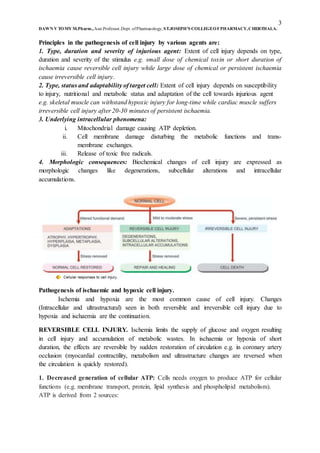
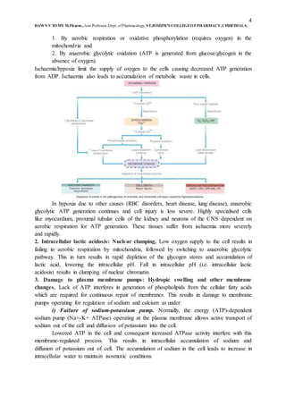
The document discusses cell injury and its causes. It defines cell injury as damage to cells from changes in their internal or external environment. There are two main types of causes of cell injury - genetic and acquired. Acquired causes are more common and include hypoxia/ischemia, physical agents, chemicals/drugs, microbes, immunological factors, nutritional imbalances, aging, and psychological stress. The severity and reversibility of cell injury depends on factors like the injurious agent, duration of exposure, cell type, and underlying intracellular changes. Reversible cell injury from short-term hypoxia/ischemia involves decreased ATP production, lactic acidosis, and damage to membrane pumps causing swelling.
Cellular adaptation
Adjustments which the cells make in response to stresses.
Adjustments may be for physiologic need or response to non-lethal pathogenic injury (Pathologic adaptation).
Adaptive responses are reversible on withdrawal of stimulus.
If the irritant stimulus persists for longer duration or is more lethal, the cell may not be able to survive.
“Survival of the Adaptable”
TYPES OF CELLULAR ADAPTATION:
Atrophy
Hypertrophy
Hyperplasia
Metaplasia
Dysplasia
Basic principles of cell injury and adaptationshivangimistry3
Shivangi Mistry, M.pharm in Pharmacology, Assistant Professor, bhagwan Mahavir College of Pharmacy, Morphology of cell injury, Phathogenesis, Intracellular accumulation, calcification
Mechanism of cell injury
Types of cell injury
Reversible and irreversible cell injury
Etiology of cell injury
Apoptosis, it's types and mechanism
Necrosis, it's types and mechanism
Cellular Adaptation
as cells encounter stresses they undergo functional or structural adaptations to maintain viability / homeostasis.
Injury - altered homeostasis
if limits of the adaptive response are exceeded or if adaptation not possible, a sequence of events called cell injury occurs.
Reversible Cell Injury
removal of stress results in complete restoration of structural & functional integrity.
b) Irreversible Cell Injury / Cell Death
if stimulus persists or is severe enough from the start, the cell suffers irreversible cell injury and death.
2 main morphologic patterns: necrosis & apoptosis.
Adaptations are reversible changes in the size, number, phenotype, metabolic activity, or functions of cells in response to changes in their environment.
Physiologic adaptations are responses of cells to normal stimulation by hormones or endogenous chemical mediators
Pathologic adaptations are responses to stress that allow cells to modulate their structure and function and thus escape injury.
Hypertrophy refers to an increase in the size of cells, that results in an increase in the size of the affected organ
The hypertrophied organ has no new cells, just larger cells.
Types:
a) physiologic b) pathologic
Causes:
a) increased functional demand b) hormonal stimulation
This report, prepared by the student at the College of Dentistry, Hassan Atheed , in the third phase discusses scientific topics, but it maybe did not be 100% complete.
Cell injury (cell death): it is the variable changes in morphological and functional properties of cell occurs due to internal or external causes (ex. Chemical, physical, infectious and genetic agents), that obligate cell to respond for preserving normal hemostasis (adaptation) or death (necrosis) when the injury factors sever cell unable to adept, cell may also killed by another pathway even when it have the ability to adept for saving other cells and tissue by programed cell death (apoptosis).
حسن عضيد
ARTIFICIAL INTELLIGENCE IN HEALTHCARE.pdfAnujkumaranit
Artificial intelligence (AI) refers to the simulation of human intelligence processes by machines, especially computer systems. It encompasses tasks such as learning, reasoning, problem-solving, perception, and language understanding. AI technologies are revolutionizing various fields, from healthcare to finance, by enabling machines to perform tasks that typically require human intelligence.
Lung Cancer: Artificial Intelligence, Synergetics, Complex System Analysis, S...Oleg Kshivets
RESULTS: Overall life span (LS) was 2252.1±1742.5 days and cumulative 5-year survival (5YS) reached 73.2%, 10 years – 64.8%, 20 years – 42.5%. 513 LCP lived more than 5 years (LS=3124.6±1525.6 days), 148 LCP – more than 10 years (LS=5054.4±1504.1 days).199 LCP died because of LC (LS=562.7±374.5 days). 5YS of LCP after bi/lobectomies was significantly superior in comparison with LCP after pneumonectomies (78.1% vs.63.7%, P=0.00001 by log-rank test). AT significantly improved 5YS (66.3% vs. 34.8%) (P=0.00000 by log-rank test) only for LCP with N1-2. Cox modeling displayed that 5YS of LCP significantly depended on: phase transition (PT) early-invasive LC in terms of synergetics, PT N0—N12, cell ratio factors (ratio between cancer cells- CC and blood cells subpopulations), G1-3, histology, glucose, AT, blood cell circuit, prothrombin index, heparin tolerance, recalcification time (P=0.000-0.038). Neural networks, genetic algorithm selection and bootstrap simulation revealed relationships between 5YS and PT early-invasive LC (rank=1), PT N0—N12 (rank=2), thrombocytes/CC (3), erythrocytes/CC (4), eosinophils/CC (5), healthy cells/CC (6), lymphocytes/CC (7), segmented neutrophils/CC (8), stick neutrophils/CC (9), monocytes/CC (10); leucocytes/CC (11). Correct prediction of 5YS was 100% by neural networks computing (area under ROC curve=1.0; error=0.0).
CONCLUSIONS: 5YS of LCP after radical procedures significantly depended on: 1) PT early-invasive cancer; 2) PT N0--N12; 3) cell ratio factors; 4) blood cell circuit; 5) biochemical factors; 6) hemostasis system; 7) AT; 8) LC characteristics; 9) LC cell dynamics; 10) surgery type: lobectomy/pneumonectomy; 11) anthropometric data. Optimal diagnosis and treatment strategies for LC are: 1) screening and early detection of LC; 2) availability of experienced thoracic surgeons because of complexity of radical procedures; 3) aggressive en block surgery and adequate lymph node dissection for completeness; 4) precise prediction; 5) adjuvant chemoimmunoradiotherapy for LCP with unfavorable prognosis.
Factory Supply Best Quality Pmk Oil CAS 28578–16–7 PMK Powder in Stockrebeccabio
Factory Supply Best Quality Pmk Oil CAS 28578–16–7 PMK Powder in Stock
Telegram: bmksupplier
signal: +85264872720
threema: TUD4A6YC
You can contact me on Telegram or Threema
Communicate promptly and reply
Free of customs clearance, Double Clearance 100% pass delivery to USA, Canada, Spain, Germany, Netherland, Poland, Italy, Sweden, UK, Czech Republic, Australia, Mexico, Russia, Ukraine, Kazakhstan.Door to door service
Hot Selling Organic intermediates
- Video recording of this lecture in English language: https://youtu.be/lK81BzxMqdo
- Video recording of this lecture in Arabic language: https://youtu.be/Ve4P0COk9OI
- Link to download the book free: https://nephrotube.blogspot.com/p/nephrotube-nephrology-books.html
- Link to NephroTube website: www.NephroTube.com
- Link to NephroTube social media accounts: https://nephrotube.blogspot.com/p/join-nephrotube-on-social-media.html
Ethanol (CH3CH2OH), or beverage alcohol, is a two-carbon alcohol
that is rapidly distributed in the body and brain. Ethanol alters many
neurochemical systems and has rewarding and addictive properties. It
is the oldest recreational drug and likely contributes to more morbidity,
mortality, and public health costs than all illicit drugs combined. The
5th edition of the Diagnostic and Statistical Manual of Mental Disorders
(DSM-5) integrates alcohol abuse and alcohol dependence into a single
disorder called alcohol use disorder (AUD), with mild, moderate,
and severe subclassifications (American Psychiatric Association, 2013).
In the DSM-5, all types of substance abuse and dependence have been
combined into a single substance use disorder (SUD) on a continuum
from mild to severe. A diagnosis of AUD requires that at least two of
the 11 DSM-5 behaviors be present within a 12-month period (mild
AUD: 2–3 criteria; moderate AUD: 4–5 criteria; severe AUD: 6–11 criteria).
The four main behavioral effects of AUD are impaired control over
drinking, negative social consequences, risky use, and altered physiological
effects (tolerance, withdrawal). This chapter presents an overview
of the prevalence and harmful consequences of AUD in the U.S.,
the systemic nature of the disease, neurocircuitry and stages of AUD,
comorbidities, fetal alcohol spectrum disorders, genetic risk factors, and
pharmacotherapies for AUD.
HOT NEW PRODUCT! BIG SALES FAST SHIPPING NOW FROM CHINA!! EU KU DB BK substit...GL Anaacs
Contact us if you are interested:
Email / Skype : kefaya1771@gmail.com
Threema: PXHY5PDH
New BATCH Ku !!! MUCH IN DEMAND FAST SALE EVERY BATCH HAPPY GOOD EFFECT BIG BATCH !
Contact me on Threema or skype to start big business!!
Hot-sale products:
NEW HOT EUTYLONE WHITE CRYSTAL!!
5cl-adba precursor (semi finished )
5cl-adba raw materials
ADBB precursor (semi finished )
ADBB raw materials
APVP powder
5fadb/4f-adb
Jwh018 / Jwh210
Eutylone crystal
Protonitazene (hydrochloride) CAS: 119276-01-6
Flubrotizolam CAS: 57801-95-3
Metonitazene CAS: 14680-51-4
Payment terms: Western Union,MoneyGram,Bitcoin or USDT.
Deliver Time: Usually 7-15days
Shipping method: FedEx, TNT, DHL,UPS etc.Our deliveries are 100% safe, fast, reliable and discreet.
Samples will be sent for your evaluation!If you are interested in, please contact me, let's talk details.
We specializes in exporting high quality Research chemical, medical intermediate, Pharmaceutical chemicals and so on. Products are exported to USA, Canada, France, Korea, Japan,Russia, Southeast Asia and other countries.
Flu Vaccine Alert in Bangalore Karnatakaaddon Scans
As flu season approaches, health officials in Bangalore, Karnataka, are urging residents to get their flu vaccinations. The seasonal flu, while common, can lead to severe health complications, particularly for vulnerable populations such as young children, the elderly, and those with underlying health conditions.
Dr. Vidisha Kumari, a leading epidemiologist in Bangalore, emphasizes the importance of getting vaccinated. "The flu vaccine is our best defense against the influenza virus. It not only protects individuals but also helps prevent the spread of the virus in our communities," he says.
This year, the flu season is expected to coincide with a potential increase in other respiratory illnesses. The Karnataka Health Department has launched an awareness campaign highlighting the significance of flu vaccinations. They have set up multiple vaccination centers across Bangalore, making it convenient for residents to receive their shots.
To encourage widespread vaccination, the government is also collaborating with local schools, workplaces, and community centers to facilitate vaccination drives. Special attention is being given to ensuring that the vaccine is accessible to all, including marginalized communities who may have limited access to healthcare.
Residents are reminded that the flu vaccine is safe and effective. Common side effects are mild and may include soreness at the injection site, mild fever, or muscle aches. These side effects are generally short-lived and far less severe than the flu itself.
Healthcare providers are also stressing the importance of continuing COVID-19 precautions. Wearing masks, practicing good hand hygiene, and maintaining social distancing are still crucial, especially in crowded places.
Protect yourself and your loved ones by getting vaccinated. Together, we can help keep Bangalore healthy and safe this flu season. For more information on vaccination centers and schedules, residents can visit the Karnataka Health Department’s official website or follow their social media pages.
Stay informed, stay safe, and get your flu shot today!
Report Back from SGO 2024: What’s the Latest in Cervical Cancer?bkling
Are you curious about what’s new in cervical cancer research or unsure what the findings mean? Join Dr. Emily Ko, a gynecologic oncologist at Penn Medicine, to learn about the latest updates from the Society of Gynecologic Oncology (SGO) 2024 Annual Meeting on Women’s Cancer. Dr. Ko will discuss what the research presented at the conference means for you and answer your questions about the new developments.
Tom Selleck Health: A Comprehensive Look at the Iconic Actor’s Wellness Journeygreendigital
Tom Selleck, an enduring figure in Hollywood. has captivated audiences for decades with his rugged charm, iconic moustache. and memorable roles in television and film. From his breakout role as Thomas Magnum in Magnum P.I. to his current portrayal of Frank Reagan in Blue Bloods. Selleck's career has spanned over 50 years. But beyond his professional achievements. fans have often been curious about Tom Selleck Health. especially as he has aged in the public eye.
Follow us on: Pinterest
Introduction
Many have been interested in Tom Selleck health. not only because of his enduring presence on screen but also because of the challenges. and lifestyle choices he has faced and made over the years. This article delves into the various aspects of Tom Selleck health. exploring his fitness regimen, diet, mental health. and the challenges he has encountered as he ages. We'll look at how he maintains his well-being. the health issues he has faced, and his approach to ageing .
Early Life and Career
Childhood and Athletic Beginnings
Tom Selleck was born on January 29, 1945, in Detroit, Michigan, and grew up in Sherman Oaks, California. From an early age, he was involved in sports, particularly basketball. which played a significant role in his physical development. His athletic pursuits continued into college. where he attended the University of Southern California (USC) on a basketball scholarship. This early involvement in sports laid a strong foundation for his physical health and disciplined lifestyle.
Transition to Acting
Selleck's transition from an athlete to an actor came with its physical demands. His first significant role in "Magnum P.I." required him to perform various stunts and maintain a fit appearance. This role, which he played from 1980 to 1988. necessitated a rigorous fitness routine to meet the show's demands. setting the stage for his long-term commitment to health and wellness.
Fitness Regimen
Workout Routine
Tom Selleck health and fitness regimen has evolved. adapting to his changing roles and age. During his "Magnum, P.I." days. Selleck's workouts were intense and focused on building and maintaining muscle mass. His routine included weightlifting, cardiovascular exercises. and specific training for the stunts he performed on the show.
Selleck adjusted his fitness routine as he aged to suit his body's needs. Today, his workouts focus on maintaining flexibility, strength, and cardiovascular health. He incorporates low-impact exercises such as swimming, walking, and light weightlifting. This balanced approach helps him stay fit without putting undue strain on his joints and muscles.
Importance of Flexibility and Mobility
In recent years, Selleck has emphasized the importance of flexibility and mobility in his fitness regimen. Understanding the natural decline in muscle mass and joint flexibility with age. he includes stretching and yoga in his routine. These practices help prevent injuries, improve posture, and maintain mobilit
Couples presenting to the infertility clinic- Do they really have infertility...Sujoy Dasgupta
Dr Sujoy Dasgupta presented the study on "Couples presenting to the infertility clinic- Do they really have infertility? – The unexplored stories of non-consummation" in the 13th Congress of the Asia Pacific Initiative on Reproduction (ASPIRE 2024) at Manila on 24 May, 2024.
Couples presenting to the infertility clinic- Do they really have infertility...
CELL INJURY AND ADAPTIONS
1. 1
DAWN V TOMY M.Pharm.,Asst.Professor,Dept. ofPharmacology, ST.JOSEPH’S COLLEGEOFPHARMACY,CHERTHALA.
CELL INJURY AND ADAPTATIONS
Cells: They are the basic unit of life. There are 2 type cells; the epithelial cells and
the mesenchymal cells.
Cellular theory of diseases states that diseases occur due to abnormalities in the cells. The
diseases begin with cell injury due to different types of stress and followed by loss of their
cellular function.
Cell injury is defined as the damage to the cell, due to different types of stress (change in its
internal/external environment) encountered. Cell may adjust itself to the changes to a certain
extend by homeostasis. The cell injury (response) depends up on:
i. The type of cell and tissue involved e.g. parenchymal cells more prone for damage
than connective tissues. Cells with functions of excretion, metabolism and
detoxification are at high risk e.g. renal tubular cells, hepatocytes, myocardium, brain
etc.
ii. Nature of injurious agents, toxicity and concentration e.g. living agents’ causes more
damage than non-living agents.
iii. The duration of stress exposure e.g. longer the duration more extensive damage is
seen and type of cell injury.
The various cellular responses to cell injury are:
a. Increased functional demands leads to cellular adaptations, and if stress is removed it
will revert back to normal.
b. Mild to moderate stress leads to reversible cell injury e.g. degenerations, sub cellular
alterations, intracellular accumulations, persistent/continuous stress leads to
irreversible cell injury e.g. cell death.
To treat a disease, it is necessary to determine the causes of cell injury, their mechanisms and
cellular adaptations.
Aetiology/Etiology of Cell Injury: (important)
The cells may be injured by 2 causes:
1. By genetic causes:
a) Developmental defects - Errors in morphogenesis (teratogenicity) occurs due to
chemicals e.g. alcohol, drugs (Thalidomide), physical and biochemical agents,
nutrition deficiency (folic acid deficiency leads to defective DNA replication).
b) Cytogenetic (karyotype) defects - Chromosomal abnormalities e.g. changes in
any of the 46 chromosomes (44 autosomes and 2 sex chromosomes) polyploidy
(e.g. triploid i.e. 3XN=69 chromosomes, tetraploid i.e. 4XN=92 chromosomes),
aneuploidy (monosomy i.e. 2N-1=45 chromosomes, trisomy i.e. 2N+1=47
chromosomes).
c) Single gene defects (G6PD deficiency anaemia) – Mendelian disorders e.g.
mutation (point mutation, stop codon/Nonsense mutation, frameshift mutation,
trinucleotide repeat mutation), inheritance pattern (dominant gene, recessive
2. 2
DAWN V TOMY M.Pharm.,Asst.Professor,Dept. ofPharmacology, ST.JOSEPH’S COLLEGEOFPHARMACY,CHERTHALA.
gene, polymorphism, autosomal dominant inheritance, autosomal recessive
inheritance, ‘X’ linked disorders).
d) Multifactorial inheritance disorders – Due to combined defects of genetic
composition and environment influence e.g. cleft lip and cleft palate, pyloric
stenosis, diabetes mellitus.
2. By acquired causes: It includes most of the common diseases in man. The acquired
causes are:
a) Hypoxia and ischemia: Oxygen is essential for metabolism and ATP production
(energy). Deficiency of oxygen leads to cell injury. Hypoxia occurs mainly due
to ischaemia (reduced blood supply) resulting from blood vessel occlusions.
Hypoxia may also results from disorders of RBC’s e.g. anaemia, CO poisoning,
heart disorders, lung diseases and increased demand of tissues.
b) Physical agents: Mechanical trauma e.g. loss of blood in road accidents,
thermal trauma e.g. by heat and cold, electricity e.g. electrocution, radiation e.g.
UV and ionising radiations, rapid changes in atmospheric pressure.
c) Chemical agents and drugs: Chemical poisons e.g. cyanide, arsenic, mercury
etc., strong acids and alkali e.g. sulphuric acid, sodium hydroxide,
environmental pollutants e.g. smoke, dust, insecticide and pesticide e.g.
organomercurials, oxygen at high concentration e.g. hyperbaric oxygen,
hypertonic glucose and salt (cause crenation of RBC), hypotonic solution (cause
haemolysis of RBC), social agents e.g. Alcohol, narcotic drugs, therapeutic
administration of drugs.
d) Microbial agents (pathogens): bacteria, virus, fungi, protozoa, metazoan and
rickettsia infections.
e) Immunologic agents: Hypersensitivity reactions, anaphylactic reactions and
autoimmune diseases.
f) Nutritional derangements: Deficiency/excess leads to nutritional imbalance,
nutritional deficiency (starvation) results in Marasmus (progressive wasting),
Kwashiorkor (protein deficiency), loss of appetite, diarrhoea leads to loss of
weight, deficiency of minerals leads to anaemia (iron/Vit B12/folic acid
deficiency), Nutritional excess results in obesity, atherosclerosis, heart disease,
hypertension.
g) Aging: Cellular aging/senescence leads to impaired ability of cells to undergo
replication and repair causing cell death.
h) Psychogenic diseases (common acquired mental disorders due to mental stress,
anxiety): mental stress, anxiety, over work, frustration leads to depression and
schizophrenia. Other reasons include problems of drug addition, alcoholism,
smoking which leads to liver damage, chronic bronchitis, lung cancer, peptic
ulcer, hypertension, ischaemia and heart disease.
i) Iatrogenic factors: Physician induced diseases e.g. occurrence of disease/death
due to error in judgement by the physician.
j) Idiopathic diseases: Disease of unknown cause (aetiology unknown) e.g.
essential hypertension or idiopathic hypertension, cancer.
3. 3
DAWN V TOMY M.Pharm.,Asst.Professor,Dept. ofPharmacology, ST.JOSEPH’S COLLEGEOFPHARMACY,CHERTHALA.
Principles in the pathogenesis of cell injury by various agents are:
1. Type, duration and severity of injurious agent: Extent of cell injury depends on type,
duration and severity of the stimulus e.g. small dose of chemical toxin or short duration of
ischaemia cause reversible cell injury while large dose of chemical or persistent ischaemia
cause irreversible cell injury.
2. Type, status and adaptability of target cell: Extent of cell injury depends on susceptibility
to injury, nutritional and metabolic status and adaptation of the cell towards injurious agent
e.g. skeletal muscle can withstand hypoxic injury for long-time while cardiac muscle suffers
irreversible cell injury after 20-30 minutes of persistent ischaemia.
3. Underlying intracellular phenomena:
i. Mitochondrial damage causing ATP depletion.
ii. Cell membrane damage disturbing the metabolic functions and trans-
membrane exchanges.
iii. Release of toxic free radicals.
4. Morphologic consequences: Biochemical changes of cell injury are expressed as
morphologic changes like degenerations, subcellular alterations and intracellular
accumulations.
Pathogenesis of ischaemic and hypoxic cell injury.
Ischemia and hypoxia are the most common cause of cell injury. Changes
(Intracellular and ultrastructural) seen in both reversible and irreversible cell injury due to
hypoxia and ischaemia are the continuation.
REVERSIBLE CELL INJURY. Ischemia limits the supply of glucose and oxygen resulting
in cell injury and accumulation of metabolic wastes. In ischaemia or hypoxia of short
duration, the effects are reversible by sudden restoration of circulation e.g. in coronary artery
occlusion (myocardial contractility, metabolism and ultrastructure changes are reversed when
the circulation is quickly restored).
1. Decreased generation of cellular ATP: Cells needs oxygen to produce ATP for cellular
functions (e.g. membrane transport, protein, lipid synthesis and phospholipid metabolism).
ATP is derived from 2 sources:
4. 4
DAWN V TOMY M.Pharm.,Asst.Professor,Dept. ofPharmacology, ST.JOSEPH’S COLLEGEOFPHARMACY,CHERTHALA.
1. By aerobic respiration or oxidative phosphorylation (requires oxygen) in the
mitochondria and
2. By anaerobic glycolytic oxidation (ATP is generated from glucose/glycogen in the
absence of oxygen).
Ischaemia/hypoxia limit the supply of oxygen to the cells causing decreased ATP generation
from ADP. Ischaemia also leads to accumulation of metabolic waste in cells.
In hypoxia due to other causes (RBC disorders, heart disease, lung disease), anaerobic
glycolytic ATP generation continues and cell injury is less severe. Highly specialised cells
like myocardium, proximal tubular cells of the kidney and neurons of the CNS dependent on
aerobic respiration for ATP generation. These tissues suffer from ischaemia more severely
and rapidly.
2. Intracellular lactic acidosis: Nuclear clumping. Low oxygen supply to the cell results in
failing in aerobic respiration by mitochondria, followed by switching to anaerobic glycolytic
pathway. This in turn results in rapid depletion of the glycogen stores and accumulation of
lactic acid, lowering the intracellular pH. Fall in intracellular pH (i.e. intracellular lactic
acidosis) results in clumping of nuclear chromatin.
3. Damage to plasma membrane pumps: Hydropic swelling and other membrane
changes. Lack of ATP interferes in generation of phospholipids from the cellular fatty acids
which are required for continuous repair of membranes. This results in damage to membrane
pumps operating for regulation of sodium and calcium as under:
i) Failure of sodium-potassium pump. Normally, the energy (ATP)-dependent
sodium pump (Na+-K+ ATPase) operating at the plasma membrane allows active transport of
sodium out of the cell and diffusion of potassium into the cell.
Lowered ATP in the cell and consequent increased ATPase activity interfere with this
membrane-regulated process. This results in intracellular accumulation of sodium and
diffusion of potassium out of cell. The accumulation of sodium in the cell leads to increase in
intracellular water to maintain isosmotic conditions
5. 5
DAWN V TOMY M.Pharm.,Asst.Professor,Dept. ofPharmacology, ST.JOSEPH’S COLLEGEOFPHARMACY,CHERTHALA.
ii) Failure of calcium pump. Membrane damage causes disturbance in the calcium
ion exchange across the cell membrane. Excess of calcium moves into the cell (i.e. calcium
influx), particularly in the mitochondria, causing its swelling and deposition of phospholipid-
rich amorphous densities.
Ultrastructural evidence of reversible cell membrane damage is seen in the form of
loss of microvilli, intramembranous particles and focal projections of the cytoplasm (blebs).
Myelin figures may be seen lying in the cytoplasm or present outside the cell, these are
derived from membranes (plasma or organellar) enclosing water and dissociated lipoproteins
between the lamellae of injured membranes.
4. Reduced protein synthesis: Dispersed ribosomes. As a result of continued hypoxia,
membranes of endoplasmic reticulum and Golgi apparatus swell up. Ribosomes are detached
from granular endoplasmic reticulum and polysomes are degraded to monosomes, thus
dispersing ribosomes in the cytoplasm and inactivating their function. Similar reduced protein
synthesis occurs in Golgi apparatus. Up to this point, withdrawal of acute stress that resulted
in reversible cell injury can restore the cell to normal state.
IRREVERSIBLE CELL INJURY:
Persistent ischemia/hypoxia results in irreversible damage to the structures and
functions of the cell (cell death). The factors which differentiate irreversible from reversible
cell injury are:
1. Inability of the cell to reverse mitochondrial dysfunction on reperfusion or
reoxygenation.
2. Disturbance in cell membrane function and plasma membrane. Other factors includes:
Reduction in ATP.
Depletion of proteins.
Reduced intracellular pH.
Leakage of lysosomal enzymes into the cytoplasm.
Ultrastructural changes during cell injury due to hypoxia-Ischemia.
6. 6
DAWN V TOMY M.Pharm.,Asst.Professor,Dept. ofPharmacology, ST.JOSEPH’S COLLEGEOFPHARMACY,CHERTHALA.
The biochemical changes and ultrastructural changes due to irreversible cell injury are:
1. Calcium influx: Mitochondrial damage:
2. Activated phospholipases: Membrane damage:
3. Intracellular proteases: Cytoskeletal damage:
4. Activated endonucleases: Nuclear damage:
5. Lysosomal hydrolytic enzymes: Lysosomal damage, cell death and phagocytosis:
1. Calcium influx: Mitochondrial damage:
Cytosolic influx of Ca2+ ions occurs after reperfusion of irreversible cell injury.
Intracellular Ca2+ ions accumulation in the mitochondria disables its function. Mitochondrial
changes occur:
Vacuoles in the mitochondria and
Deposits of amorphous calcium salts.
2. Activated phospholipases: Membrane damage:
Influx of calcium in the cell activates endogenous phospholipases which degrades
membrane phospholipids. Lytic enzyme ATPase is also activated, it results in depletion of
ATP. Decreased replacement/synthesis of membrane phospholipids due to reduced ATP.
3. Intracellular proteases: Cytoskeletal damage:
Cytoskeleton of the cell (i.e. microfilaments, microtubules and intermediate filaments)
is damaged (degraded) by activated intracellular proteases or cell swelling.
4. Activated endonucleases: Nuclear damage:
Nucleoproteins are damaged by the activated lysosomal enzymes such as proteases
and endonucleases. Irreversible damage to the nucleus can be in 3 forms:
1. Pyknosis: Condensation and clumping of nucleus (dark basophilic).
2. Karyorrhexis: Nuclear fragmentation in to small bits and dispersed in the cytoplasm.
3. Karyolysis: Dissolution of the nucleus.
5. Lysosomal hydrolytic enzymes: Lysosomal damage, cell death and phagocytosis:
Damage of lysosome releases lysosomal hydrolytic enzymes, which are activated due
to lack of oxygen and acidic pH.
Common enzyme markers of different type’s cell death are:
(Hydrolytic enzymes includes: Hydrolase, RNAase, DNAase, protease, glycosidase,
phosphatase, lipase, amylase, cathepsin etc).
7. 7
DAWN V TOMY M.Pharm.,Asst.Professor,Dept. ofPharmacology, ST.JOSEPH’S COLLEGEOFPHARMACY,CHERTHALA.
Activation leads to enzymatic digestion of the cell inclusions and results in cell death.
Enzymes leak across the cell membrane into the serum and the estimation of which is used as
clinical parameters of cell death. Example, in myocardial infarction, estimation of elevated
Serum Glutamic Oxaloacetic Transaminase (SGOT), Lactate Dehydrogenase (LDH),
isoenzyme of Creatine Kinase (CK-MB), and cardiac Troponins (cTn) are markers for death
of heart muscle.
Ischemia-Reperfusion Injury and Free Radical-Mediated Cell Injury:
Duration of Ischemia/hypoxia and restoration of blood flow may result in 3 different
conditions:
1. From Ischemia to reversible injury: If period of Ischemia is of short duration,
reperfusion with resupply of oxygen restores the structure and functions of the injured
cell i.e. Reversible cell injury.
2. From Ischemia to reperfusion injury: If Ischemia is for longer duration before
reperfusion; damage occurs in already injured cell i.e. Ischemia-reperfusion injury.
3. From Ischemia to irreversible injury: Long period of Ischemia produces irreversible
cell injury. Blood flow restoration doesn’t help recovery. No reperfusion injury
occurs. Cell death due to excess of intracellular sodium and calcium ions (due to cell
membrane damage).
The mechanism of reperfusion injury and free radical mediated injury follows 3 main steps:
1. Calcium overload.
2. Generation of reactive oxygen radicals (superoxide, H2O2, hydroxyl radicals).
3. Subsequent inflammatory reaction.
1. CALCIUM OVERLOAD:
Restoration of blood supply provides more calcium ions but the ATP store of the cell
is low resulting in intracellular calcium accumulation. It triggers lipid peroxidation causing
membrane damage.
2. GENERATION OF REACTIVE OXYGEN RADICALS:
Molecular forms as reactive oxygen free radicals or reactive oxygen species can
damage the cells.
Mechanism of oxygen free radical generation: Metabolism in the cell involves
generation of ATP by oxidative process in which bi-radical oxygen (O2) combines with
hydrogen atom (H) and forms water (H2O) cytochrome oxidase catalyses the reaction. It
involves 4 steps and 1 electron donation at each step. Oxygen free radicals are intermediate
chemicals having unpaired electrons generated in mitochondria. Three intermediate
molecules of partially reduced oxygen are usually formed during cell injury:
Superoxide oxygen (O’2): one electron.
Hydrogen peroxide (H2O2): two electrons.
Hydroxyl radical (OH–): three electrons.
These are generated from enzymatic and non-enzymatic reactions:
1. Superoxide (O’2): Superoxide anion O’2 may be generated by direct auto-oxidation of
O2 during mitochondrial electron transport reaction, O’2 is also produced
8. 8
DAWN V TOMY M.Pharm.,Asst.Professor,Dept. ofPharmacology, ST.JOSEPH’S COLLEGEOFPHARMACY,CHERTHALA.
enzymatically by xanthine oxidase and cytochrome P450 in the mitochondria/cytosol.
O’2 converted to H2O2 by superoxide dismutase (SOD).
2. Hydrogen peroxide (H2O2): H2O2 is reduced to water enzymatically by catalase (in the
peroxisomes) and glutathione peroxidase GSH (both in the cytosol and
mitochondria).
3. Hydroxyl radical (OH– most reactive): OH– radical is formed by 2 ways in biologic
processes—by radiolysis of water and by reaction of H2O2 with ferrous (Fe2+) ions
(Fenton reaction).
Mechanisms of generation of free radicals by four electron step reduction of oxygen. (SOD =
superoxide dismutase; GSH = glutathione peroxidase).
Other oxygen free radicals formed in the body are:
1. Superoxide free radical in Fenton reaction.
2. Nitric oxide (NO), generated by body cells (endothelial cells, neurons, macrophages
etc), combines with superoxide to form peroxynitrate (ONOO a potent free radical).
3. Halide reagent (chlorine or chloride) in the leucocytes reacts with superoxide to form
hypochlorous acid (HOCl a cytotoxic free radical).
4. Exogenous sources of free radicals include tobacco and industrial pollutants.
Free radicals are formed in physiological/pathological processes. Oxygen free radicals are
unstable and are destroyed spontaneously by catalytic action of enzymes such as superoxide
dismutase (SOD), catalase and glutathione peroxidase. The net effect of free radical injury
depends upon the rate of free radical formation and rate of destruction. Free radicals are
highly destructive to the cell as they have electron-free residue and binds to all molecules of
the cell (termed oxidative stress). Out of various free radicals, hydroxyl radical is the most
reactive species.
Free radicals may produce membrane damage by:
i) Lipid peroxidation. Polyunsaturated fatty acids (PUFA) of membrane are attacked
repeatedly and severely by oxygen derived free radicals to yield highly destructive
PUFA radicals—lipid hydroperoxy radicals and lipid hypoperoxides. This reaction is
termed lipid peroxidation. The lipid peroxides are decomposed by transition metals
such as iron. Lipid peroxidation is propagated to other sites causing widespread
membrane damage and destruction of organelles.
ii) Oxidation of proteins. Oxygen-derived free radicals cause cell injury by oxidation of
protein macromolecules of the cells, cross-linkages of labile amino acids as well as by
9. 9
DAWN V TOMY M.Pharm.,Asst.Professor,Dept. ofPharmacology, ST.JOSEPH’S COLLEGEOFPHARMACY,CHERTHALA.
fragmentation of polypeptides directly. The end-result is degradation of cytosolic
neutral proteases and cell destruction.
iii) DNA damage. Free radicals cause breaks in the single strands of the nuclear and
mitochondrial DNA. This results in cell injury; it may also cause malignant
transformation of cells.
iv) Cytoskeletal damage. Reactive oxygen species are also known to interact with
cytoskeletal elements and interfere in mitochondrial aerobic phosphorylation and thus
cause ATP depletion.
Conditions causing free radical cell injury are:
i) Ischaemic reperfusion injury
ii) Ionising radiation by causing radiolysis of water
iii) Chemical toxicity
iv) Chemical carcinogenesis
v) Hyperoxia (toxicity due to oxygen therapy)
vi) Cellular aging
vii) Killing of microbial agents
viii) Inflammatory damage
ix) Destruction of tumour cells
x) Atherosclerosis.
Antioxidants: Antioxidants are endogenous or exogenous substances which inactivate the
free radicals and includes: Vitamins E, A and C (ascorbic acid) Sulfhydryl-containing
compounds e.g. cysteine and glutathione. Serum proteins e.g. ceruloplasmin and transferrin.
3. SUBSEQUENT INFLAMMATORY REACTION.
Ischemia-reperfusion event is followed by inflammatory reaction. Incoming activated
neutrophils utilise oxygen quickly (oxygen burst) and release a lot of oxygen free radicals.
Ischemia is also associated with accumulation of precursors of ATP, namely ADP and
pyruvate, which further build-up generation of free radicals.
Pathogenesis of Chemical Injury
Chemicals induce cell injury by one of the two mechanisms:
1. By direct cytotoxicity or
2. By conversion of chemicals into reactive metabolites.
Direct cytotoxic effects.
Some chemicals combine with components of the cell and produce direct cytotoxicity
without requiring metabolic activation. The cytotoxic damage is usually greatest to cells
which are involved in the metabolism of such chemicals e.g. in mercuric chloride the greatest
damage occurs to cells of the alimentary tract where it is absorbed and kidney where it is
excreted.
Cyanide kills the cell by poisoning mitochondrial cytochrome oxidase thus blocking
oxidative phosphorylation. Other examples of directly cytotoxic chemicals include
chemotherapeutic agents used in treatment of cancer, toxic heavy metals such as mercury,
lead and iron.
10. 10
DAWN V TOMY M.Pharm.,Asst.Professor,Dept. ofPharmacology, ST.JOSEPH’S COLLEGEOFPHARMACY,CHERTHALA.
Conversion to reactive toxic metabolites.
This mechanism involves metabolic activation to yield ultimate toxin that interacts
with the target cells. The target cells in this group of chemicals may not be the same cell that
metabolised the toxin. Example of cell injury by conversion of reactive metabolites is toxic
liver necrosis caused by carbon tetrachloride (CCl4), acetaminophen (commonly used
analgesic and antipyretic) and bromobenzene. Cell injury by CCl4 is classic example of an
industrial toxin (earlier used in dry-cleaning industry) that produces cell injury by conversion
to a highly toxic free radical, CCl3, in the body’s drug-metabolising P450 enzyme system in
the liver cells. Thus, it produces profound liver cell injury by free radical generation. Other
mechanism of cell injury includes direct toxic effect on cell membrane and nucleus.
Pathogenesis of Physical Injury
Injuries caused by mechanical force are of medicolegal significance. But they may
lead to a state of shock. Radiation injury to human by accidental or therapeutic exposure is of
importance in treatment of persons with malignant tumours as well as may have carcinogenic
influences.
Killing of cells by ionising radiation is the result of direct formation of hydroxyl
radicals from radiolysis of water. These hydroxyl radicals damage the cell membrane as well
as may interact with DNA of the target cell. In proliferating cells, there is inhibition of DNA
replication and eventual cell death by apoptosis (e.g. epithelial cells). In non-proliferating
cells there is no effect of inhibition of DNA synthesis and in these cells there is cell
membrane damage followed by cell death by necrosis (e.g. neurons).
MORPHOLOGY OF CELL INJURY
Depending upon the severity of cell injury, degree of damage and residual effects on
cells and tissues are variable.
MORPHOLOGY OF REVERSIBLE CELL INJURY
Morphologic forms of reversible cell injury are:
1. Hydropic change (cloudy swelling, or vacuolar degeneration)
2. Fatty change
3. Hyaline change
4. Mucoid change
1. Hydropic Change
Hydropic change means accumulation of water within the cytoplasm of the cell. Other
synonyms used are cloudy swelling (for gross appearance of the affected organ) and vacuolar
degeneration (due to cytoplasmic vacuolation).
ETIOLOGY. This is the commonest and earliest form of cell injury from almost all causes.
The common causes include acute and subacute cell injury from various etiologic agents such
as bacterial toxins, chemicals, poisons, burns, high fever, intravenous administration of
hypertonic glucose or saline etc.
PATHOGENESIS. Cloudy swelling results from impaired regulation of sodium and
potassium at the level of cell membrane. This results in intracellular accumulation of sodium
and escape of potassium. This, in turn, leads to rapid flow of water into the cell to maintain
iso-osmotic conditions and hence cellular swelling occurs. In addition, influx of calcium too
11. 11
DAWN V TOMY M.Pharm.,Asst.Professor,Dept. ofPharmacology, ST.JOSEPH’S COLLEGEOFPHARMACY,CHERTHALA.
occurs. Hydropic swelling is an entirely reversible change upon removal of the injurious
agent.
MORPHOLOGIC FEATURES. Grossly, the affected organ such as kidney, liver, pancreas,
or heart muscle is enlarged due to swelling. The cut surface bulges outwards and is slightly
opaque.
Microscopically, it is characterised by the following features.
i) The cells are swollen and the microvasculature compressed.
ii) Small clear vacuoles are seen in the cells and hence the term vacuolar
degeneration. These vacuoles represent distended cisternae of the endoplasmic
reticulum.
iii) Small cytoplasmic blebs may be seen.
iv) The nucleus may appear pale.
2. Hyaline Change
The word ‘hyaline’ means glassy (hyalos = glass). Hyaline is a descriptive histologic
term for glassy, homogeneous, eosinophilic appearance of material in haematoxylin and
eosin-stained sections and does not refer to any specific substance. Though fibrin and
amyloid have hyaline appearance, they have distinctive features and staining reactions and
can be distinguished from non-specific hyaline material. Hyaline change is associated with
heterogeneous pathologic conditions. It may be intracellular or extracellular.
INTRACELLULAR HYALINE. Intracellular hyaline is mainly seen in epithelial cells. A
few examples are as follows:
1. Hyaline droplets in the proximal tubular epithelial cells in cases of excessive
reabsorption of plasma proteins.
2. Hyaline degeneration of rectus abdominalis muscle called Zenker’s degeneration,
occurring in typhoid fever. The muscle loses its fibrillar staining and becomes glassy
and hyaline.
3. Mallory’s hyaline represents aggregates of intermediate filaments in the hepatocytes in
alcoholic liver cell injury.
4. Nuclear or cytoplasmic hyaline inclusions seen in some viral infections.
5. Russell’s bodies representing excessive immunoglobulins in the rough endoplasmic
reticulum of the plasma cells.
EXTRACELLULAR HYALINE. Extracellular hyaline is seen in connective tissues. A few
examples of extracellular hyaline change are as under:
1. Hyaline degeneration in leiomyomas of the uterus.
2. Hyalinised old scar of fibrocollagenous tissues.
3. Hyaline arteriolosclerosis in renal vessels in hypertension and diabetes mellitus.
4. Hyalinised glomeruli in chronic glomerulonephritis.
5. Corpora amylacea are rounded masses of concentric hyaline laminae seen in the
prostate in the elderly, in the brain and in the spinal cord in old age, and in old infarcts
of the lung.
3. Mucoid Change
Mucus secreted by mucous glands is a combination of proteins complexed with
mucopolysaccharides. Mucin, a glycoprotein, is its chief constituent. Mucin is normally
12. 12
DAWN V TOMY M.Pharm.,Asst.Professor,Dept. ofPharmacology, ST.JOSEPH’S COLLEGEOFPHARMACY,CHERTHALA.
produced by epithelial cells of mucous membranes and mucous glands, as well as by some
connective tissues like in the umbilical cord. By convention, connective tissue mucin is
termed myxoid (mucus like). Both types of mucin are stained by alcian blue. However,
epithelial mucin stains positively with periodic acid-Schiff (PAS), while connective tissue
mucin is PAS negative but is stained positively with colloidal iron.
EPITHELIAL MUCIN. Following are some examples of functional excess of epithelial
mucin:
1. Catarrhal inflammation of mucous membrane (e.g. of respiratory tract, alimentary
tract, uterus).
2. Obstruction of duct leading to mucocele in the oral cavity and gallbladder.
3. Cystic fibrosis of the pancreas.
4. Mucin-secreting tumours (e.g. of ovary, stomach, large bowel etc).
CONNECTIVE TISSUE MUCIN. A few examples of disturbances of connective tissue
mucin are as under:
1. Mucoid or myxoid degeneration in some tumours e.g. myxomas, neurofibromas,
fibroadenoma, soft tissue sarcomas etc.
2. Dissecting aneurysm of the aorta due to Erdheim’s medial degeneration and Marfan’s
syndrome.
3. Myxomatous change in the dermis in myxoedema.
4. Myxoid change in the synovium in ganglion on the wrist.
SUBCELLULAR ALTERATIONS IN CELL INJURY
Certain morphologically distinct alterations at subcellular level occur at the level of
cytoskeleton, lysosomes, endoplasmic reticulum and mitochondria:
1. CYTOSKELETAL CHANGES. Components of cytoskeleton may show the following
morphologic abnormalities:
i) Defective microtubules:
In Chédiak-Higashi syndrome characterised by poor phagocytic activity of neutrophils.
Poor sperm motility causing sterility. Immotile cilia syndrome (Kartagener’s syndrome)
characterised by immotile cilia of respiratory tract and consequent chronic infection due
to defective clearance of inhaled bacteria. Defects in leucocyte function of phagocytes
such as migration and chemotaxis.
ii) Defective microfilaments:
1. In myopathies
2. Muscular dystrophies
iii) Accumulation of intermediate filaments: Various classes of intermediate filaments
(cytokeratin, desmin, vimentin, glial fibrillary acidic protein, and neurofilament) may
accumulate in the cytosol. For example:
Mallory’s body or alcoholic hyaline as intracytoplasmic eosinophilic inclusion seen in
alcoholic liver disease which is collection of cytokeratin intermediate filaments.
Neurofibrillary tangles, neurities and senile plaques in Alzheimer’s disease are composed
of neurofilaments and paired helical filaments.
13. 13
DAWN V TOMY M.Pharm.,Asst.Professor,Dept. ofPharmacology, ST.JOSEPH’S COLLEGEOFPHARMACY,CHERTHALA.
2. LYSOSOMAL CHANGES. Lysosomes contain powerful hydrolytic enzymes.
Heterophagy and autophagy are the two ways by which lysosomes show morphologic
changes of phagocytic function.
i) Heterophagy. Phagocytosis (cell eating) and pinocytosis (cell drinking) are the two
forms by which material from outside is taken up by the lysosomes of cells such as
polymorphs and macrophages to form phagolysosomes. This is termed heterophagy.
Microbial agents and foreign particulate material are eliminated by this mechanism.
ii) Autophagy. This is the process by which worn out intracellular organelles and other
cytoplasmic material form autophagic vacuole that fuses with lysosome to form
autophagolysosome.
iii) Indigestible material. Some indigestible exogenous particles such as carbon or
endogenous substances such as lipofuscin may persist in the lysosomes of the cells for
a long time as residual bodies.
iv) Storage diseases. Lysosomal storage diseases due to hereditary deficiency of
enzymes may result in abnormal collection of metabolites in the lysosomes of cells.
3. SER CHANGES. Hypertrophy of smooth endoplasmic reticulum of liver cells as an
adaptive change may occur in response to prolonged use of barbiturates.
4. MITOCHONDRIAL CHANGES. Mitochondrial injury plays an important role in cell
injury. Morphologic changes of cell injury in mitochondria may be seen in the following
conditions:
i) Megamitochondria. Megamitochondria consisting of unusually big mitochondria are
seen in alcoholic liver disease and nutritional deficiency conditions.
ii) Alterations in the number of mitochondria may occur. Their number increases in
hypertrophy and decreases in atrophy.
iii) Oncocytoma in the salivary glands, thyroid and kidneys consists of tumour cells
having very large mitochondria.
iv) Myopathies having defect in mitochondria have abnormal cristae.
MORPHOLOGY OF IRREVERSIBLE CELL INJURY (CELL DEATH)
Cell death is a state of irreversible injury. It may occur in the living body as a local or
focal change (i.e. autolysis, necrosis and apoptosis) and the changes that follow it (i.e.
gangrene and pathologic calcification), or result in end of the life (somatic death).
AUTOLYSIS
Autolysis (i.e. self-digestion) is disintegration of the cell by its own hydrolytic enzymes
liberated from lysosomes. Autolysis can occur in the living body when it is surrounded by
inflammatory reaction (vital reaction), but the term is generally used for postmortem change
in which there is complete absence of surrounding inflammatory response. Autolysis is rapid
in some tissues rich in hydrolytic enzymes such as in the pancreas, and gastric mucosa;
intermediate in tissues like the heart, liver and kidney; and slow in fibrous tissue.
Morphologically, autolysis is identified by homogeneous and eosinophilic cytoplasm with
loss of cellular details and remains of cell as debris.
14. 14
DAWN V TOMY M.Pharm.,Asst.Professor,Dept. ofPharmacology, ST.JOSEPH’S COLLEGEOFPHARMACY,CHERTHALA.
NECROSIS
Necrosis is defined as a localised area of death of tissue followed by degradation of tissue by
hydrolytic enzymes liberated from dead cells; it is invariably accompanied by inflammatory
reaction. Necrosis can be caused by various agents such as hypoxia, chemical and physical
agents, microbial agents, immunological injury, etc. Two essential changes characterise
irreversible cell injury in necrosis of all types:
i) Cell digestion by lytic enzymes. Morphologically this change is identified as
homogeneous and intensely eosinophilic cytoplasm. Occasionally, it may show
cytoplasmic vacuolation or dystrophic calcification.
ii) Denaturation of proteins. This process is morphologically seen as characteristic
nuclear changes in necrotic cell. These nuclear changes may include: condensation of
nuclear chromatin (pyknosis) which may either undergo dissolution (karyolysis) or
fragmentation into many granular clumps (karyorrhexis).
Types of Necrosis
Morphologically, there are five types of necrosis: coagulative, liquefaction (colliquative),
caseous, fat, and fibrinoid necrosis.
1. COAGULATIVE NECROSIS. This is the most common type of necrosis caused by
irreversible focal injury, mostly from sudden cessation of blood flow (ischaemia), and less
often from bacterial and chemical agents. The organs commonly affected are the heart,
kidney, and spleen.
Grossly, foci of coagulative necrosis in the early stage are pale, firm, and slightly swollen.
With progression, they become more yellowish, softer, and shrunken.
Microscopically, the hallmark of coagulative necrosis is the conversion of normal cells into
their ‘tombstones’ i.e. outlines of the cells are retained so that the cell type can still be
recognised but their cytoplasmic and nuclear details are lost. The necrosed cells are swollen
and appear more eosinophilic than the normal, along with nuclear changes described above.
But cell digestion and liquefaction fail to occur (c.f. liquefaction necrosis). Eventually, the
necrosed focus is infiltrated by inflammatory cells and the dead cells are phagocytosed
leaving granular debris and fragments of cells.
2. LIQUEFACTION (COLLIQUATIVE) NECROSIS. Liquefaction or colliquative
necrosis occurs commonly due to ischaemic injury and bacterial or fungal infections. It
occurs due to degradation of tissue by the action of powerful hydrolytic enzymes. The
common examples are infarct brain and abscess cavity.
Grossly, the affected area is soft with liquefied centre containing necrotic debris. Later, a cyst
wall is formed.
Microscopically, the cystic space contains necrotic cell debris and macrophages filled with
phagocytosed material. The cyst wall is formed by proliferating capillaries, inflammatory
cells, and gliosis (proliferating glial cells) in the case of brain and proliferating fibroblasts in
the case of abscess cavity.
3. CASEOUS NECROSIS. Caseous necrosis is found in the centre of foci of tuberculous
infections. It combines features of both coagulative and liquefactive necrosis.
Grossly, foci of caseous necrosis, as the name implies, resemble dry cheese and are soft,
granular and yellowish. This appearance is partly attributed to the histotoxic effects of
15. 15
DAWN V TOMY M.Pharm.,Asst.Professor,Dept. ofPharmacology, ST.JOSEPH’S COLLEGEOFPHARMACY,CHERTHALA.
lipopolysaccharides present in the capsule of the tubercle bacilli, Mycobacterium
tuberculosis.
Microscopically, the necrosed foci are structureless, eosinophilic, and contain granular
debris. The surrounding tissue shows characteristic granulomatous inflammatory reaction
consisting of epithelioid cells with interspersed giant cells of Langhans’ or foreign body type
and peripheral mantle of lymphocytes.
4. FAT NECROSIS. Fat necrosis is a special form of cell death occurring at two
anatomically different locations but morphologically similar lesions. These are: following
acute pancreatic necrosis, and traumatic fat necrosis commonly in breasts.
In the case of pancreas, there is liberation of pancreatic lipases from injured or inflamed
tissue that results in necrosis of the pancreas as well as of the fat depots throughout the
peritoneal cavity, and sometimes, even affecting the extraabdominal adipose tissue.
Fat necrosis hydrolyses neutral fat present in adipose cells into glycerol and free fatty acids.
The damaged adipose cells assume cloudy appearance. The leaked out free fatty acids
complex with calcium to form calcium soaps (saponification) discussed later under
dystrophic calcification.
Grossly, fat necrosis appears as yellowish-white and firm deposits. Formation of calcium
soaps imparts the necrosed foci firmer and chalky white appearance.
Microscopically, the necrosed fat cells have cloudy appearance and are surrounded by an
inflammatory reaction. Formation of calcium soaps is identified in the tissue sections as
amorphous, granular and basophilic material.
5. FIBRINOID NECROSIS. Fibrinoid necrosis is characterised by deposition of fibrin-like
material which has the staining properties of fibrin. It is encountered in various examples of
immunologic tissue injury (e.g. in immune complex vasculitis, autoimmune diseases, Arthus
reaction etc), arterioles in hypertension, peptic ulcer etc.
Microscopically, fibrinoid necrosis is identified by brightly eosinophilic, hyaline-like
deposition in the vessel wall. Necrotic focus is surrounded by nuclear debris of neutrophils
(leucocytoclasis). Local haemorrhage may occur due to rupture of the blood vessel.
APOPTOSIS
Apoptosis is a form of ‘coordinated and internally programmed cell death’ having
significance in a variety of physiologic and pathologic conditions (apoptosis is a Greek word
meaning ‘falling off’ or ‘dropping off’). Distinct from necrosis by being a form of cell death
which is controlled and regulated by the rate of cell division; when the cell is not needed,
pathway of cell death is activated (‘cell suicide’) and is unaccompanied by any inflammation
and collateral tissue damage.
APOPTOSIS IN BIOLOGIC PROCESSES. Apoptosis is responsible for mediating cell
death in a wide variety of physiologic and pathologic processes as under:
Physiologic Processes:
1. Organised cell destruction in sculpting of tissues during development of embryo.
2. Physiologic involution of cells in hormone-dependent tissues e.g. endometrial shedding,
regression of lactating breast after withdrawal of breast-feeding.
3. Normal cell destruction followed by replacement proliferation such as in intestinal
epithelium.
16. 16
DAWN V TOMY M.Pharm.,Asst.Professor,Dept. ofPharmacology, ST.JOSEPH’S COLLEGEOFPHARMACY,CHERTHALA.
4. Involution of the thymus in early age.
Pathologic Processes:
1. Cell death in tumours exposed to chemotherapeutic agents.
2. Cell death by cytotoxic T cells in immune mechanisms such as in graft-versus-host
disease and rejection reactions.
3. Progressive depletion of CD4+T cells in the pathogenesis of AIDS.
4. Cell death in viral infections e.g. formation of Councilman bodies in viral hepatitis.
5. Pathologic atrophy of organs and tissues on withdrawal of stimuli e.g. prostatic atrophy
after orchiectomy, atrophy of kidney or salivary gland on obstruction of ureter or ducts,
respectively.
6. Cell death in response to injurious agents involved in causation of necrosis e.g. radiation,
hypoxia and mild thermal injury.
7. In degenerative diseases of CNS e.g. in Alzheimer’s disease, Parkinson’s disease, and
chronic infective dementias.
8. Heart diseases e.g. heart failure, acute myocardial infarction (20% necrosis and 80%
apoptosis).
MORPHOLOGIC FEATURES. The characteristic morphologic changes in apoptosis seen in
histologic and electron microscopic examination are:
1. Involvement of single cells or small clusters of cells in the background of viable cells.
2. The apoptotic cells are round to oval shrunken masses of intensely eosinophilic
cytoplasm (mummified cell) containing shrunken or almost-normal organelles.
3. The nuclear chromatin is condensed or fragmented (pyknosis or karyorrehexis).
4. The cell membrane may show convolutions or projections on the surface.
5. There may be formation of membrane-bound nearspherical bodies on or around the cell
called apoptotic bodies containing compacted organelles.
6. Characteristically, unlike necrosis, there is no acute inflammatory reaction around
apoptosis.
7. Phagocytosis of apoptotic bodies by macrophages takes place at varying speed. There
may be swift phagocytosis, or loosely floating apoptotic cells after losing contact, with
each other and basement membrane as single cells, or may result in major cell loss in
the tissue without significant change in the overall tissue structure.
Techniques to identify and count apoptotic cells. In addition to routine H & E stain,
apoptotic cells can be identified and counted by following methods:
1. Staining of chromatin condensation (haematoxylin, Feulgen, acridine orange).
2. Flow cytometry to visualise rapid cell shrinkage.
3. DNA changes detected by in situ techniques or by gel electrophoresis.
4. Annexin V as marker for apoptotic cell membrane having phosphatidylserine on the cell
exterior.
BIOCHEMICAL CHANGES. Biochemical processes underlying the morphologic changes
are as under:
1. Proteolysis of cytoskeletal proteins.
2. Protein-protein cross linking.
3. Fragmentation of nuclear chromatin by activation of nuclease.
4. Appearance of phosphatidylserine on the outer surface of cell membrane.
17. 17
DAWN V TOMY M.Pharm.,Asst.Professor,Dept. ofPharmacology, ST.JOSEPH’S COLLEGEOFPHARMACY,CHERTHALA.
5. In some forms of apoptosis, appearance of an adhesive glycoprotein thrombospondin on
the outer surface of apoptotic bodies.
6. Appearance of phosphatidylserine and thrombospondin on the outer surface of apoptotic
cell facilitates early recognition by macrophages for phagocytosis prior to appearance of
inflammatory cells.
MOLECULAR MECHANISMS OF APOPTOSIS. Several physiologic and pathologic
processes activate apoptosis in a variety of ways. However, in general the following events
sum up the sequence involved in apoptosis:
1. Initiators of apoptosis. Triggers for signalling programmed cell death act at the cell
membrane, either intracellularly or extracellularly. These include the following:
v) Withdrawal of signals required for normal cell survival (e.g. absence of
certain hormones, growth factors, cytokines).
vi) Extracellular signals triggering of programmed cell death (e.g. activation
of FAS receptor belonging to TNF-R family).
vii) Intracellular stimuli e.g. heat, radiation, hypoxia etc.
2. Process of programmed cell death. After the cell has�been initiated�into self-destruct
mode, the programme inbuilt in the cell gets activated as under:
i) Activation of caspases. Caspases are a series of proteolytic or protein-splitting
enzymes which act on nuclear proteins and organelles containing protein
components. The term ‘caspase’ is derived from: c for cystein protease; asp for
aspartic acid; and ase is used for naming an enzyme. Caspases get activated either
by coming in contact with some etiologic agent of cell injury agent or by unknown
mechanism.
ii) Activation of death receptors. Activated caspases set in activation of FAS receptor
(CD 95), a cell surface receptor present on cytotoxic (CD 8+) T cells, belonging to
the family of tumour necrosis factor receptors (TNF-R). FAS receptor is
appropriately called a death receptor because on coming in contact with the
specific binding site on the target cell, it activates specific growth controlling
genes, BCL-2 and p53.
iii) Activation of growth controlling genes (BCL-2 and p53). BCL- 2 gene is a human
counterpart of CED-9 (cell death) gene found in programmed cell death of
nematode worm Caenorabditis elegans. BCL-2 gene family is located in the outer
mitochondrial membrane and includes both activators and inhibitors of apoptosis.
Thus, it may regulate the apoptotic process by binding to some related proteins
(e.g. to BAX and BAD) for promoting apoptosis, or to BCL-XL for inhibiting
apoptosis. The net effect on the mitochondrial membrane is thus based on the pro-
apoptotic and anti-apoptotic actions of BCL-2 gene family. Besides BCL-2, the
apoptotic pathway is partly also governed by p53 molecule which promotes
apoptosis.
iv) Cell death. The above mechanisms lead to proteolytic actions on nucleus,
chromatin clumping, cytoskeletal damage, disruption of endoplasmic reticulum,
mitochondrial damage, and disturbed cell membrane.
3. Phagocytosis. The dead apoptotic cells develop membrane changes which promote their
phagocytosis. Phosphatidylserine and thrombospondin molecules which are normally present
18. 18
DAWN V TOMY M.Pharm.,Asst.Professor,Dept. ofPharmacology, ST.JOSEPH’S COLLEGEOFPHARMACY,CHERTHALA.
on the inside of the cell membrane, appear on the outer surface of the cells in apoptosis,
which facilitate their identification by adjacent phagocytes and promotes phagocytosis. The
phagocytosis is unaccompanied by any other inflammatory cells.
GANGRENE
Gangrene is a form of necrosis of tissue with superadded putrefaction. The type of necrosis is
usually coagulative due to ischaemia (e.g. in gangrene of the bowel, gangrene of limb). On
the other hand, gangrenous or necrotising inflammation is characterised by primarily
inflammation provoked by virulent bacteria resulting in massive tissue necrosis. Thus, the
end-result of necrotising inflammation and gangrene is the same but the way the two are
produced, is different. The examples of necrotising inflammation are: gangrenous
appendicitis, gangrenous stomatitis (noma, cancrum oris). There are 2 main forms of
gangrene—dry and wet, and a variant form of wet gangrene called gas gangrene. In all types
of gangrene, necrosis undergoes liquefaction by the action of putrefactive bacteria.
Dry Gangrene
This form of gangrene begins in the distal part of a limb due to ischaemia. The typical
example is the dry gangrene in the toes and feet of an old patient due to arteriosclerosis.
Other causes of dry gangrene foot include thromboangiitis obliterans (Buerger’s disease),
Raynaud’s disease, trauma, ergot poisoning. It is usually initiated in one of the toes which is
farthest from the blood supply, containing so little blood that even the invading bacteria find
it hard to grow in the necrosed tissue. The gangrene spreads slowly upwards until it reaches a
point where the blood supply is adequate to keep the tissue viable. A line of separation is
formed at this point between the gangrenous part and the viable part.
MORPHOLOGIC FEATURES. Grossly, the affected part is dry, shrunken and dark black,
resembling the foot of a mummy. It is black due to liberation of haemoglobin from
haemolysed red blood cells which is acted upon by hydrogen disulfide (H2S) produced by
bacteria resulting in formation of black iron sulfide. The line of separation usually brings
about complete separation with eventual falling off of the gangrenous tissue if it is not
removed surgically.
Histologically, there is necrosis with smudging of the tissue. The line of separation consists
of inflammatory granulation tissue.
Wet Gangrene
Wet gangrene occurs in naturally moist tissues and organs such as the mouth, bowel, lung,
cervix, vulva etc. Diabetic foot is another example of wet gangrene due to high sugar content
in the necrosed tissue which favours growth of bacteria. Bed sores occurring in a bed-ridden
patient due to pressure on sites like the sacrum, buttocks and heels are the other important
clinical conditions included in wet gangrene. Wet gangrene usually develops rapidly due to
blockage of venous, and less commonly, arterial blood flow from thrombosis or embolism.
The affected part is stuffed with blood which favours the rapid growth of putrefactive
bacteria. The toxic products formed by bacteria are absorbed causing profound systemic
manifestations of septicaemia, and finally death. The spreading wet gangrene generally lacks
clear-cut line of demarcation and may spread to peritoneal cavity causing peritonitis.
MORPHOLOGIC FEATURES.
19. 19
DAWN V TOMY M.Pharm.,Asst.Professor,Dept. ofPharmacology, ST.JOSEPH’S COLLEGEOFPHARMACY,CHERTHALA.
Grossly, the affected part is soft, swollen, putrid, rotten and dark. The classic example is
gangrene of bowel, commonly due to strangulated hernia, volvulus or intussusception.
Histologically, there is coagulative necrosis with stuffing of affected part with blood. There is
ulceration of the mucosa and intense inflammatory infiltration. Lumen of the bowel contains
mucus and blood. The line of demarcation between gangrenous segment and viable bowel is
generally not clear-cut.
GAS GANGRENE. It is a special form of wet gangrene caused by gas-forming clostridia
(gram-positive anaerobic bacteria) which gain entry into the tissues through open
contaminated wounds, especially in the muscles, or as a complication of operation on colon
which normally contains clostridia. Clostridia produce various toxins which produce necrosis
and oedema locally and are also absorbed producing profound systemic manifestations.
MORPHOLOGIC FEATURES. Grossly, the affected area is swollen, oedematous, painful
and crepitant due to accumulation of gas bubbles within the tissues. Subsequently, the
affected tissue becomes dark black and foul smelling.
Microscopically, the muscle fibres undergo coagulative necrosis with liquefaction. Large
number of gram-positive bacilli can be identified. At the periphery, a zone of leucocytic
infiltration, oedema and congestion are found. Capillary and venous thrombi are common.
PATHOLOGIC CALCIFICATION
Deposition of calcium salts in tissues other than osteoid or enamel is called pathologic or
heterotopic calcification. Two distinct types of pathologic calcification are:
1. Dystrophic calcification, which is characterised by deposition of calcium salts in dead
or degenerated tissues with normal calcium metabolism and normal serum calcium
levels.
2. Metastatic calcification, on the other hand, occurs in apparently normal tissues and is
associated with deranged calcium metabolism and hypercalcaemia.
Etiology and pathogenesis of the two are different but morphologically the deposits in both
resemble normal minerals of the bone.
Histologically, in routine H and E stained sections, calcium salts appear as deeply basophilic,
irregular and granular clumps. The deposits may be intracellular, extracellular, or at both
locations. Occasionally, heterotopic bone formation (ossification) may occur. Calcium
deposits can be confirmed by special stains like silver impregnation method of von-Kossa
producing black colour, and alizarin red S that produces red staining. Pathologic calcification
is often accompanied by diffuse or granular deposits of iron giving positive Prussian blue
reaction in Perl’s stain.
Etiopathogenesis
The two types of pathologic calcification result from distinctly different etiologies and
mechanisms.
DYSTROPHIC CALCIFICATION. As apparent from definition, dystrophic calcification
may occur due to 2 types of causes:
1. Calcification in dead tissue
2. Calcification of degenerated tissue.
Calcification in dead tissue
20. 20
DAWN V TOMY M.Pharm.,Asst.Professor,Dept. ofPharmacology, ST.JOSEPH’S COLLEGEOFPHARMACY,CHERTHALA.
1. Caseous necrosis in tuberculosis is the most common site for dystrophic calcification.
Living bacilli may be present even in calcified tuberculous lesions, lymph nodes,
lungs, etc.
2. Liquefaction necrosis in chronic abscesses may get calcified.
3. Fat necrosis following acute pancreatitis or traumatic fat necrosis in the breast results
in deposition of calcium soaps.
4. Gamna-Gandy bodies in chronic venous congestion (CVC) of the spleen is
characterised by calcific deposits admixed with haemosiderin on fibrous tissue.
5. Infarcts may sometimes undergo dystrophic calcification.
6. Thrombi, especially in the veins, may produce phleboliths.
7. Haematomas in the vicinity of bones may undergo dystrophic calcification.
8. Dead parasites like in hydatid cyst, Schistosoma eggs, and cysticercosis are some of
the examples showing dystrophic calcification.
9. Calcification in breast cancer detected by mammography.
10. Congenital toxoplasmosis involving the central nervous system visualised by
calcification in the infant brain.
Calcification in degenerated tissues
1. Dense old scars may undergo hyaline degeneration and subsequent calcification.
2. Atheromas in the aorta and coronaries frequently undergo calcification.
3. Mönckeberg’s sclerosis shows calcification in the tunica media of muscular arteries in
elderly people.
4. Stroma of tumours such as uterine fibroids, breast cancer, thyroid adenoma, goitre etc
show calcification.
5. Some tumours show characteristic spherules of calcification called psammoma bodies
or calcospherites such as in meningioma, papillary serous cystadenocarcinoma of the
ovary and papillary carcinoma of the thyroid.
6. Cysts which have been present for a long time may show calcification of their walls e.g.
epidermal and pilar cysts.
7. Calcinosis cutis is a condition of unknown cause in which there are irregular nodular
deposits of calcium salts in the skin and subcutaneous tissue.
8. Senile degenerative changes may be accompanied by dystrophic calcification such as in
costal cartilages, tracheal or bronchial cartilages, and pineal gland in the brain etc.
Pathogenesis of dystrophic calcification. It is not quite clear as to how dystrophic
calcification takes place. Since serum calcium levels are within normal limits, the denatured
proteins in necrotic or degenerated tissue bind phosphate ions, which react with calcium ions
to form precipitates of calcium phosphate. The process of dystrophic calcification has been
likened to the formation of normal hydroxyapatite in the bone involving 2 phases: initiation
and propagation. Initiation is the phase in which precipitates of calcium phosphate begin to
accumulate intracellularly in the mitochondria, or extracellularly in membrane-bound
vesicles. Propagation is the phase in which minerals deposited in the initiation phase are
propagated to form mineral crystals.
METASTATIC CALCIFICATION. Since metastatic calcification occurs in normal tissues
due to hypercalcaemia, its causes would include one of the following two conditions:
1. Excessive mobilisation of calcium from the bone.
21. 21
DAWN V TOMY M.Pharm.,Asst.Professor,Dept. ofPharmacology, ST.JOSEPH’S COLLEGEOFPHARMACY,CHERTHALA.
2. Excessive absorption of calcium from the gut.
Excessive mobilisation of calcium from the bone. These causes are more common and
include the following:
1. Hyperparathyroidism which may be primary such as due to parathyroid adenoma, or
secondary such as from parathyroid hyperplasia, chronic renal failure etc.
2. Bony destructive lesions such as multiple myeloma, metastatic carcinoma.
3. Prolonged immobilisation of a patient results in disuse atrophy of the bones and
hypercalcaemia.
Excessive absorption of calcium from the gut. Less often, excess calcium may be absorbed
from the gut causing hypercalcaemia and metastatic calcification. These causes are as under:
1. Hypervitaminosis D results in increased calcium absorption.
2. Milk-alkali syndrome caused by excessive oral intake of calcium in the form of milk
and administration of calcium carbonate in the treatment of peptic ulcer.
3. Hypercalcaemia of infancy is another condition in which metastatic calcification may
occur.
Sites of metastatic calcification. Metastatic calcification may occur in any normal tissue of
the body but affects the following organs more commonly:
1. Kidneys, especially at the basement membrane of tubular epithelium and in the
tubular lumina causing nephrocalcinosis.
2. Lungs, especially in the alveolar walls.
3. Stomach, on the acid-secreting fundal glands.
4. Blood vessels, especially on the internal elastic lamina.
5. Cornea is another site affected by metastatic calcification.
6. Synovium of the joint causing pain and dysfunction.
Pathogenesis of metastatic calcification. Metasatic calcification at the above-mentioned
sites occurs due to excessive binding of inorganic phosphate ions with calcium ions, which
are elevated due to underlying metabolic derangement. This leads to formation of precipitates
of calcium phosphate at the preferential sites. Metastatic calcification is reversible upon
correction of underlying metabolic disorder.
CELLULAR ADAPTATIONS
For survival on exposure to stress, the cells make adjustments with the changes in their
environment (i.e. adapt) to the physiologic needs (physiologic adaptation) and to non-lethal
pathologic injury (pathologic adaptation). Such physiologic and pathologic adaptations occur
by:
1. Decreasing or increasing their size i.e. atrophy and hypertrophy respectively, or by
increasing their number i.e. hyperplasia.
2. Changing the pathway of phenotypic differentiation of cells i.e. metaplasia and
dysplasia (prefix word meta- means transformation; dys- means bad development).
In general, the adaptive responses are reversible on withdrawal of stimulus. However, if the
irritant stimulus persists for long time, the cell may not be able to survive and may either die
or progress further e.g. cell death may occur in sustained atrophy; dysplasia may progress
into carcinoma in situ.
22. 22
DAWN V TOMY M.Pharm.,Asst.Professor,Dept. ofPharmacology, ST.JOSEPH’S COLLEGEOFPHARMACY,CHERTHALA.
Various mechanisms which may be involved in adaptive cellular responses include the
following:
i) Altered cell surface receptor binding.
ii) Alterations in signal for protein synthesis.
iii) Synthesis of new proteins by the target cell such as heatshock proteins (HSPs).
ATROPHY
Reduction of the number and size of parenchymal cells of an organ or its parts which was
once normal is called atrophy (compared from hypoplasia which is the term used for
developmentally small size, and aplasia for extreme failure of development so that only
rudimentary tissue is present).
CAUSES. Atrophy may occur from physiologic or pathologic causes:
A. Physiologic atrophy. Atrophy is a normal process of aging in some tissues, which could
be due to loss of endocrine stimulation or arteriosclerosis. For example:
i) Atrophy of lymphoid tissue in lymph nodes, appendix and thymus.
ii) Atrophy of gonads after menopause.
iii) Atrophy of brain with aging.
B. Pathologic atrophy. The causes are as under:
1. Starvation atrophy. In starvation, there is first depletion of carbohydrate and fat stores
followed by protein catabolism. There is general weakness, emaciation and anaemia referred
to as cachexia seen in cancer and severely ill patients.
2. Ischaemic atrophy. Gradual diminution of blood supply due to atherosclerosis may result
in shrinkage of the affected organ e.g.
i) Small atrophic kidney in atherosclerosis of renal artery.
ii) Atrophy of brain in cerebral atherosclerosis.
3. Disuse atrophy. Prolonged diminished functional activity is associated with disuse atrophy
of the organ e.g.
i) Wasting of muscles of limb immobilised in cast.
ii) Atrophy of .the pancreas in obstruction of pancreatic duct.
4. Neuropathic atrophy. Interruption in nerve supply leads to wasting of muscles e.g.
i) Poliomyelitis
23. 23
DAWN V TOMY M.Pharm.,Asst.Professor,Dept. ofPharmacology, ST.JOSEPH’S COLLEGEOFPHARMACY,CHERTHALA.
ii) Motor neuron disease
iii) Nerve section.
5. Endocrine atrophy. Loss of endocrine regulatory mechanism results in reduced metabolic
activity of tissues and hence atrophy e.g.
i) Hypopituitarism may lead to atrophy of thyroid, adrenal and gonads.
ii) Hypothyroidism may cause atrophy of the skin and its adnexal structures.
6. Pressure atrophy. Prolonged pressure from benign tumours or cyst or aneurysm may cause
compression and atrophy of the tissues e.g.
i) Erosion of spine by tumour in nerve root.
ii) Erosion of skull by meningioma arising from piaarachnoid.
iii) Erosion of sternum by aneurysm of arch of aorta.
7. Idiopathic atrophy. There are some examples of atrophy where no obvious cause is present
e.g.
i) Myopathies.
ii) Testicular atrophy.
MORPHOLOGIC FEATURES. Irrespective of the underlying cause for atrophy, the
pathologic changes are similar. The organ is small, often shrunken. The cells become smaller
in size but are not dead cells. Shrinkage in cell size is due to reduction in cell organelles,
chiefly mitochondria, myofilaments and endoplasmic reticulum.
There is often increase in the number of autophagic vacuoles containing cell debris. These
autophagic vacuoles may persist to form ‘residual bodies’ in the cell cytoplasm e.g.
lipofuscin pigment granules in brown atrophy.
HYPERTROPHY
An increase in the size of parenchymal cells resulting in enlargement of the organ or tissue,
without any change in the number of cells.
CAUSES. Hypertrophy may be physiologic or pathologic.
In both cases, it is caused either by increased functional demand or by hormonal stimulation.
Hypertrophy without accompanying hyperplasia affects mainly muscles. In non-dividing cells
too, only hypertrophy occurs.
A. Physiologic hypertrophy. Enlarged size of the uterus in pregnancy is an excellent
example of physiologic hypertrophy as well as hyperplasia.
B. Pathologic hypertrophy. Examples of certain diseases associated with hypertrophy are as
under:
1. Hypertrophy of cardiac muscle may occur in a number of cardiovascular diseases. A few
conditions producing left ventricular hypertrophy are as under:
i) Systemic hypertension
ii) Aortic valve disease (stenosis and insufficiency)
iii) Mitral insufficiency
2. Hypertrophy of smooth muscle e.g.
i) Cardiac achalasia (in oesophagus)
ii) Pyloric stenosis (in stomach)
iii) Intestinal strictures
iv) Muscular arteries in hypertension.
24. 24
DAWN V TOMY M.Pharm.,Asst.Professor,Dept. ofPharmacology, ST.JOSEPH’S COLLEGEOFPHARMACY,CHERTHALA.
3. Hypertrophy of skeletal muscle e.g. hypertrophied muscles in athletes and manual
labourers.
4. Compensatory hypertrophy may occur in an organ when the contralateral organ is removed
e.g.
i) Following nephrectomy on one side in a young patient, there is compensatory
hypertrophy as well as hyperplasia of the nephrons of the other kidney.
ii) Adrenal hyperplasia following removal of one adrenal gland.
MORPHOLOGIC FEATURES. The affected organ is enlarged and heavy. For example, a
hypertrophied heart of a patient with systemic hypertension may weigh 700-800 g as
compared to average normal adult weight of 350 g. There is enlargement of muscle fibres as
well as of nuclei. At ultrastructural level, there is increased synthesis of DNA and RNA,
increased protein synthesis and increased number of organelles like mitochondria,
endoplasmic reticulum and myofibrils.
HYPERPLASIA
An increase in the number of parenchymal cells resulting in enlargement of the organ
or tissue. Hyperplasia occurs due to increased recruitment of cells from G0 (resting) phase of
the cell cycle to undergo mitosis, when stimulated. Labile cells (e.g. epithelial cells of the
skin and mucous membranes, cells of the bone marrow and lymph nodes) and stable cells
(e.g. parenchymal cells of the liver, pancreas, kidney, adrenal, and thyroid) can undergo
hyperplasia, while permanent cells (e.g. neurons, cardiac and skeletal muscle) have little or
no capacity for regenerative hyperplastic growth.
Neoplasia differs from hyperplasia in having hyperplastic growth with loss of growth-
regulatory mechanism due to change in genetic composition of the cell.
Hyperplasia is of 2 types:
A. Physiologic hyperplasia. Two types are:
1. Hormonal hyperplasia i.e. hyperplasia occurring under the influence of hormonal
stimulation e.g.
i) Hyperplasia of female breast at puberty, during pregnancy and lactation.
ii) Hyperplasia of pregnant uterus.
iii) Proliferative activity of normal endometrium after a normal menstrual cycle.
iv) Prostatic hyperplasia in old age.
2. Compensatory hyperplasia i.e. hyperplasia occurring due to removal of part of an organ or
a contralateral organ in paired organ e.g.
i) Regeneration of the liver following partial hepatectomy.
ii) Regeneration of epidermis after skin abrasion.
iii) Following nephrectomy on one side, there is hyperplasia of nephrons of the other
kidney.
B. Pathologic hyperplasia are due to excessive stimulation of hormones or growth factors
e.g.
i) Endometrial hyperplasia following oestrogen excess.
ii) In wound healing, there is formation of granulation tissue due to proliferation of
fibroblasts and endothelial cells.
iii) Formation of skin warts from hyperplasia of epidermis due to human papilloma virus.
25. 25
DAWN V TOMY M.Pharm.,Asst.Professor,Dept. ofPharmacology, ST.JOSEPH’S COLLEGEOFPHARMACY,CHERTHALA.
iv) Pseudocarcinomatous hyperplasia of the skin.
v) Intraductal epithelial hyperplasia in the breast in fibrocystic breast disease.
PATHOLOGIC FEATURES. Enlargement of the affected organ or tissue and increase in the
number of cells due to increased rate of DNA synthesis and increased mitoses of the cells.
METAPLASIA
It is the reversible change of one type of epithelial/mesenchymal adult cells to another
type of adult epithelial/mesenchymal cells in response to abnormal stimuli.
Metaplasia is divided into 2 types as epithelial and mesenchymal.
A. EPITHELIAL METAPLASIA: The metaplastic change may be patchy or diffuse,
results in decreased mucus secretion resulting in infection. The 2 types of epithelial
metaplasia are:
1. Squamous metaplasia. Specialised epithelium undergoes squamous metaplastic change
due to chronic irritation (mechanical, chemical or infective in origin). Examples of
squamous metaplasia are seen in:
i) In bronchus (normally lined by pseudostratified columnar ciliated epithelium) in
chronic smokers.
ii) In uterine endocervix (normally lined by simple columnar epithelium) in prolapse
of the uterus and in old age.
iii) In gallbladder (normally lined by simple columnar epithelium) in chronic
cholecystitis with cholelithiasis.
iv) In prostate (ducts normally lined by simple columnar epithelium) in chronic
prostatitis and oestrogen therapy.
v) In renal pelvis and urinary bladder (normally lined by transitional epithelium) in
chronic infection and stones.
vi) In vitamin A deficiency, apart from xerophthalmia, there is squamous metaplasia
in the nose, bronchi, urinary tract, lacrimal and salivary glands.
2. Columnar metaplasia. There are some conditions in which there is transformation to
columnar epithelium. Example:
i) Intestinal metaplasia in healed chronic gastric ulcer.
ii) Columnar metaplasia in Barrett’s oesophagus, in which there is change of normal
squamous epithelium to columnar epithelium.
iii) Conversion of pseudostratified ciliated columnar epithelium in chronic bronchitis and
bronchiectasis to columnar type.
iv) In cervical erosion (congenital and adult type), there is variable area of endocervical
glandular mucosa everted into the vagina.
B. MESENCHYMAL METAPLASIA. Transformation of one adult type of mesenchymal
tissue to another. Two types:
1. Osseous metaplasia. Osseous metaplasia is formation of bone in fibrous tissue, cartilage
and myxoid tissue. Examples are:
i) In arterial wall in old age (Mönckeberg’s medial calcific sclerosis).
ii) In soft tissues in myositis ossificans.
iii) In cartilage of larynx and bronchi in elderly people.
iv) In scar of chronic inflammation of prolonged duration.
26. 26
DAWN V TOMY M.Pharm.,Asst.Professor,Dept. ofPharmacology, ST.JOSEPH’S COLLEGEOFPHARMACY,CHERTHALA.
v) In the fibrous stroma of tumour.
2. Cartilaginous metaplasia. Occurs in healing of fractures when bones not moved.
DYSPLASIA
Disordered cellular development with metaplasia and hyperplasia characterised by cellular
proliferation and cytological damage. Changes are:
1. Increased number of layers of epithelial cells (scaling).
2. Disordered arrangement of cells.
3. Loss of basal polarity i.e. nuclei lying away from basement membrane.
4. Cellular and nuclear pleomorphism.
5. Increased nucleocytoplasmic ratio.
6. Nuclear hyperchromatism (deposition of chromium) e.g. uterine cervix and respiratory
tract.
7. Increased mitotic activity.