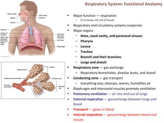
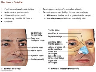
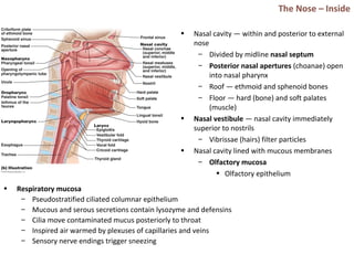
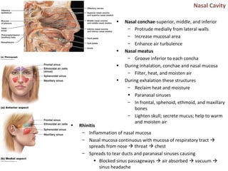
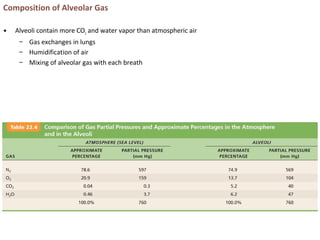
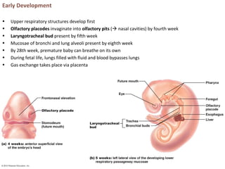
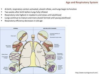
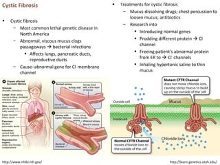
Respiratory System: Functional Anatomy
• Major function — respiration
– O2 in tissues, CO2 out of tissues
• Respiratory and circulatory systems cooperate
• Major organs
– Nose, nasal cavity, and paranasal sinuses
– Pharynx
– Larynx
– Trachea
– Bronchi and their branches
– Lungs and alveoli
• Respiratory zone — gas exchange
– Respiratory bronchioles, alveolar ducts, and alveoli
• Conducting zone — gas transport
– Everything else; cleanses, warms, humidifies air
• Diaphragm and intercostal muscles promote ventilation
• Pulmonary ventilation — air into and out of lungs
• External respiration — gasexchange between lungs and
blood
• Transport — gases in blood
• Internal respiration — gasexchange between blood and
tissues
The Nose – Outside
• Provides an airway for respiration
• Moistens and warms the air
• Filters and cleans the air
• Resonating chamber for speech
• Olfaction
• Two regions — external nose and nasal cavity
• External nose — root, bridge, dorsum nasi, and apex
– Philtrum — shallow vertical groove inferior to apex
– Nostrils (nares) — bounded laterally by alae
The Nose – Inside
• Nasal cavity — within and posterior to external
nose
– Divided by midline nasal septum
– Posterior nasal apertures (choanae) open
into nasal pharynx
– Roof — ethmoid and sphenoid bones
– Floor — hard (bone) and soft palates
(muscle)
• Nasal vestibule — nasal cavity immediately
superior to nostrils
– Vibrissae (hairs) filter particles
• Nasal cavity lined with mucous membranes
– Olfactory mucosa
• Olfactory epithelium
• Respiratory mucosa
– Pseudostratified ciliated columnar epithelium
– Mucous and serous secretions contain lysozyme and defensins
– Cilia move contaminated mucus posteriorly to throat
– Inspired air warmed by plexuses of capillaries and veins
– Sensory nerve endings trigger sneezing
Nasal Cavity
• Nasal conchae-superior, middle, and inferior
– Protrude medially from lateral walls
– Increase mucosal area
– Enhance air turbulence
• Nasal meatus
– Groove inferior to each concha
• During inhalation, conchae and nasal mucosa
– Filter, heat, and moisten air
• During exhalation these structures
– Reclaim heat and moisture
• Paranasal sinuses
– In frontal, sphenoid, ethmoid, and maxillary
bones
– Lighten skull; secrete mucus; help to warm
and moisten air
• Rhinitis
– Inflammation of nasal mucosa
– Nasal mucosa continuous with mucosa of respiratory tract 
spreads from nose  throat  chest
– Spreads to tear ducts and paranasal sinuses causing
• Blocked sinus passageways  air absorbed  vacuum 
sinus headache
Pharynx
• Muscular tube from base of skull to C6
– Connects nasal cavity and mouth to larynx and esophagus
– Composed of skeletal muscle
• Three regions
• Nasopharynx
– Air passageway posterior to nasal cavity
– Lining - pseudostratified columnar epithelium
– Soft palate and uvula close nasopharynx during swallowing
– Pharyngeal tonsil (adenoids) on posterior wall
– Pharyngotympanic (auditory) tubes drain and equalize pressure in middle ear; open into lateral
walls
• Oropharynx
– Passageway for food and air from level of soft palate to epiglottis
– Lining of stratified squamous epithelium
– Isthmus of fauces-opening to oral cavity
– Palatine tonsils-in lateral walls of fauces
– Lingual tonsil-on posterior surface of tongue
• Laryngopharynx
– Passageway for food and air
– Extends to larynx, where continuous with esophagus
– Lined with stratified squamous epithelium
Larynx
• Attaches to hyoid bone; opens into
laryngopharynx; continuous with trachea
• Functions
– Provides patent airway
– Routes air and food into proper channels
– Voice production
• Houses vocal folds
• Superior portion–stratified squamous epithelium
• Inferior to vocal folds–pseudostratified ciliated
columnar epithelium
• Nine cartilages of larynx
– All hyaline cartilage except epiglottis
– Thyroid cartilage with laryngeal
prominence (Adam's apple)
– Ring-shaped cricoid cartilage
– Paired arytenoid, cuneiform, and
corniculate cartilages
– Epiglottis-elastic cartilage; covers laryngeal
inlet during swallowing; covered in taste
bud-containing mucosa
Vocal folds
• Vocal ligaments-deep to laryngeal mucosa
– Attach arytenoid cartilages to thyroid
cartilage
– Contain elastic fibers
– Form core of vocal folds (true vocal cords)
• Glottis-opening between vocal folds
• Folds vibrate to produce sound as air
rushes up from lungs
• Vestibular folds (false vocal cords)
– Superior to vocal folds
– No part in sound production
– Help to close glottis during swallowing
• Speech-intermittent release of expired air while
opening and closing glottis
• Pitch — length and tension of vocal cords
• Loudness — force of air
• Pharynx, oral, nasal, and sinus cavities amplify
and enhance sound
• Language — muscles of pharynx, tongue, soft
palate, and lips
• Vocal folds may act as sphincter to prevent air
passage
• Example-Valsalva's maneuver
– Glottis closes to prevent exhalation
– Abdominal muscles contract
– Intra-abdominal pressure rises
– Emptying rectum or stabilizes trunk
Trachea
• Windpipe–from larynx into mediastinum
• Wall composed of three layers
– Mucosa-ciliated pseudostratified epithelium with
goblet cells
– Submucosa-connective tissue with seromucous glands
– Adventitia-outermost layer made of connective tissue;
encases C-shaped rings of hyaline cartilage
• Trachealis muscle
– Connects posterior parts of cartilage rings
– Contracts during coughing to expel mucus
• Carina
– Spar of cartilage on last, expanded tracheal cartilage
– Point where trachea branches into two main bronchi
Bronchi and Subdivisions
• Air passages undergo 23 orders of branching  bronchial (respiratory) tree
• From tips of bronchial tree  conducting zone structures  respiratory zone structures
• Trachea → right and left main (primary) bronchi
• Each main bronchus enters hilum of one lung
– Right main bronchus wider, shorter, more vertical than left
• Each main bronchus branches into lobar (secondary) bronchi (three on right, two on left)
– Each lobar bronchus supplies one lobe
• Each lobar bronchus branches into segmental (tertiary) bronchi
– Segmental bronchi divide repeatedly
• Branches become smaller and smaller 
– Bronchioles-less than 1 mm in diameter
– Terminal bronchioles-smallest-less than
0.5 mm diameter
• From bronchi through bronchioles, structural changes occur
– Cartilage rings become irregular plates; in bronchioles elastic fibers replace cartilage
– Epithelium changes from pseudostratified columnar to cuboidal; cilia and goblet cells become sparse
– Relative amount of smooth muscle increases
• Allows constriction
Respiratory zone
• Begins as terminal bronchioles  respiratory bronchioles  alveolar ducts  alveolar sacs
– Alveolar sacs contain clusters of alveoli
• ~300 million alveoli make up most of lung volume
• Sites of gas exchange
Conducting and Respiratory Zones
Respiratory Membrane. Alveoli
• Alveolar and capillary walls and their fused basement
membranes
– ~0.5-µ m-thick; gas exchange across membrane by
simple diffusion
• Alveolar walls
– Single layer of squamous epithelium (type I alveolar
cells)
• Scattered cuboidal type II alveolar cells secrete surfactant
and antimicrobial proteins
• Surrounded by fine elastic fibers and pulmonary capillaries
• Alveolar pores connect adjacent alveoli
• Equalize air pressure throughout lung
• Alveolar macrophages keep alveolar surfaces sterile
– 2 million dead macrophages/hour carried by cilia 
throat  swallowed
Alveolar Anatomy
Lungs
• Occupy all thoracic cavity except mediastinum
• Root — site of vascular and bronchial
attachment to mediastinum
• Costal surface — anterior, lateral, and posterior
surfaces
• Composed primarily of alveoli
• Stroma — elastic connective tissue  elasticity
• Apex-superior tip; deep to clavicle
• Base-inferior surface; rests on diaphragm
• Hilum-on mediastinal surface; site for entry/exit of blood
vessels, bronchi, lymphatic vessels, and nerves
• Left lung smaller than right
– Cardiac notch-concavity for heart
– Separated into superior and inferior lobes by oblique
fissure
Lungs
• Right lung
– Superior, middle, inferior lobes separated by
oblique and horizontal fissures
• Bronchopulmonary segments (10 right, 8–10
left) separated by connective tissue septa
– If diseased can be individually removed
• Lobules-smallest subdivisions visible to naked
eye; served by bronchioles and their branches
Pleurae
• Thin, double-layered serosa
• Parietal pleura – walls of pleural
cavity
• Visceral pleura – external lung
surface
• Pleural fluid fills pleural cavity
– Lubrication and surface tension
 lungs’ expansion and recoil
• Pulmonary circulation (low pressure, high volume)
– Pulmonary arteries — systemic venous blood to lungs
• Branch and feed into pulmonary capillaries
– Pulmonary veins — oxygenated blood to the heart
• Bronchial arteries — blood to lung tissue
– From aorta; enter lungs at hilum
– Part of systemic circulation
– Supply all lung tissue except alveoli
– Bronchial veins anastomose with pulmonary veins
Mechanics of Breathing. Pressures
• Pulmonary ventilation
– Inspiration-gases flow into lungs
– Expiration-gases exit lungs
• Atmospheric pressure (Patm)
– Pressure exerted by air surrounding body
– 760 mm Hg at sea level = 1 atmosphere
• Respiratory pressures
– Negative respiratory pressure < Patm
– Positive respiratory pressure > Patm
– Zero respiratory pressure = Patm
• Intrapulmonary (intra-alveolar) pressure (Ppul)
– Pressure in alveoli
– Fluctuates with breathing
– Always eventually equalizes with Patm
• Intrapleural pressure (Pip)
– Pressure in pleural cavity
– Fluctuates with breathing
– Always a negative pressure (<Patm and <Ppul)
– Fluid level must be minimal
• If accumulates  positive Pip pressure
• Negative Pip caused by
– Inward forces → lung collapse
• Elastic recoil of lungs ↓ lung size
• Surface tension of alveolar fluid ↓
alveolar size
– Outward force → enlarge lungs
• Elastic chest wall ↑ thorax outward
Pressure Relationships
• If Pip = Ppul or Patm  lungs collapse
• (Ppul – Pip) = transpulmonary pressure
– Keeps airways open
– Greater transpulmonary pressure  larger
lungs
• Atelectasis (lung collapse) due to
– Plugged bronchioles → collapse of alveoli
– Pneumothorax – air in pleural cavity
• Rupture in parietal or visceral pleura
• Air removed with chest tubes; pleurae
heal  lung reinflates
Pulmonary Ventilation. Gas Laws
• Inspiration and expiration
• Depends on volume changes in thoracic cavity
– Volume changes → pressure changes
– Pressure changes → gases flow to equalize pressure
Boyle’s Law
•Relationship between pressure and volume of a gas
– Gases fill container; if container size reduced 
increased pressure
•Pressure (P) varies inversely with volume (V):
– P1V1 = P2V2
Inspiration • Active process
– Inspiratory muscles (diaphragm and external intercostals) contract
– Thoracic volume increases  intrapulmonary pressure drops (to −1 mm Hg)
– Lungs stretched and intrapulmonary volume increases
– Air flows into lungs, down its pressure gradient, until Ppul = Patm
• Forced inspiration
• Vigorous exercise, COPD  accessory muscles (scalenes, sternocleidomastoid,
pectoralis minor)  further increase in thoracic cage size
Expiration• Quiet expiration normally passive process
– Inspiratory muscles relax
– Thoracic cavity volume decreases
– Elastic lungs recoil and intrapulmonary volume decreases  pressure increases (Ppul rises to +1 mm
Hg) 
– Air flows out of lungs down its pressure gradient until Ppul = 0
• Note: forced expiration-active process; uses abdominal (oblique and transverse) and internal intercostal
muscles
Physical Factors Influencing Pulmonary Ventilation
• Three factors hinder air passage and pulmonary ventilation;
require energy to overcome
• Airway resistance
– Friction in airways – major nonelastic source of
resistance
– F=ΔP/R
• ∆P - pressure gradient between atmosphere and
alveoli
– Resistance usually insignificant
• Large airway diameters
• Progressive branching of airways ↑ total cross-
sectional area
• Resistance greatest in medium-sized bronchi
– Resistance disappears at terminal bronchioles where
diffusion drives gas movement
• As airway resistance rises, breathing movements become more strenuous
• Severe constriction or obstruction of bronchioles
– Can prevent life-sustaining ventilation
– Can occur during acute asthma attacks; stops ventilation
• Epinephrine dilates bronchioles, reduces air resistance
• Surfactant
– Detergent-like lipid and protein complex produced by type II alveolar cells
– ↓ surface tension of alveolar fluid → no alveolar collapse
– Insufficient quantity in premature infants causes infant respiratory distress syndrome
  alveoli collapse after each breath
Alveolar Surface Tension
• Surface tension
– Attracts liquid molecules to one
another at gas-liquid interface
– Resists any increase of the surface
area of liquid
– Water – high surface tension; coats
alveolar walls  reduces them to
smallest size
Lung Compliance
• Measure of change in lung volume
that occurs with given change in
transpulmonary pressure
• Higher lung compliance  easier to
expand lungs
• Normally high due to
– Distensibility of lung tissue
– Alveolar surface tension
• Diminished by
– Nonelastic scar tissue replacing lung tissue (fibrosis)
– Reduced production of surfactant
– Decreased flexibility of thoracic cage
• Homeostatic imbalances that reduce compliance
– Deformities of thorax
– Ossification of costal cartilage
– Paralysis of intercostal muscles
Respiratory Volumes and Capacities
• Used to assess respiratory status
– Tidal volume (TV)
– Inspiratory reserve volume (IRV)
– Expiratory reserve volume (ERV)
– Residual volume (RV)
• Combinations of respiratory volumes
– Inspiratory capacity (IC)
– Functional residual capacity (FRC)
– Vital capacity (VC)
– Total lung capacity (TLC)
Dead Space. Pulmonary Function Tests
• Anatomical dead space
– No contribution to gas exchange
– Air remaining in passageways; ~150 ml
• Alveolar dead space–non-functional alveoli due
to collapse or obstruction
• Total dead space-sum of anatomical and
alveolar dead space
• Spirometer measures respiratory volumes and
capacities
• Spirometry can distinguish between
– Obstructive pulmonary disease—increased
airway resistance (e.g., bronchitis)
• TLC, FRC, RV ↑
– Restrictive disorders — VC, TLC, FRC, RV ↓
(disease or fibrosis)
• To measure rate of gas movement
– Forced vital capacity (FVC) — gas forcibly
expelled after taking deep breath
– Forced expiratory volume (FEV) — amount
of gas expelled during specific time
intervals of FVC
Alveolar Ventilation
• Minute ventilation-total amount of gas flow into or out of respiratory tract in one minute
– Normal at rest = ~ 6 L/min
– Normal with exercise = up to 200 L/min
– Only rough estimate of respiratory efficiency
• Good indicator of effective ventilation
• Alveolar ventilation rate (AVR)-flow of gases into and out of alveoli during a particular time
• Dead space normally constant
• Rapid, shallow breathing decreases AVR
AVR = frequency X (TV – dead space)
(ml/min) (breaths/min) (ml/breath)
Nonrespiratory Air Movements
• May modify normal respiratory rhythm
• Most result from reflex action; some voluntary
• Examples include-cough, sneeze, crying, laughing, hiccups, and yawns
Gas Exchanges – Blood, Lungs, and Tissues
• External respiration – diffusion of gases in lungs
• Internal respiration – diffusion of gases at body tissues
• Both involve
– Physical properties of gases
– Composition of alveolar gas
Dalton’s Law
• For mixture of gases: Ptotal = Σ Pindividual
• Partial pressure
– Pi = k C● i
Henry’s Law
• Gas mixtures in contact with liquid
– Cwater = ks P● i (each gas dissolves in proportion to its
partial pressure)
– At equilibrium, pressure in both phases equal:
• Pwater= Pair
– Amount of each gas that will dissolve depends on
• Solubility – CO2 20 times more soluble in water
than O2; little N2 dissolves in water
• Temperature – T ↑, solubility ↓
Composition of Alveolar Gas
• Alveoli contain more CO2 and water vapor than atmospheric air
– Gas exchanges in lungs
– Humidification of air
– Mixing of alveolar gas with each breath
External Respiration
• Exchange of O2 and CO2 across respiratory membrane Influenced by
– Thickness and surface area of respiratory membrane
• 0.5 to 1 µ m thick
• Large total surface area (40 times that of skin) for gas exchange
• Thicken if lungs become waterlogged and edematous  gas exchange inadequate
• Reduced surface area in emphysema (walls of adjacent alveoli break down), tumors,
inflammation, mucus
External Respiration
• Exchange of O2 and CO2 across respiratory membrane
Influenced by
– Partial pressure gradients and gas solubilities
• O2 gradient in lungs
– Venous blood Po2 = 40 mm Hg
– Alveolar Po2 = 104 mm Hg
– Oxygen to blood
• CO2 gradient in lungs
– Venous blood Pco2 = 45 mm Hg
– Alveolar Pco2 = 40 mm Hg
– CO2 to lungs
• Though gradient not as steep, CO2 diffuses in equal
amounts with oxygen
– CO2 20 times more soluble in plasma than
oxygen
Ventilation-Perfusion Coupling
• Exchange of O2 and CO2 across respiratory membrane Influenced by
– Ventilation-perfusion coupling
• Perfusion-blood flow reaching alveoli
• Ventilation-amount of gas reaching alveoli
• Ventilation and perfusion matched (coupled) for efficient gas exchange
– Never balanced for all alveoli due to
• Regional variations due to effect of gravity on blood and air flow
• Some alveolar ducts plugged with mucus
Ventilation-Perfusion Coupling
• Perfusion
– Changes in Po2 in alveoli cause changes in
diameters of arterioles
• Where alveolar O2 is high, arterioles
dilate
• Where alveolar O2 is low, arterioles
constrict
• Directs most blood where alveolar
oxygen high
• Changes in Pco2 in alveoli cause changes in
diameters of bronchioles
– Where alveolar CO2 is high, bronchioles
dilate
– Where alveolar CO2 is low, bronchioles
constrict
– Allows elimination of CO2 more rapidly
Internal Respiration
• Capillary gas exchange in body tissues
• Partial pressures and diffusion gradients reversed
compared to external respiration
– Tissue Po2 always lower than in systemic arterial blood
 oxygen from blood to tissues
– CO2  from tissues to blood
– Venous blood Po2 40 mm Hg and Pco2
45 mm Hg
O2 Transport
• Molecular O2 carried in blood
– 1.5% dissolved in plasma
– 98.5% loosely bound to each Fe of hemoglobin (Hb) in
RBCs
• 4 O2 per Hb
• Oxyhemoglobin (HbO2)
• Deoxyhemoglobin (HHb – no O2)
• Loading and unloading of O2 → change in shape of Hb
– As O2 binds, Hb affinity for O2 increases and vice versa
– Cooperative binding
• All four heme groups carry O2 – full saturation
• Rate of loading and unloading of O2 regulated to ensure
adequate oxygen delivery to cells
– Po2
– Temperature
– Blood pH
– Pco2
– Concentration of BPG–produced by RBCs during
glycolysis; levels rise when oxygen levels chronically
low
Hemoglobin Dissociation Curve
• Oxygen-hemoglobin dissociation curve
• Hemoglobin saturation plotted against Po2 is S-
shaped curve
• Binding and release of O2 influenced by Po2
• In arterial blood
– Po2 = 100 mm Hg
– Contains 20 ml oxygen per 100 ml blood (20 vol %)
– Hb is 98% saturated
• Further increases in Po2 (e.g., breathing deeply) produce minimal increases in O2 binding
• In venous blood
– Po2 = 40 mm Hg
– Contains 15 vol % oxygen
– Hb is 75% saturated
• Venous reserve
• Oxygen remaining in venous blood
Factors Influencing Hb Saturation
• Increases in temperature, H+
, Pco2, and BPG
– Modify structure of hemoglobin; its affinity for O2 ↓
– Occur in systemic capillaries
– ↑ O2 unloading from blood
– Shift O2-Hb curve to right
• Decreases in these factors shift curve to left
– Decreases oxygen unloading from blood
• As cells metabolize glucose and use O2
– Pco2 and H+
↑ in capillary blood  blood pH ↓, Pco2 ↑
– Heat ↑  Hb affinity for O2 ↓  oxygen unloading ↑
– Bohr effect: oxygen unloading in tissues that use O2
• Hypoxia
– Inadequate O2 delivery to tissues  cyanosis
– Anemic hypoxia–too few RBCs; abnormal or too little Hb
– Ischemic hypoxia–impaired/blocked circulation
– Histotoxic hypoxia–cells unable to use O2, as in metabolic
poisons
– Hypoxemic hypoxia–abnormal ventilation; pulmonary disease
– Carbon monoxide poisoning–especially from fire; 200X
greater affinity for Hb than oxygen
CO2 Transport
• CO2 transported in blood in three forms
– 7 to 10% dissolved in plasma
– 20% bound to globin of hemoglobin (carbaminohemoglobin)
– 70% transported as bicarbonate ions (HCO3
–
) in plasma
• CO2 combines with water to form carbonic acid (H2CO3), which quickly dissociates
• Occurs primarily in RBCs, where carbonic anhydrase reversibly and rapidly catalyzes reaction
• In systemic capillaries
– HCO3
–
quickly diffuses from RBCs into plasma
• Chloride shift occurs
– Outrush of HCO3
–
from RBCs balanced as Cl–
moves into RBCs from plasma
• In pulmonary capillaries
– HCO3
–
moves into RBCs (while Cl-
move out); binds with H+
to form H2CO3
– H2CO3 split by carbonic anhydrase into CO2 and water
– CO2 diffuses into alveoli
Haldane Effect
• Amount of CO2 transported affected by Po2
– HHb forms carbaminohemoglobin and buffers H+
more
easily 
– Po2 and hemoglobin saturation ↓; CO2 in blood ↑
• ↑ CO2 exchange in tissues and lungs
• At tissues, as more CO2 enters blood
– O2 unloading ↑ (Bohr effect)
– HbO2 releases O2and readily forms
carbaminohemoglobin
• Carbonic acid–bicarbonate buffer system–resists changes in blood pH
– If H+
concentration in blood rises, excess H+
is removed by combining with HCO3
–
 H2CO3
– If H+
concentration begins to drop, H2CO3 dissociates, releasing H+
– HCO3
–
is alkaline reserve of carbonic acid-bicarbonate buffer system
• Changes in respiratory rate and depth affect blood pH
– Slow, shallow breathing  increased CO2 in blood drop in pH
– Rapid, deep breathing  decreased CO2 in blood  rise in pH
• Higher brain centers, chemoreceptors, and other reflexes
• Neural controls
– Neurons in reticular formation of medulla and pons
– Clustered neurons in medulla important
• Ventral respiratory group
• Dorsal respiratory group
• Ventral respiratory group (VRG)
– Rhythm-generating and integrative center
– Sets eupnea (12–15 breaths/minute)
• Normal respiratory rate and rhythm
– Its inspiratory neurons excite inspiratory muscles via
phrenic (diaphragm) and intercostal nerves (external
intercostals)
– Expiratory neurons inhibit inspiratory neurons
• Dorsal respiratory group (DRG)
– Near root of cranial nerve IX
– Integrates input from peripheral stretch and
chemoreceptors; sends information  VRG
Control of Respiration. Medullary Respiratory Centers
Pontine Respiratory Centers
• Influence and modify activity of VRG
• Smooth out transition between inspiration and
expiration and vice versa
• Transmit impulses to VRG  modify and fine-
tune breathing rhythms during vocalization,
sleep, exercise
Respiratory Rhythm
•Not well understood
•One hypothesis
– Pacemaker neurons with intrinsic
rhythmicity
•Most widely accepted hypothesis
– Reciprocal inhibition of two sets of
interconnected pacemaker neurons in
medulla that generate rhythm
Factors influencing Breathing Rate and Depth
• Depth determined by how actively respiratory center stimulates respiratory muscles
• Rate determined by how long inspiratory center active
• Both modified in response to changing body demands
– Most important are changing levels of CO2, O2, and H+
– Sensed by central and peripheral chemoreceptors
Chemical Factors
• Influence of Pco2 (most potent; most closely
controlled)
– blood Pco2 levels ↑ (hypercapnia), CO2
accumulates in brain 
– CO2 in brain hydrated  carbonic acid 
dissociates, releasing H+
 pH ↓
– H+
stimulates central chemoreceptors of
brain stem
– Chemoreceptors synapse with respiratory
regulatory centers  depth and rate of
breathing ↑  blood Pco2 ↓  pH ↑
• Hyperventilation –depth/rate of breathing ↑;
exceeds body's need to remove CO2
  blood CO2 levels ↓ (hypocapnia) 
cerebral vasoconstriction and cerebral
ischemia  dizziness, fainting
• Apnea – breathing cessation from abnormally
low Pco2
Chemical Factors
• Influence of Po2
– Peripheral chemoreceptors in aortic and
carotid bodies – arterial O2 level sensors
• When excited, cause respiratory
centers to increase ventilation
– ↓ Po2 – normally slight effect on ventilation
• O2 reservoir in HbO2
• Requires substantial drop in arterial
Po2 (to 60 mm Hg) to stimulate
increased ventilation
• Influence of arterial pH
– Can modify respiratory rate and rhythm
even if CO2 and O2 levels normal
– Mediated by peripheral chemoreceptors
– ↓ pH may reflect
• CO2 retention; accumulation of lactic
acid; excess ketone bodies
– Respiratory system controls ↑ pH by ↑
respiratory rate and depth
• ↑CO2 levels most powerful respiratory stimulant
• Normally blood Po2 affects breathing only
indirectly by influencing peripheral
chemoreceptor sensitivity to changes in Pco2
• When arterial Po2 falls below 60 mm Hg, it
becomes major stimulus for respiration (via
peripheral chemoreceptors)
• Changes in arterial pH resulting from CO2
retention or metabolic factors act indirectly
through peripheral chemoreceptors
Influence of Higher Brain Centers
• Hypothalamic controls act through limbic system to modify rate and depth of respiration
– Example-breath holding that occurs in anger or gasping with pain
• Rise in body temperature increases respiratory rate
• Cortical controls-direct signals from cerebral motor cortex that bypass medullary controls
– Example-voluntary breath holding
• Brain stem reinstates breathing when blood CO2 critical
Inflation Reflex
• Hering-Breuer Reflex (inflation reflex)
– Stretch receptors in pleurae and airways
stimulated by lung inflation
• Inhibitory signals to medullary
respiratory centers end inhalation and
allow expiration
• Acts as protective response more than
normal regulatory mechanism
Pulmonary Irritant Reflexes
• Receptors in bronchioles respond to irritants
– Communicate with respiratory centers via
vagal nerve afferents
• Promote reflexive constriction of air passages
• Same irritant  cough in trachea or bronchi;
sneeze in nasal cavity
Respiratory Adjustments: Exercise and High Altitude
• Adjustments geared to both intensity and
duration of exercise
• Hyperpnea
– ↑ ventilation (10 to 20 fold)
• Pco2, Po2, and pH remain during exercise≡
• Three neural factors cause increase in
ventilation as exercise begins
– Psychological stimuli
– Simultaneous cortical motor activation of
skeletal muscles and respiratory centers
– Excitatory impulses to respiratory centers
from proprioceptors
• Ventilation declines suddenly as exercise ends
because the three neural factors shut off
• Gradual decline to baseline because of decline in
CO2 flow after exercise ends
• Exercise  anaerobic respiration  lactic acid
– Not from poor respiratory function; from
insufficient cardiac output or skeletal
muscle inability to increase oxygen uptake
• Quick travel to altitudes above 2400 meters
(8000 feet) may  symptoms of acute mountain
sickness (AMS)
– Atmospheric pressure and Po2 levels ↓
– Headaches, shortness of breath, nausea,
and dizziness
– (Lethal) cerebral and pulmonary edema
• Acclimatization-respiratory and hematopoietic
adjustments to high altitude
– Chemoreceptors – more responsive to Pco2
when Po2 ↓
– ↓ in Po2 directly stimulates peripheral
chemoreceptors
– Minute ventilation ↑ and stabilizes in few
days to 2–3 L/min higher than at sea level
• Always lower-than-normal Hb saturation levels
– Less O2 available
• ↓ blood O2 → ↑ production of EPO in kidneys
• RBC ↑ slowly → long-term compensation
COPD. Emphysema
• Chronic obstructive pulmonary disease (COPD)
– Exemplified by chronic bronchitis and emphysema
– Irreversible decrease in ability to force air out of lungs
– Other common features
• History of smoking in 80% of patients
• Dyspnea - labored breathing ("air hunger")
• Coughing and frequent pulmonary infections
• Most develop respiratory failure
(hypoventilation) accompanied by respiratory
acidosis, hypoxemia
• Emphysema
– Permanent enlargement of alveoli; destruction of
alveolar walls; decreased lung elasticity 
• Accessory muscles necessary for breathing
  exhaustion from energy usage
• Hyperinflation  flattened diaphragm  reduced
ventilation efficiency
• Damaged pulmonary capillaries  enlarged right
ventricle
Homeostatic Imbalance
• Chronic bronchitis
– Inhaled irritants  chronic excessive mucus 
inflamed and fibrosed lower respiratory passageways
 obstructed airways  impaired lung ventilation and
gas exchange  frequent pulmonary infections
• COPD symptoms and treatment
– Strength of innate respiratory drive  different
symptoms in patients
• "Pink puffers"–thin; near-normal blood gases
• "Blue bloaters"–stocky, hypoxic
– Treated with bronchodilators, corticosteroids, oxygen,
sometimes surgery
• Asthma – reversible COPD
– Characterized by coughing, dyspnea, wheezing, and
chest tightness
– Active inflammation of airways precedes
bronchospasms
– Airway inflammation is immune response caused by
release of interleukins, production of IgE, and
recruitment of inflammatory cells
– Airways thickened with inflammatory exudate magnify
effect of bronchospasms
Tuberculosis. Lung Cancer.
• Tuberculosis (TB)
– Infectious disease caused by bacterium
Mycobacterium tuberculosis
– Symptoms-fever, night sweats, weight loss,
racking cough, coughing up blood
– Treatment- 12-month course of antibiotics
• Are antibiotic resistant strains
• Lung cancer
– Leading cause of cancer deaths in North
America
– 90% of all cases result of smoking
– Three most common types
• Adenocarcinoma (~40% of cases)
peripheral lung areas (bronchial
glands, alveolar cells)
• Squamous cell carcinoma (20–40% of
cases) in bronchial epithelium
• Small cell carcinoma (~20% of cases) -
lymphocyte-like cells; originate in
primary bronchi; metastasize
http://www.lung.org/
Lung Cancer
• Lung cancer
– Early detection key to survival
– Helical CT scan better than chest X ray
– Developing breath test of gold nanoparticles
– If no metastasis  surgery to remove diseased lung
tissue
– If metastasis  radiation and chemotherapy
• Potential new therapies for lung cancer
– Antibodies targeting growth factors required by tumor;
or deliver toxic agents to tumor
– Cancer vaccines to stimulate immune system
– Gene therapy to replace defective genes
http://www.aboutcancer.com/
Early Development
• Upper respiratory structures develop first
• Olfactory placodes invaginate into olfactory pits ( nasal cavities) by fourth week
• Laryngotracheal bud present by fifth week
• Mucosae of bronchi and lung alveoli present by eighth week
• By 28th week, premature baby can breathe on its own
• During fetal life, lungs filled with fluid and blood bypasses lungs
• Gas exchange takes place via placenta
Age and Respiratory System
• At birth, respiratory centers activated, alveoli inflate, and lungs begin to function
• Two weeks after birth before lungs fully inflated
• Respiratory rate highest in newborns and slows until adulthood
• Lungs continue to mature and more alveoli formed until young adulthood
• Respiratory efficiency decreases in old age
http://www.nursingconsult.com/
Cystic Fibrosis
• Cystic fibrosis
– Most common lethal genetic disease in
North America
– Abnormal, viscous mucus clogs
passageways  bacterial infections
• Affects lungs, pancreatic ducts,
reproductive ducts
– Cause–abnormal gene for Cl-
membrane
channel
http://learn.genetics.utah.edu/http://www.nhlbi.nih.gov/
• Treatments for cystic fibrosis
– Mucus-dissolving drugs; chest percussion to
loosen mucus; antibiotics
– Research into
• Introducing normal genes
• Prodding different protein  Cl-
channel
• Freeing patient's abnormal protein
from ER to  Cl-
channels
• Inhaling hypertonic saline to thin
mucus

The respiratory system

  • 1.
    Respiratory System: FunctionalAnatomy • Major function — respiration – O2 in tissues, CO2 out of tissues • Respiratory and circulatory systems cooperate • Major organs – Nose, nasal cavity, and paranasal sinuses – Pharynx – Larynx – Trachea – Bronchi and their branches – Lungs and alveoli • Respiratory zone — gas exchange – Respiratory bronchioles, alveolar ducts, and alveoli • Conducting zone — gas transport – Everything else; cleanses, warms, humidifies air • Diaphragm and intercostal muscles promote ventilation • Pulmonary ventilation — air into and out of lungs • External respiration — gasexchange between lungs and blood • Transport — gases in blood • Internal respiration — gasexchange between blood and tissues
  • 2.
    The Nose –Outside • Provides an airway for respiration • Moistens and warms the air • Filters and cleans the air • Resonating chamber for speech • Olfaction • Two regions — external nose and nasal cavity • External nose — root, bridge, dorsum nasi, and apex – Philtrum — shallow vertical groove inferior to apex – Nostrils (nares) — bounded laterally by alae
  • 3.
    The Nose –Inside • Nasal cavity — within and posterior to external nose – Divided by midline nasal septum – Posterior nasal apertures (choanae) open into nasal pharynx – Roof — ethmoid and sphenoid bones – Floor — hard (bone) and soft palates (muscle) • Nasal vestibule — nasal cavity immediately superior to nostrils – Vibrissae (hairs) filter particles • Nasal cavity lined with mucous membranes – Olfactory mucosa • Olfactory epithelium • Respiratory mucosa – Pseudostratified ciliated columnar epithelium – Mucous and serous secretions contain lysozyme and defensins – Cilia move contaminated mucus posteriorly to throat – Inspired air warmed by plexuses of capillaries and veins – Sensory nerve endings trigger sneezing
  • 4.
    Nasal Cavity • Nasalconchae-superior, middle, and inferior – Protrude medially from lateral walls – Increase mucosal area – Enhance air turbulence • Nasal meatus – Groove inferior to each concha • During inhalation, conchae and nasal mucosa – Filter, heat, and moisten air • During exhalation these structures – Reclaim heat and moisture • Paranasal sinuses – In frontal, sphenoid, ethmoid, and maxillary bones – Lighten skull; secrete mucus; help to warm and moisten air • Rhinitis – Inflammation of nasal mucosa – Nasal mucosa continuous with mucosa of respiratory tract  spreads from nose  throat  chest – Spreads to tear ducts and paranasal sinuses causing • Blocked sinus passageways  air absorbed  vacuum  sinus headache
  • 5.
    Pharynx • Muscular tubefrom base of skull to C6 – Connects nasal cavity and mouth to larynx and esophagus – Composed of skeletal muscle • Three regions • Nasopharynx – Air passageway posterior to nasal cavity – Lining - pseudostratified columnar epithelium – Soft palate and uvula close nasopharynx during swallowing – Pharyngeal tonsil (adenoids) on posterior wall – Pharyngotympanic (auditory) tubes drain and equalize pressure in middle ear; open into lateral walls • Oropharynx – Passageway for food and air from level of soft palate to epiglottis – Lining of stratified squamous epithelium – Isthmus of fauces-opening to oral cavity – Palatine tonsils-in lateral walls of fauces – Lingual tonsil-on posterior surface of tongue • Laryngopharynx – Passageway for food and air – Extends to larynx, where continuous with esophagus – Lined with stratified squamous epithelium
  • 6.
    Larynx • Attaches tohyoid bone; opens into laryngopharynx; continuous with trachea • Functions – Provides patent airway – Routes air and food into proper channels – Voice production • Houses vocal folds • Superior portion–stratified squamous epithelium • Inferior to vocal folds–pseudostratified ciliated columnar epithelium • Nine cartilages of larynx – All hyaline cartilage except epiglottis – Thyroid cartilage with laryngeal prominence (Adam's apple) – Ring-shaped cricoid cartilage – Paired arytenoid, cuneiform, and corniculate cartilages – Epiglottis-elastic cartilage; covers laryngeal inlet during swallowing; covered in taste bud-containing mucosa
  • 7.
    Vocal folds • Vocalligaments-deep to laryngeal mucosa – Attach arytenoid cartilages to thyroid cartilage – Contain elastic fibers – Form core of vocal folds (true vocal cords) • Glottis-opening between vocal folds • Folds vibrate to produce sound as air rushes up from lungs • Vestibular folds (false vocal cords) – Superior to vocal folds – No part in sound production – Help to close glottis during swallowing • Speech-intermittent release of expired air while opening and closing glottis • Pitch — length and tension of vocal cords • Loudness — force of air • Pharynx, oral, nasal, and sinus cavities amplify and enhance sound • Language — muscles of pharynx, tongue, soft palate, and lips • Vocal folds may act as sphincter to prevent air passage • Example-Valsalva's maneuver – Glottis closes to prevent exhalation – Abdominal muscles contract – Intra-abdominal pressure rises – Emptying rectum or stabilizes trunk
  • 8.
    Trachea • Windpipe–from larynxinto mediastinum • Wall composed of three layers – Mucosa-ciliated pseudostratified epithelium with goblet cells – Submucosa-connective tissue with seromucous glands – Adventitia-outermost layer made of connective tissue; encases C-shaped rings of hyaline cartilage • Trachealis muscle – Connects posterior parts of cartilage rings – Contracts during coughing to expel mucus • Carina – Spar of cartilage on last, expanded tracheal cartilage – Point where trachea branches into two main bronchi
  • 9.
    Bronchi and Subdivisions •Air passages undergo 23 orders of branching  bronchial (respiratory) tree • From tips of bronchial tree  conducting zone structures  respiratory zone structures • Trachea → right and left main (primary) bronchi • Each main bronchus enters hilum of one lung – Right main bronchus wider, shorter, more vertical than left • Each main bronchus branches into lobar (secondary) bronchi (three on right, two on left) – Each lobar bronchus supplies one lobe • Each lobar bronchus branches into segmental (tertiary) bronchi – Segmental bronchi divide repeatedly • Branches become smaller and smaller  – Bronchioles-less than 1 mm in diameter – Terminal bronchioles-smallest-less than 0.5 mm diameter
  • 10.
    • From bronchithrough bronchioles, structural changes occur – Cartilage rings become irregular plates; in bronchioles elastic fibers replace cartilage – Epithelium changes from pseudostratified columnar to cuboidal; cilia and goblet cells become sparse – Relative amount of smooth muscle increases • Allows constriction Respiratory zone • Begins as terminal bronchioles  respiratory bronchioles  alveolar ducts  alveolar sacs – Alveolar sacs contain clusters of alveoli • ~300 million alveoli make up most of lung volume • Sites of gas exchange Conducting and Respiratory Zones
  • 11.
    Respiratory Membrane. Alveoli •Alveolar and capillary walls and their fused basement membranes – ~0.5-µ m-thick; gas exchange across membrane by simple diffusion • Alveolar walls – Single layer of squamous epithelium (type I alveolar cells) • Scattered cuboidal type II alveolar cells secrete surfactant and antimicrobial proteins • Surrounded by fine elastic fibers and pulmonary capillaries • Alveolar pores connect adjacent alveoli • Equalize air pressure throughout lung • Alveolar macrophages keep alveolar surfaces sterile – 2 million dead macrophages/hour carried by cilia  throat  swallowed
  • 12.
  • 13.
    Lungs • Occupy allthoracic cavity except mediastinum • Root — site of vascular and bronchial attachment to mediastinum • Costal surface — anterior, lateral, and posterior surfaces • Composed primarily of alveoli • Stroma — elastic connective tissue  elasticity • Apex-superior tip; deep to clavicle • Base-inferior surface; rests on diaphragm • Hilum-on mediastinal surface; site for entry/exit of blood vessels, bronchi, lymphatic vessels, and nerves • Left lung smaller than right – Cardiac notch-concavity for heart – Separated into superior and inferior lobes by oblique fissure
  • 14.
    Lungs • Right lung –Superior, middle, inferior lobes separated by oblique and horizontal fissures • Bronchopulmonary segments (10 right, 8–10 left) separated by connective tissue septa – If diseased can be individually removed • Lobules-smallest subdivisions visible to naked eye; served by bronchioles and their branches
  • 15.
    Pleurae • Thin, double-layeredserosa • Parietal pleura – walls of pleural cavity • Visceral pleura – external lung surface • Pleural fluid fills pleural cavity – Lubrication and surface tension  lungs’ expansion and recoil • Pulmonary circulation (low pressure, high volume) – Pulmonary arteries — systemic venous blood to lungs • Branch and feed into pulmonary capillaries – Pulmonary veins — oxygenated blood to the heart • Bronchial arteries — blood to lung tissue – From aorta; enter lungs at hilum – Part of systemic circulation – Supply all lung tissue except alveoli – Bronchial veins anastomose with pulmonary veins
  • 16.
    Mechanics of Breathing.Pressures • Pulmonary ventilation – Inspiration-gases flow into lungs – Expiration-gases exit lungs • Atmospheric pressure (Patm) – Pressure exerted by air surrounding body – 760 mm Hg at sea level = 1 atmosphere • Respiratory pressures – Negative respiratory pressure < Patm – Positive respiratory pressure > Patm – Zero respiratory pressure = Patm • Intrapulmonary (intra-alveolar) pressure (Ppul) – Pressure in alveoli – Fluctuates with breathing – Always eventually equalizes with Patm • Intrapleural pressure (Pip) – Pressure in pleural cavity – Fluctuates with breathing – Always a negative pressure (<Patm and <Ppul) – Fluid level must be minimal • If accumulates  positive Pip pressure • Negative Pip caused by – Inward forces → lung collapse • Elastic recoil of lungs ↓ lung size • Surface tension of alveolar fluid ↓ alveolar size – Outward force → enlarge lungs • Elastic chest wall ↑ thorax outward
  • 17.
    Pressure Relationships • IfPip = Ppul or Patm  lungs collapse • (Ppul – Pip) = transpulmonary pressure – Keeps airways open – Greater transpulmonary pressure  larger lungs • Atelectasis (lung collapse) due to – Plugged bronchioles → collapse of alveoli – Pneumothorax – air in pleural cavity • Rupture in parietal or visceral pleura • Air removed with chest tubes; pleurae heal  lung reinflates
  • 18.
    Pulmonary Ventilation. GasLaws • Inspiration and expiration • Depends on volume changes in thoracic cavity – Volume changes → pressure changes – Pressure changes → gases flow to equalize pressure Boyle’s Law •Relationship between pressure and volume of a gas – Gases fill container; if container size reduced  increased pressure •Pressure (P) varies inversely with volume (V): – P1V1 = P2V2
  • 19.
    Inspiration • Activeprocess – Inspiratory muscles (diaphragm and external intercostals) contract – Thoracic volume increases  intrapulmonary pressure drops (to −1 mm Hg) – Lungs stretched and intrapulmonary volume increases – Air flows into lungs, down its pressure gradient, until Ppul = Patm • Forced inspiration • Vigorous exercise, COPD  accessory muscles (scalenes, sternocleidomastoid, pectoralis minor)  further increase in thoracic cage size
  • 20.
    Expiration• Quiet expirationnormally passive process – Inspiratory muscles relax – Thoracic cavity volume decreases – Elastic lungs recoil and intrapulmonary volume decreases  pressure increases (Ppul rises to +1 mm Hg)  – Air flows out of lungs down its pressure gradient until Ppul = 0 • Note: forced expiration-active process; uses abdominal (oblique and transverse) and internal intercostal muscles
  • 22.
    Physical Factors InfluencingPulmonary Ventilation • Three factors hinder air passage and pulmonary ventilation; require energy to overcome • Airway resistance – Friction in airways – major nonelastic source of resistance – F=ΔP/R • ∆P - pressure gradient between atmosphere and alveoli – Resistance usually insignificant • Large airway diameters • Progressive branching of airways ↑ total cross- sectional area • Resistance greatest in medium-sized bronchi – Resistance disappears at terminal bronchioles where diffusion drives gas movement • As airway resistance rises, breathing movements become more strenuous • Severe constriction or obstruction of bronchioles – Can prevent life-sustaining ventilation – Can occur during acute asthma attacks; stops ventilation • Epinephrine dilates bronchioles, reduces air resistance
  • 23.
    • Surfactant – Detergent-likelipid and protein complex produced by type II alveolar cells – ↓ surface tension of alveolar fluid → no alveolar collapse – Insufficient quantity in premature infants causes infant respiratory distress syndrome   alveoli collapse after each breath Alveolar Surface Tension • Surface tension – Attracts liquid molecules to one another at gas-liquid interface – Resists any increase of the surface area of liquid – Water – high surface tension; coats alveolar walls  reduces them to smallest size
  • 24.
    Lung Compliance • Measureof change in lung volume that occurs with given change in transpulmonary pressure • Higher lung compliance  easier to expand lungs • Normally high due to – Distensibility of lung tissue – Alveolar surface tension • Diminished by – Nonelastic scar tissue replacing lung tissue (fibrosis) – Reduced production of surfactant – Decreased flexibility of thoracic cage • Homeostatic imbalances that reduce compliance – Deformities of thorax – Ossification of costal cartilage – Paralysis of intercostal muscles
  • 25.
    Respiratory Volumes andCapacities • Used to assess respiratory status – Tidal volume (TV) – Inspiratory reserve volume (IRV) – Expiratory reserve volume (ERV) – Residual volume (RV) • Combinations of respiratory volumes – Inspiratory capacity (IC) – Functional residual capacity (FRC) – Vital capacity (VC) – Total lung capacity (TLC)
  • 26.
    Dead Space. PulmonaryFunction Tests • Anatomical dead space – No contribution to gas exchange – Air remaining in passageways; ~150 ml • Alveolar dead space–non-functional alveoli due to collapse or obstruction • Total dead space-sum of anatomical and alveolar dead space • Spirometer measures respiratory volumes and capacities • Spirometry can distinguish between – Obstructive pulmonary disease—increased airway resistance (e.g., bronchitis) • TLC, FRC, RV ↑ – Restrictive disorders — VC, TLC, FRC, RV ↓ (disease or fibrosis) • To measure rate of gas movement – Forced vital capacity (FVC) — gas forcibly expelled after taking deep breath – Forced expiratory volume (FEV) — amount of gas expelled during specific time intervals of FVC
  • 27.
    Alveolar Ventilation • Minuteventilation-total amount of gas flow into or out of respiratory tract in one minute – Normal at rest = ~ 6 L/min – Normal with exercise = up to 200 L/min – Only rough estimate of respiratory efficiency • Good indicator of effective ventilation • Alveolar ventilation rate (AVR)-flow of gases into and out of alveoli during a particular time • Dead space normally constant • Rapid, shallow breathing decreases AVR AVR = frequency X (TV – dead space) (ml/min) (breaths/min) (ml/breath)
  • 28.
    Nonrespiratory Air Movements •May modify normal respiratory rhythm • Most result from reflex action; some voluntary • Examples include-cough, sneeze, crying, laughing, hiccups, and yawns
  • 29.
    Gas Exchanges –Blood, Lungs, and Tissues • External respiration – diffusion of gases in lungs • Internal respiration – diffusion of gases at body tissues • Both involve – Physical properties of gases – Composition of alveolar gas Dalton’s Law • For mixture of gases: Ptotal = Σ Pindividual • Partial pressure – Pi = k C● i Henry’s Law • Gas mixtures in contact with liquid – Cwater = ks P● i (each gas dissolves in proportion to its partial pressure) – At equilibrium, pressure in both phases equal: • Pwater= Pair – Amount of each gas that will dissolve depends on • Solubility – CO2 20 times more soluble in water than O2; little N2 dissolves in water • Temperature – T ↑, solubility ↓
  • 30.
    Composition of AlveolarGas • Alveoli contain more CO2 and water vapor than atmospheric air – Gas exchanges in lungs – Humidification of air – Mixing of alveolar gas with each breath
  • 31.
    External Respiration • Exchangeof O2 and CO2 across respiratory membrane Influenced by – Thickness and surface area of respiratory membrane • 0.5 to 1 µ m thick • Large total surface area (40 times that of skin) for gas exchange • Thicken if lungs become waterlogged and edematous  gas exchange inadequate • Reduced surface area in emphysema (walls of adjacent alveoli break down), tumors, inflammation, mucus
  • 32.
    External Respiration • Exchangeof O2 and CO2 across respiratory membrane Influenced by – Partial pressure gradients and gas solubilities • O2 gradient in lungs – Venous blood Po2 = 40 mm Hg – Alveolar Po2 = 104 mm Hg – Oxygen to blood • CO2 gradient in lungs – Venous blood Pco2 = 45 mm Hg – Alveolar Pco2 = 40 mm Hg – CO2 to lungs • Though gradient not as steep, CO2 diffuses in equal amounts with oxygen – CO2 20 times more soluble in plasma than oxygen
  • 33.
    Ventilation-Perfusion Coupling • Exchangeof O2 and CO2 across respiratory membrane Influenced by – Ventilation-perfusion coupling • Perfusion-blood flow reaching alveoli • Ventilation-amount of gas reaching alveoli • Ventilation and perfusion matched (coupled) for efficient gas exchange – Never balanced for all alveoli due to • Regional variations due to effect of gravity on blood and air flow • Some alveolar ducts plugged with mucus
  • 34.
    Ventilation-Perfusion Coupling • Perfusion –Changes in Po2 in alveoli cause changes in diameters of arterioles • Where alveolar O2 is high, arterioles dilate • Where alveolar O2 is low, arterioles constrict • Directs most blood where alveolar oxygen high • Changes in Pco2 in alveoli cause changes in diameters of bronchioles – Where alveolar CO2 is high, bronchioles dilate – Where alveolar CO2 is low, bronchioles constrict – Allows elimination of CO2 more rapidly
  • 35.
    Internal Respiration • Capillarygas exchange in body tissues • Partial pressures and diffusion gradients reversed compared to external respiration – Tissue Po2 always lower than in systemic arterial blood  oxygen from blood to tissues – CO2  from tissues to blood – Venous blood Po2 40 mm Hg and Pco2 45 mm Hg
  • 36.
    O2 Transport • MolecularO2 carried in blood – 1.5% dissolved in plasma – 98.5% loosely bound to each Fe of hemoglobin (Hb) in RBCs • 4 O2 per Hb • Oxyhemoglobin (HbO2) • Deoxyhemoglobin (HHb – no O2) • Loading and unloading of O2 → change in shape of Hb – As O2 binds, Hb affinity for O2 increases and vice versa – Cooperative binding • All four heme groups carry O2 – full saturation • Rate of loading and unloading of O2 regulated to ensure adequate oxygen delivery to cells – Po2 – Temperature – Blood pH – Pco2 – Concentration of BPG–produced by RBCs during glycolysis; levels rise when oxygen levels chronically low
  • 37.
    Hemoglobin Dissociation Curve •Oxygen-hemoglobin dissociation curve • Hemoglobin saturation plotted against Po2 is S- shaped curve • Binding and release of O2 influenced by Po2
  • 38.
    • In arterialblood – Po2 = 100 mm Hg – Contains 20 ml oxygen per 100 ml blood (20 vol %) – Hb is 98% saturated • Further increases in Po2 (e.g., breathing deeply) produce minimal increases in O2 binding
  • 39.
    • In venousblood – Po2 = 40 mm Hg – Contains 15 vol % oxygen – Hb is 75% saturated • Venous reserve • Oxygen remaining in venous blood
  • 40.
    Factors Influencing HbSaturation • Increases in temperature, H+ , Pco2, and BPG – Modify structure of hemoglobin; its affinity for O2 ↓ – Occur in systemic capillaries – ↑ O2 unloading from blood – Shift O2-Hb curve to right • Decreases in these factors shift curve to left – Decreases oxygen unloading from blood • As cells metabolize glucose and use O2 – Pco2 and H+ ↑ in capillary blood  blood pH ↓, Pco2 ↑ – Heat ↑  Hb affinity for O2 ↓  oxygen unloading ↑ – Bohr effect: oxygen unloading in tissues that use O2 • Hypoxia – Inadequate O2 delivery to tissues  cyanosis – Anemic hypoxia–too few RBCs; abnormal or too little Hb – Ischemic hypoxia–impaired/blocked circulation – Histotoxic hypoxia–cells unable to use O2, as in metabolic poisons – Hypoxemic hypoxia–abnormal ventilation; pulmonary disease – Carbon monoxide poisoning–especially from fire; 200X greater affinity for Hb than oxygen
  • 41.
    CO2 Transport • CO2transported in blood in three forms – 7 to 10% dissolved in plasma – 20% bound to globin of hemoglobin (carbaminohemoglobin) – 70% transported as bicarbonate ions (HCO3 – ) in plasma • CO2 combines with water to form carbonic acid (H2CO3), which quickly dissociates • Occurs primarily in RBCs, where carbonic anhydrase reversibly and rapidly catalyzes reaction
  • 42.
    • In systemiccapillaries – HCO3 – quickly diffuses from RBCs into plasma • Chloride shift occurs – Outrush of HCO3 – from RBCs balanced as Cl– moves into RBCs from plasma
  • 43.
    • In pulmonarycapillaries – HCO3 – moves into RBCs (while Cl- move out); binds with H+ to form H2CO3 – H2CO3 split by carbonic anhydrase into CO2 and water – CO2 diffuses into alveoli
  • 44.
    Haldane Effect • Amountof CO2 transported affected by Po2 – HHb forms carbaminohemoglobin and buffers H+ more easily  – Po2 and hemoglobin saturation ↓; CO2 in blood ↑ • ↑ CO2 exchange in tissues and lungs • At tissues, as more CO2 enters blood – O2 unloading ↑ (Bohr effect) – HbO2 releases O2and readily forms carbaminohemoglobin • Carbonic acid–bicarbonate buffer system–resists changes in blood pH – If H+ concentration in blood rises, excess H+ is removed by combining with HCO3 –  H2CO3 – If H+ concentration begins to drop, H2CO3 dissociates, releasing H+ – HCO3 – is alkaline reserve of carbonic acid-bicarbonate buffer system • Changes in respiratory rate and depth affect blood pH – Slow, shallow breathing  increased CO2 in blood drop in pH – Rapid, deep breathing  decreased CO2 in blood  rise in pH
  • 45.
    • Higher braincenters, chemoreceptors, and other reflexes • Neural controls – Neurons in reticular formation of medulla and pons – Clustered neurons in medulla important • Ventral respiratory group • Dorsal respiratory group • Ventral respiratory group (VRG) – Rhythm-generating and integrative center – Sets eupnea (12–15 breaths/minute) • Normal respiratory rate and rhythm – Its inspiratory neurons excite inspiratory muscles via phrenic (diaphragm) and intercostal nerves (external intercostals) – Expiratory neurons inhibit inspiratory neurons • Dorsal respiratory group (DRG) – Near root of cranial nerve IX – Integrates input from peripheral stretch and chemoreceptors; sends information  VRG Control of Respiration. Medullary Respiratory Centers
  • 46.
    Pontine Respiratory Centers •Influence and modify activity of VRG • Smooth out transition between inspiration and expiration and vice versa • Transmit impulses to VRG  modify and fine- tune breathing rhythms during vocalization, sleep, exercise Respiratory Rhythm •Not well understood •One hypothesis – Pacemaker neurons with intrinsic rhythmicity •Most widely accepted hypothesis – Reciprocal inhibition of two sets of interconnected pacemaker neurons in medulla that generate rhythm
  • 47.
    Factors influencing BreathingRate and Depth • Depth determined by how actively respiratory center stimulates respiratory muscles • Rate determined by how long inspiratory center active • Both modified in response to changing body demands – Most important are changing levels of CO2, O2, and H+ – Sensed by central and peripheral chemoreceptors
  • 48.
    Chemical Factors • Influenceof Pco2 (most potent; most closely controlled) – blood Pco2 levels ↑ (hypercapnia), CO2 accumulates in brain  – CO2 in brain hydrated  carbonic acid  dissociates, releasing H+  pH ↓ – H+ stimulates central chemoreceptors of brain stem – Chemoreceptors synapse with respiratory regulatory centers  depth and rate of breathing ↑  blood Pco2 ↓  pH ↑ • Hyperventilation –depth/rate of breathing ↑; exceeds body's need to remove CO2   blood CO2 levels ↓ (hypocapnia)  cerebral vasoconstriction and cerebral ischemia  dizziness, fainting • Apnea – breathing cessation from abnormally low Pco2
  • 49.
    Chemical Factors • Influenceof Po2 – Peripheral chemoreceptors in aortic and carotid bodies – arterial O2 level sensors • When excited, cause respiratory centers to increase ventilation – ↓ Po2 – normally slight effect on ventilation • O2 reservoir in HbO2 • Requires substantial drop in arterial Po2 (to 60 mm Hg) to stimulate increased ventilation • Influence of arterial pH – Can modify respiratory rate and rhythm even if CO2 and O2 levels normal – Mediated by peripheral chemoreceptors – ↓ pH may reflect • CO2 retention; accumulation of lactic acid; excess ketone bodies – Respiratory system controls ↑ pH by ↑ respiratory rate and depth • ↑CO2 levels most powerful respiratory stimulant • Normally blood Po2 affects breathing only indirectly by influencing peripheral chemoreceptor sensitivity to changes in Pco2 • When arterial Po2 falls below 60 mm Hg, it becomes major stimulus for respiration (via peripheral chemoreceptors) • Changes in arterial pH resulting from CO2 retention or metabolic factors act indirectly through peripheral chemoreceptors
  • 50.
    Influence of HigherBrain Centers • Hypothalamic controls act through limbic system to modify rate and depth of respiration – Example-breath holding that occurs in anger or gasping with pain • Rise in body temperature increases respiratory rate • Cortical controls-direct signals from cerebral motor cortex that bypass medullary controls – Example-voluntary breath holding • Brain stem reinstates breathing when blood CO2 critical
  • 51.
    Inflation Reflex • Hering-BreuerReflex (inflation reflex) – Stretch receptors in pleurae and airways stimulated by lung inflation • Inhibitory signals to medullary respiratory centers end inhalation and allow expiration • Acts as protective response more than normal regulatory mechanism Pulmonary Irritant Reflexes • Receptors in bronchioles respond to irritants – Communicate with respiratory centers via vagal nerve afferents • Promote reflexive constriction of air passages • Same irritant  cough in trachea or bronchi; sneeze in nasal cavity
  • 52.
    Respiratory Adjustments: Exerciseand High Altitude • Adjustments geared to both intensity and duration of exercise • Hyperpnea – ↑ ventilation (10 to 20 fold) • Pco2, Po2, and pH remain during exercise≡ • Three neural factors cause increase in ventilation as exercise begins – Psychological stimuli – Simultaneous cortical motor activation of skeletal muscles and respiratory centers – Excitatory impulses to respiratory centers from proprioceptors • Ventilation declines suddenly as exercise ends because the three neural factors shut off • Gradual decline to baseline because of decline in CO2 flow after exercise ends • Exercise  anaerobic respiration  lactic acid – Not from poor respiratory function; from insufficient cardiac output or skeletal muscle inability to increase oxygen uptake • Quick travel to altitudes above 2400 meters (8000 feet) may  symptoms of acute mountain sickness (AMS) – Atmospheric pressure and Po2 levels ↓ – Headaches, shortness of breath, nausea, and dizziness – (Lethal) cerebral and pulmonary edema • Acclimatization-respiratory and hematopoietic adjustments to high altitude – Chemoreceptors – more responsive to Pco2 when Po2 ↓ – ↓ in Po2 directly stimulates peripheral chemoreceptors – Minute ventilation ↑ and stabilizes in few days to 2–3 L/min higher than at sea level • Always lower-than-normal Hb saturation levels – Less O2 available • ↓ blood O2 → ↑ production of EPO in kidneys • RBC ↑ slowly → long-term compensation
  • 53.
    COPD. Emphysema • Chronicobstructive pulmonary disease (COPD) – Exemplified by chronic bronchitis and emphysema – Irreversible decrease in ability to force air out of lungs – Other common features • History of smoking in 80% of patients • Dyspnea - labored breathing ("air hunger") • Coughing and frequent pulmonary infections • Most develop respiratory failure (hypoventilation) accompanied by respiratory acidosis, hypoxemia • Emphysema – Permanent enlargement of alveoli; destruction of alveolar walls; decreased lung elasticity  • Accessory muscles necessary for breathing   exhaustion from energy usage • Hyperinflation  flattened diaphragm  reduced ventilation efficiency • Damaged pulmonary capillaries  enlarged right ventricle
  • 54.
    Homeostatic Imbalance • Chronicbronchitis – Inhaled irritants  chronic excessive mucus  inflamed and fibrosed lower respiratory passageways  obstructed airways  impaired lung ventilation and gas exchange  frequent pulmonary infections • COPD symptoms and treatment – Strength of innate respiratory drive  different symptoms in patients • "Pink puffers"–thin; near-normal blood gases • "Blue bloaters"–stocky, hypoxic – Treated with bronchodilators, corticosteroids, oxygen, sometimes surgery • Asthma – reversible COPD – Characterized by coughing, dyspnea, wheezing, and chest tightness – Active inflammation of airways precedes bronchospasms – Airway inflammation is immune response caused by release of interleukins, production of IgE, and recruitment of inflammatory cells – Airways thickened with inflammatory exudate magnify effect of bronchospasms
  • 55.
    Tuberculosis. Lung Cancer. •Tuberculosis (TB) – Infectious disease caused by bacterium Mycobacterium tuberculosis – Symptoms-fever, night sweats, weight loss, racking cough, coughing up blood – Treatment- 12-month course of antibiotics • Are antibiotic resistant strains • Lung cancer – Leading cause of cancer deaths in North America – 90% of all cases result of smoking – Three most common types • Adenocarcinoma (~40% of cases) peripheral lung areas (bronchial glands, alveolar cells) • Squamous cell carcinoma (20–40% of cases) in bronchial epithelium • Small cell carcinoma (~20% of cases) - lymphocyte-like cells; originate in primary bronchi; metastasize http://www.lung.org/
  • 56.
    Lung Cancer • Lungcancer – Early detection key to survival – Helical CT scan better than chest X ray – Developing breath test of gold nanoparticles – If no metastasis  surgery to remove diseased lung tissue – If metastasis  radiation and chemotherapy • Potential new therapies for lung cancer – Antibodies targeting growth factors required by tumor; or deliver toxic agents to tumor – Cancer vaccines to stimulate immune system – Gene therapy to replace defective genes http://www.aboutcancer.com/
  • 57.
    Early Development • Upperrespiratory structures develop first • Olfactory placodes invaginate into olfactory pits ( nasal cavities) by fourth week • Laryngotracheal bud present by fifth week • Mucosae of bronchi and lung alveoli present by eighth week • By 28th week, premature baby can breathe on its own • During fetal life, lungs filled with fluid and blood bypasses lungs • Gas exchange takes place via placenta
  • 58.
    Age and RespiratorySystem • At birth, respiratory centers activated, alveoli inflate, and lungs begin to function • Two weeks after birth before lungs fully inflated • Respiratory rate highest in newborns and slows until adulthood • Lungs continue to mature and more alveoli formed until young adulthood • Respiratory efficiency decreases in old age http://www.nursingconsult.com/
  • 59.
    Cystic Fibrosis • Cysticfibrosis – Most common lethal genetic disease in North America – Abnormal, viscous mucus clogs passageways  bacterial infections • Affects lungs, pancreatic ducts, reproductive ducts – Cause–abnormal gene for Cl- membrane channel http://learn.genetics.utah.edu/http://www.nhlbi.nih.gov/ • Treatments for cystic fibrosis – Mucus-dissolving drugs; chest percussion to loosen mucus; antibiotics – Research into • Introducing normal genes • Prodding different protein  Cl- channel • Freeing patient's abnormal protein from ER to  Cl- channels • Inhaling hypertonic saline to thin mucus