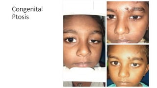
PTOSIS
DEFINITION
• Greek word means to “Fall”
• Abnormal drooping of upper eyelid is called ptosis
• Normally upper eyelid covers 1/6th of cornea 2mm
Types of Ptosis
• Congenital Ptosis
• Aquired ptosis
1. Neurogenic ptosis
2. Myogenic ptosis
3. Aponeurotic ptosis
4. Mechanical ptosis
Aponeurotic Ptosis
Congenital
Ptosis
Mechanical Ptosis
Simple congenital ptosis
• Developmental dystrophy of levator muscle
• occasionally Associated with weakness of SR
• Lid crease absent in congenital ptosis
LPS
• Orginates from lesser wing of sphenoid
• Fibres directed above SR
• Anterior fibres expand to form aponeurosis
• Fibres pass though orbicularis Oculi inserted in upper eyelid
• Some fibres gain attachment to lower half of the anterior surface of
tarsal plate
Aponeurotic fibres form medial and lateral horninserted into frontal
pocess of maxilla and latera orbital tubercle
Deep fibres and smooth muscle fibres attached to superior tarsal plate
Some fibres gain attachment to fornix of conjunctiva
3rd Nerve paralysis ====== COMPLETE PTOSIS
SYMPATHETIC PARALYSIS ==== PARTIAL PTOSIS
Marcus gunn jaw winking syndrome
• Accounts for 5% all congenital Ptosis
• Retraction of ptotic lid in conjunction with ipsilateral pterygoid
muscle
Examination of Ptosis
HISTORY
•Involvement of one eye after the other/both eyes simultaneously
•Precipitating factors – history of trauma, eye surgeries, previous eye
diseases such as dry eye/thyroid eye disease, pregnancy, delivery,
and medical conditions
•Associated conditions – Jaw winking, diplopia, dysphagia, tiredness
•Family history – Congenital or hereditary ptosis, ocular myopathies,
blepharophimosis, etc.
MEASUREMENTS
•Margin-reflex distance 1 (MRD1): The distance between the central
corneal light reflex and upper eyelid margin with eyes in primary
position. Normal MRD 1 is 4–5 mm
•Margin-reflex distance 2 (MRD2): The distance between the central
corneal light reflex and lower eyelid margin with eyes in primary
position
•Palpebral fissure height (PFH): It is the distance between the upper
and lower eyelid margins at the axis of the pupil. The sum of the
MRD1 and the MRD2 should equal the vertical PFH[5],[6]
•
•Levator function: Berke's method estimated by
measuring the upper eyelid excursion, from downgaze
to upgaze with frontalis muscle function negated and
with the head positioned in the frontal or Frankfort
plane. The amount of lid elevation is recorded in
millimeters (mm) of levator function. The classification
of levator function:
•Poor: 0–4 mm lid elevation
•Fair: 5–11 mm lid elevation
•Good: 12–14 mm lid elevation
•Normal: >15 mm lid elevation.
•Iliff test: It is used to assess levator function in infants. Upper eyelid of
the child is everted as the child looks down. If the levator action is good,
lid reverts on its own.
•Margin crease distance: Upper eyelid crease position is the distance
from the upper eyelid crease to the eyelid margin. It is normally 7–8 mm
in males and 9–10 mm in females. High skin crease suggests
aponeurotic defect. The depth of skin crease is a guide to determine the
levator function in young children
•Margin limbal distance: Putterman's method-measurement of the
distance between the middle of upper eyelid margin to the 6 o'clock
limbus in extreme upgaze. Normal is about 9 mm
•Pretarsal show: It is an important aspect of finding out the symmetry
of eyelids. It is the distance between the lid margin and the skin fold
with the eyes in primary position.
TESTS
• Fatigue test: MRD1 should be measured first. Then the patient should
be asked to look up for 2 min after which the MRD 1 is to be
measured again. Worsening of ptosis is seen in myopathies,
myasthenia as well as senile aponeurotic ptosis
• Ice test: Glove containing ice pack is applied on the closed ptotic eye
for 2 min. If the lid elevates by 2 mm or more, it is suggestive of
myasthenia
• Tensilon test: In cases of suspected myasthenia, 2 mg of
edrophonium is injected slowly in 15–30 s. The needle is left in situ,
and the remaining 8 mg is injected slowly if no adverse reaction is
observed within 1 min. If myasthenia is the cause, ptosis improves
after the injection[1]
Phenylephrine test: Sympathomimetic agents,
such as phenylephrine or apraclonidine, can be
instilled under the eyelid to test the function of
Muller's muscle
Schirmer's test: To evaluate tear function
Tear breakup time in individuals suspected of
having dry eyes due to the potential risk of
incomplete eyelid closure and exposure
keratopathy following surgical correction
Corneal sensitivity should be tested in all cases.
Treatment
• Fasanella–Servat procedure
Upper border of the tarsus is excised with lower part of Muller's
muscle and the overlying conjunctiva. Indications are mild congenital
or acquired ptosis with good levator function, Horner's syndrome and
minor contour adjustment after any ptosis surgery.[2]
• Muller's muscle-conjunctival resection
This involves the excision of Muller's muscle and overlying
conjunctiva with reattachment of resected edges. This procedure can
achieve a maximal elevation of 2–3 mm.
• Levator advancement
• Levator resection
• Frontalis sling
In a nutshell
• Mild ptosis with good LPS action === Fasanella–Servat procedure
• Ptosis with Moderate LPS action==== LPS RESECTION
• Severe Ptosis with severe LPS dysfunction=== Froontalis sling

Ptosis.pptx

  • 1.
  • 2.
    DEFINITION • Greek wordmeans to “Fall” • Abnormal drooping of upper eyelid is called ptosis • Normally upper eyelid covers 1/6th of cornea 2mm
  • 3.
    Types of Ptosis •Congenital Ptosis • Aquired ptosis 1. Neurogenic ptosis 2. Myogenic ptosis 3. Aponeurotic ptosis 4. Mechanical ptosis
  • 4.
  • 5.
  • 6.
  • 7.
    Simple congenital ptosis •Developmental dystrophy of levator muscle • occasionally Associated with weakness of SR • Lid crease absent in congenital ptosis
  • 8.
    LPS • Orginates fromlesser wing of sphenoid • Fibres directed above SR • Anterior fibres expand to form aponeurosis • Fibres pass though orbicularis Oculi inserted in upper eyelid • Some fibres gain attachment to lower half of the anterior surface of tarsal plate
  • 9.
    Aponeurotic fibres formmedial and lateral horninserted into frontal pocess of maxilla and latera orbital tubercle Deep fibres and smooth muscle fibres attached to superior tarsal plate Some fibres gain attachment to fornix of conjunctiva 3rd Nerve paralysis ====== COMPLETE PTOSIS SYMPATHETIC PARALYSIS ==== PARTIAL PTOSIS
  • 12.
    Marcus gunn jawwinking syndrome • Accounts for 5% all congenital Ptosis • Retraction of ptotic lid in conjunction with ipsilateral pterygoid muscle
  • 16.
    Examination of Ptosis HISTORY •Involvementof one eye after the other/both eyes simultaneously •Precipitating factors – history of trauma, eye surgeries, previous eye diseases such as dry eye/thyroid eye disease, pregnancy, delivery, and medical conditions •Associated conditions – Jaw winking, diplopia, dysphagia, tiredness •Family history – Congenital or hereditary ptosis, ocular myopathies, blepharophimosis, etc.
  • 17.
    MEASUREMENTS •Margin-reflex distance 1(MRD1): The distance between the central corneal light reflex and upper eyelid margin with eyes in primary position. Normal MRD 1 is 4–5 mm •Margin-reflex distance 2 (MRD2): The distance between the central corneal light reflex and lower eyelid margin with eyes in primary position •Palpebral fissure height (PFH): It is the distance between the upper and lower eyelid margins at the axis of the pupil. The sum of the MRD1 and the MRD2 should equal the vertical PFH[5],[6] •
  • 18.
    •Levator function: Berke'smethod estimated by measuring the upper eyelid excursion, from downgaze to upgaze with frontalis muscle function negated and with the head positioned in the frontal or Frankfort plane. The amount of lid elevation is recorded in millimeters (mm) of levator function. The classification of levator function: •Poor: 0–4 mm lid elevation •Fair: 5–11 mm lid elevation •Good: 12–14 mm lid elevation •Normal: >15 mm lid elevation.
  • 19.
    •Iliff test: Itis used to assess levator function in infants. Upper eyelid of the child is everted as the child looks down. If the levator action is good, lid reverts on its own. •Margin crease distance: Upper eyelid crease position is the distance from the upper eyelid crease to the eyelid margin. It is normally 7–8 mm in males and 9–10 mm in females. High skin crease suggests aponeurotic defect. The depth of skin crease is a guide to determine the levator function in young children •Margin limbal distance: Putterman's method-measurement of the distance between the middle of upper eyelid margin to the 6 o'clock limbus in extreme upgaze. Normal is about 9 mm •Pretarsal show: It is an important aspect of finding out the symmetry of eyelids. It is the distance between the lid margin and the skin fold with the eyes in primary position.
  • 20.
    TESTS • Fatigue test:MRD1 should be measured first. Then the patient should be asked to look up for 2 min after which the MRD 1 is to be measured again. Worsening of ptosis is seen in myopathies, myasthenia as well as senile aponeurotic ptosis • Ice test: Glove containing ice pack is applied on the closed ptotic eye for 2 min. If the lid elevates by 2 mm or more, it is suggestive of myasthenia • Tensilon test: In cases of suspected myasthenia, 2 mg of edrophonium is injected slowly in 15–30 s. The needle is left in situ, and the remaining 8 mg is injected slowly if no adverse reaction is observed within 1 min. If myasthenia is the cause, ptosis improves after the injection[1]
  • 21.
    Phenylephrine test: Sympathomimeticagents, such as phenylephrine or apraclonidine, can be instilled under the eyelid to test the function of Muller's muscle Schirmer's test: To evaluate tear function Tear breakup time in individuals suspected of having dry eyes due to the potential risk of incomplete eyelid closure and exposure keratopathy following surgical correction Corneal sensitivity should be tested in all cases.
  • 22.
    Treatment • Fasanella–Servat procedure Upperborder of the tarsus is excised with lower part of Muller's muscle and the overlying conjunctiva. Indications are mild congenital or acquired ptosis with good levator function, Horner's syndrome and minor contour adjustment after any ptosis surgery.[2]
  • 23.
    • Muller's muscle-conjunctivalresection This involves the excision of Muller's muscle and overlying conjunctiva with reattachment of resected edges. This procedure can achieve a maximal elevation of 2–3 mm. • Levator advancement • Levator resection • Frontalis sling
  • 24.
    In a nutshell •Mild ptosis with good LPS action === Fasanella–Servat procedure • Ptosis with Moderate LPS action==== LPS RESECTION • Severe Ptosis with severe LPS dysfunction=== Froontalis sling