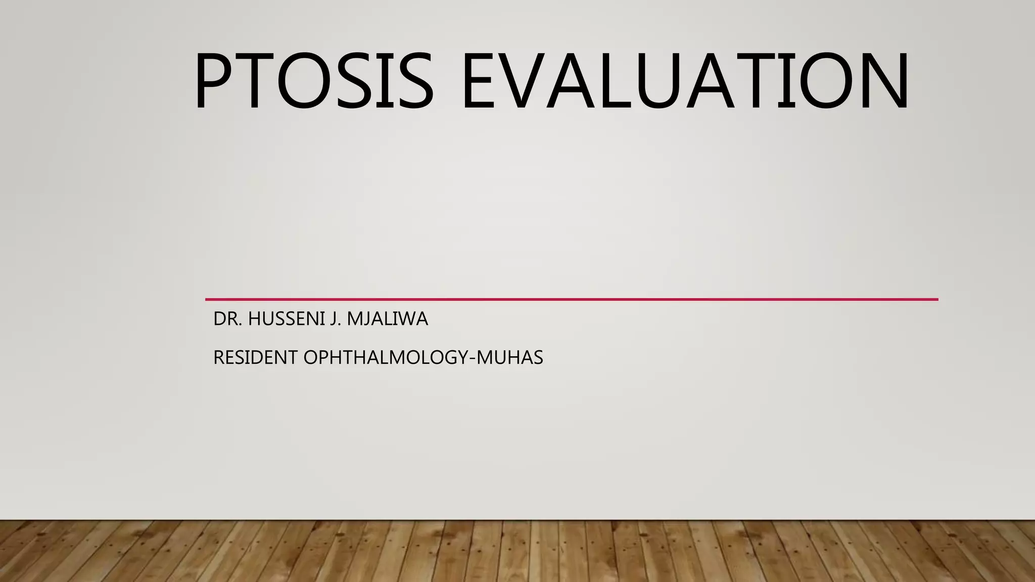
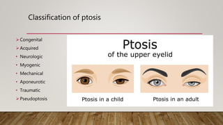
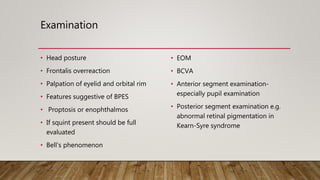
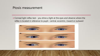
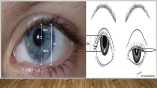
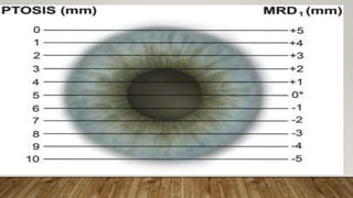
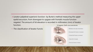
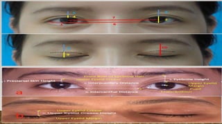
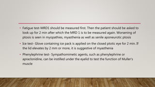
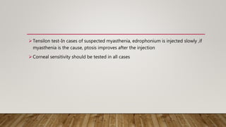
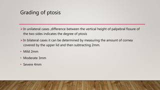
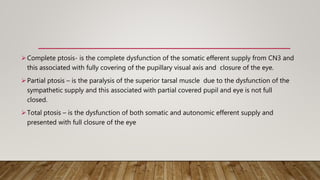
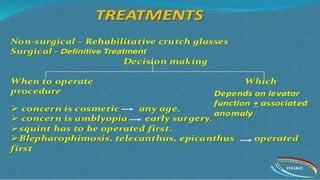
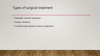
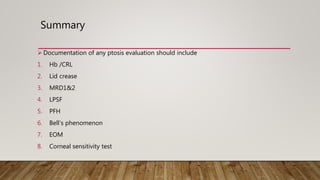
PTOSIS EVALUATION
DR. HUSSENI J. MJALIWA
RESIDENT OPHTHALMOLOGY-MUHAS
Layout
• Introduction
• Classification
• History taking
• Examination and measurement
• Grading of ptosis
• Treatment
• Reference
INTRODUCTION
• Ptosis – is the abnormal drooping of the upper eye lid in a primary position of
gaze
• Normally upper eyelid covers 1/6th of the cornea ,its about 1.5- 2mm
• In ptosis it covers more than 2mm
• Before counting to any thing you have to role out pseudoptosis on inspection
Pseudoptosis -Microphthalmia ,Phthisis bulbs, Enophthalmos, dermatochalasis ,
eyelid retraction
Classification of ptosis
Congenital
Acquired
• Neurologic
• Myogenic
• Mechanical
• Aponeurotic
• Traumatic
Pseudoptosis
Congenital
• Simple congenital ptosis- not associated with a any anomaly, congenital
associated with weakness of levator palpebral superior muscle(maldevelopment)
• Blepharophimosis syndrome – congenital
ptosis+blephrophimosis+telecanthus+epicathus inversus.
• Congenital synkinetic ptosis – Marcus Gunn jaw phenomenon,(condition of
misdirection of 3rd nerve)
Acquired
 Neurogenic
• 3rd nerve palsy
• 3rd nerve misdirection
• Horner’s syndrome
 Myogenic
• Myasthenia gravis
• Ocular myopathies
• Myotonic dystrophy
 Mechanical
• Excessive weight to upper lid –tumor, excess skin,
multiple chalazion, lid edema
 Traumatic
• Trauma or post surgery
 Aponeurotic
LPS function is good, but aponeurosis is stretched
or thinning due to repetitive stress and effect
gravity and aging causes drooping of lid
History taking
• Onset and duration of ptosis
• Variability
• Unilateral/bilateral
• Severity of ptosis - Whether the activities of daily living are affected
• Precipitating factors – history of trauma, eye surgeries, previous eye diseases such as dry
eye/thyroid eye disease, pregnancy, delivery, and medical conditions
• Associated conditions – Jaw winking, diplopia, dysphagia, tiredness
• Family history – Congenital or hereditary ptosis, ocular myopathies, blepharophimosis
Examination
• Head posture
• Frontalis overreaction
• Palpation of eyelid and orbital rim
• Features suggestive of BPES
• Proptosis or enophthalmos
• If squint present should be full
evaluated
• Bell’s phenomenon
• EOM
• BCVA
• Anterior segment examination-
especially pupil examination
• Posterior segment examination e.g.
abnormal retinal pigmentation in
Kearn-Syre syndrome
Ptosis measurement
Corneal light reflex test- you shine a light at the eyes and observe where the
reflex is located in reference to pupil.- central, eccentric, inward or outward
Marginal reflex distance 1- distance between central corneal reflex and upper lid
margin with primary position
• Normal is 4-5mm
Marginal reflex distance 2 – distance between the central corneal reflex to margin
of lower lid with eye in primary position
• Normal 5-5.5mm
Marginal reflex distance 3- distance from ocular light reflex to central of upper lid
margin when patient looks in extreme upward gaze
PFH- distance between the upper lid margin and lower lid margin at the pupil axis
• PFH=MRD1+MRD2
Levator palpebral superioris function- by Burke's method measuring the upper
eyelid excursion, from downgaze to upgaze with frontalis muscle function
negated. The amount of lid elevation is recorded in millimeters (mm) of levator
function.
• The classification of levator function:
• Iliff test- It is used to assess levator function in infants. Upper eyelid of the child is
everted as the child looks down. If the levator action is good, lid reverts on its
own
Margin crease distance- Upper eyelid crease position is the distance from the
upper eyelid crease to the eyelid margin in downward gaze.
• It is normally 7–8 mm in males and 9–10 mm in females.
pretarsal show- It is an important aspect of finding out the symmetry of eyelids. It
is the distance between the lid margin and the skin fold with the eyes in primary
position.
• Fatigue test-MRD1 should be measured first. Then the patient should be asked to
look up for 2 min after which the MRD 1 is to be measured again. Worsening of
ptosis is seen in myopathies, myasthenia as well as senile aponeurotic ptosis
• Ice test- Glove containing ice pack is applied on the closed ptotic eye for 2 min. If
the lid elevates by 2 mm or more, it is suggestive of myasthenia
• Phenylephrine test- Sympathomimetic agents, such as phenylephrine or
apraclonidine, can be instilled under the eyelid to test the function of Muller's
muscle
Tensilon test-In cases of suspected myasthenia, edrophonium is injected slowly ,if
myasthenia is the cause, ptosis improves after the injection
Corneal sensitivity should be tested in all cases
Grading of ptosis
In unilateral cases ,difference between the vertical height of palpebral fissure of
the two sides indicates the degree of ptosis
In bilateral cases it can be determined by measuring the amount of cornea
covered by the upper lid and then subtracting 2mm.
• Mild 2mm
• Moderate 3mm
• Severe 4mm
Complete ptosis- is the complete dysfunction of the somatic efferent supply from CN3 and
this associated with fully covering of the pupillary visual axis and closure of the eye.
Partial ptosis – is the paralysis of the superior tarsal muscle due to the dysfunction of the
sympathetic supply and this associated with partial covered pupil and eye is not full
closed.
Total ptosis – is the dysfunction of both somatic and autonomic efferent supply and
presented with full closure of the eye
Types of surgical treatment
• Fasanella –servant operation
• Levator resection
• Frontalis sling operation /brow suspension
Summary
 Documentation of any ptosis evaluation should include
1. Hb /CRL
2. Lid crease
3. MRD1&2
4. LPSF
5. PFH
6. Bell’s phenomenon
7. EOM
8. Corneal sensitivity test
References
• KANSKI’S clinical Ophthalmology Systametic Aproach 9th ED
• AAO- Acquired Ptosis: Evaluation and Management- By Carrie L. Morris, MD, and
David A
• Pauly M, Sruthi R. Ptosis: Evaluation and management. Kerala J Ophthalmol
2019;31:11-6.
• Internet

Ptosis Evaluation.pptx

  • 1.
    PTOSIS EVALUATION DR. HUSSENIJ. MJALIWA RESIDENT OPHTHALMOLOGY-MUHAS
  • 2.
    Layout • Introduction • Classification •History taking • Examination and measurement • Grading of ptosis • Treatment • Reference
  • 3.
    INTRODUCTION • Ptosis –is the abnormal drooping of the upper eye lid in a primary position of gaze • Normally upper eyelid covers 1/6th of the cornea ,its about 1.5- 2mm • In ptosis it covers more than 2mm
  • 4.
    • Before countingto any thing you have to role out pseudoptosis on inspection Pseudoptosis -Microphthalmia ,Phthisis bulbs, Enophthalmos, dermatochalasis , eyelid retraction
  • 6.
    Classification of ptosis Congenital Acquired •Neurologic • Myogenic • Mechanical • Aponeurotic • Traumatic Pseudoptosis
  • 7.
    Congenital • Simple congenitalptosis- not associated with a any anomaly, congenital associated with weakness of levator palpebral superior muscle(maldevelopment) • Blepharophimosis syndrome – congenital ptosis+blephrophimosis+telecanthus+epicathus inversus. • Congenital synkinetic ptosis – Marcus Gunn jaw phenomenon,(condition of misdirection of 3rd nerve)
  • 8.
    Acquired  Neurogenic • 3rdnerve palsy • 3rd nerve misdirection • Horner’s syndrome  Myogenic • Myasthenia gravis • Ocular myopathies • Myotonic dystrophy  Mechanical • Excessive weight to upper lid –tumor, excess skin, multiple chalazion, lid edema  Traumatic • Trauma or post surgery  Aponeurotic LPS function is good, but aponeurosis is stretched or thinning due to repetitive stress and effect gravity and aging causes drooping of lid
  • 9.
    History taking • Onsetand duration of ptosis • Variability • Unilateral/bilateral • Severity of ptosis - Whether the activities of daily living are affected • Precipitating factors – history of trauma, eye surgeries, previous eye diseases such as dry eye/thyroid eye disease, pregnancy, delivery, and medical conditions • Associated conditions – Jaw winking, diplopia, dysphagia, tiredness • Family history – Congenital or hereditary ptosis, ocular myopathies, blepharophimosis
  • 10.
    Examination • Head posture •Frontalis overreaction • Palpation of eyelid and orbital rim • Features suggestive of BPES • Proptosis or enophthalmos • If squint present should be full evaluated • Bell’s phenomenon • EOM • BCVA • Anterior segment examination- especially pupil examination • Posterior segment examination e.g. abnormal retinal pigmentation in Kearn-Syre syndrome
  • 11.
    Ptosis measurement Corneal lightreflex test- you shine a light at the eyes and observe where the reflex is located in reference to pupil.- central, eccentric, inward or outward
  • 12.
    Marginal reflex distance1- distance between central corneal reflex and upper lid margin with primary position • Normal is 4-5mm Marginal reflex distance 2 – distance between the central corneal reflex to margin of lower lid with eye in primary position • Normal 5-5.5mm Marginal reflex distance 3- distance from ocular light reflex to central of upper lid margin when patient looks in extreme upward gaze
  • 13.
    PFH- distance betweenthe upper lid margin and lower lid margin at the pupil axis • PFH=MRD1+MRD2
  • 16.
    Levator palpebral superiorisfunction- by Burke's method measuring the upper eyelid excursion, from downgaze to upgaze with frontalis muscle function negated. The amount of lid elevation is recorded in millimeters (mm) of levator function. • The classification of levator function:
  • 17.
    • Iliff test-It is used to assess levator function in infants. Upper eyelid of the child is everted as the child looks down. If the levator action is good, lid reverts on its own
  • 18.
    Margin crease distance-Upper eyelid crease position is the distance from the upper eyelid crease to the eyelid margin in downward gaze. • It is normally 7–8 mm in males and 9–10 mm in females. pretarsal show- It is an important aspect of finding out the symmetry of eyelids. It is the distance between the lid margin and the skin fold with the eyes in primary position.
  • 20.
    • Fatigue test-MRD1should be measured first. Then the patient should be asked to look up for 2 min after which the MRD 1 is to be measured again. Worsening of ptosis is seen in myopathies, myasthenia as well as senile aponeurotic ptosis • Ice test- Glove containing ice pack is applied on the closed ptotic eye for 2 min. If the lid elevates by 2 mm or more, it is suggestive of myasthenia • Phenylephrine test- Sympathomimetic agents, such as phenylephrine or apraclonidine, can be instilled under the eyelid to test the function of Muller's muscle
  • 21.
    Tensilon test-In casesof suspected myasthenia, edrophonium is injected slowly ,if myasthenia is the cause, ptosis improves after the injection Corneal sensitivity should be tested in all cases
  • 22.
    Grading of ptosis Inunilateral cases ,difference between the vertical height of palpebral fissure of the two sides indicates the degree of ptosis In bilateral cases it can be determined by measuring the amount of cornea covered by the upper lid and then subtracting 2mm. • Mild 2mm • Moderate 3mm • Severe 4mm
  • 23.
    Complete ptosis- isthe complete dysfunction of the somatic efferent supply from CN3 and this associated with fully covering of the pupillary visual axis and closure of the eye. Partial ptosis – is the paralysis of the superior tarsal muscle due to the dysfunction of the sympathetic supply and this associated with partial covered pupil and eye is not full closed. Total ptosis – is the dysfunction of both somatic and autonomic efferent supply and presented with full closure of the eye
  • 25.
    Types of surgicaltreatment • Fasanella –servant operation • Levator resection • Frontalis sling operation /brow suspension
  • 26.
    Summary  Documentation ofany ptosis evaluation should include 1. Hb /CRL 2. Lid crease 3. MRD1&2 4. LPSF 5. PFH 6. Bell’s phenomenon 7. EOM 8. Corneal sensitivity test
  • 28.
    References • KANSKI’S clinicalOphthalmology Systametic Aproach 9th ED • AAO- Acquired Ptosis: Evaluation and Management- By Carrie L. Morris, MD, and David A • Pauly M, Sruthi R. Ptosis: Evaluation and management. Kerala J Ophthalmol 2019;31:11-6. • Internet

Editor's Notes

  • #8 Retraction of the upper eyelid with various ocular movements
  • #9 Horner’s syndrome includes partial ptosis,miosisand loss of hemifacial sweating(anhidrosis)+enopthalmos
  • #13 MRD2- useful in rervesal ptois or lowerlid retraction MRD3- Used to determine how much levator to be resected in patient with congenital ptosis who have a vertical strabismus associated with ptosis whom strabismus surgery is not indicated
  • #19 High skin crease suggests aponeurotic defect. The depth of skin crease is a guide to determine the levator function in young children
  • #20 Items measured in the upper eyelids. 1 Horizontal distance of palpebral fissure; 2 vertical distance of palpebral fissure; 3 height of the opened upper eyelid; 4, height of double fold; 5 height of double fold to lower margin of eyebrow; 6 intercanthal distance; 7 interpupillary distance; 8 height of eyelid crease to lower margin of eyebrow; 9 height of eyelid crease; 10 height of the closed upper eyelid
  • #22 The needle is left in situ, and the remaining 8 mg is injected slowly if no adverse reaction is observed within 1 min. Since 2018 no longer used in USA due to false positive result and other reliable test like serology antibody and repetitive stress test.