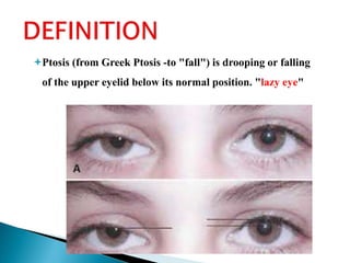
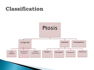
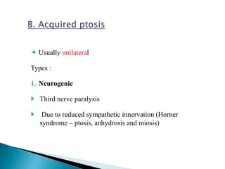
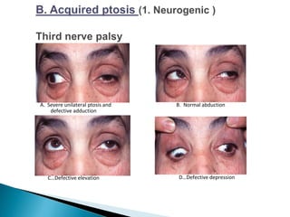
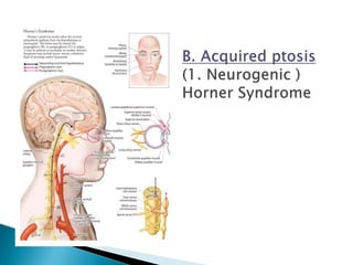
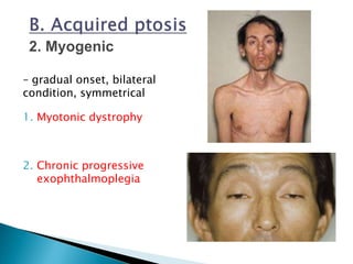
By Dr. Sulaiman
Oral And
Maxillofacial Surgery
Definition
Functional anatomy
Classification
Managment
Ptosis (from Greek Ptosis -to "fall") is drooping or falling
of the upper eyelid below its normal position. "lazy eye"
Levator Palpebrae Superioris
Primary muscle for lid elevation.
Supplied by the superior division of the oculomotor nerve.
Arises from the back of the orbit and extends forwards over the
cone of eye muscles.
Inserts into the eyelid and the tarsal plate, a fibrous semicircular
structure which gives the upper eyelid its shape.
Ptosis
Congenital
Simple
congenital
With abnormal
movements
With lid
abnormalities
Acquired
Myogenic Neurogenic Aponeurotic Mechanical
Pseudoptosis
1. Simple congenital ptosis
 not associated with any other anomaly.
2. Congenital ptosis with associated weakness of
superior rectus muscle.
3.Blephrophimosis
◦ which comprises congenital
ptosis, blepharophimosis,
telecanthus and epicanthus
inversus .
Clincal Features
 Dominent inheritance
 Moderate to severe symmetrical ptosis
 Short horizontal palpebral aperture
 Telecanthus (lateral displacement of medial canthus)
 Epicanthus inversus (lower lid fold larger than upper)
 Poorly developed nasal bridge
and hypoplasia of superior orbital
rims
4. Congenital synkinetic ptosis
(Marcus Gunn jaw winking ptosis).
In this condition there occurs retraction of the
ptotic lid with jaw movements i.e., with
stimulation of ipsilateral pterygoid muscle.
 Usually unilateral
Types :
1. Neurogenic
 Third nerve paralysis
 Due to reduced sympathetic innervation (Horner
syndrome – ptosis, anhydrosis and miosis)
C…Defective elevation D…Defective depression
B. Normal abductionA. Severe unilateral ptosis and
defective adduction
– gradual onset, bilateral
condition, symmetrical
1. Myotonic dystrophy
2. Chronic progressive
exophthalmoplegia
3. Mysthenia Gravis
Signs – bilateral ptosis, increases by prolonged
fixation or attempt to look up , external
ophthalmoplegia – partial or complete
Conformation by edrophonium injection test
1. Post Operative ptosis due to disinsertion of
Levator Palpebrae Superioris aponeurosis
from ant surface of tarsal plate.
2. Posttraumatic Dehiscence or Disinsertion
3. Involutional (snile) ptosis due to weakness.
Clincal sign
High fold with good Levator palpebrae superioris
function.
High upper lid crease Good levator function
Deep sulcusAbsent upper lid crease
Mild
Severe
Ptosis is caused by the gravitational effect
of a mass or by scarring
Tumour or inflammation weight down the lid
Dermatochalasis Large tumours
Anterior orbital lesionsSevere lid oedema
5. Snake Venom
(Black Mumba)
6. Drugs
1. Opioid
(morphine, oxycodone,
heroin)
2. anticonvulsant
(Pregabalin)
False impression of ptosis may be caused
by the following
1. Lack of support of the lids by the globe
may be due to an orbital volume deficit
associated with an artificial eye
1. Diagnosis is based on
 history
 examination,
 eye measurements,
 clinical and laboratory tests and
 imaging studies
2. Treatment
1. Age of onset
2. Duration
3. One/both eye
4. Associated history
5. Diplopia
6. Dysphagia
7. Muscle weakness
8. Vision
9. Jaw movements
10.Abnormal ocular
movements
11.Abnormal head
posture
12.Previous history
13.Trauma
14.Poisoning
15.Bleeding tendency
16.Previous photographs
17.Family history
 NORMAL POSITION OF EYELID
Usually 1-2mm below the limbus
 Vertical Palpebral fissures are normally 9-12mm.
Check for
1. Unilateral or bilateral , Complete or incomplete
2. Ocular motility
3. Visual acuity
4. Pupillary examination
5. Chin elevation
1.Vertical fissure height
2.Margin reflex distance
3.Levator palpebrae Superioris action
4.Lid crease level
5.Lid level on down gaze
 Normal – 9-
10mm in
primary gaze
 Seen in up gaze,
down gaze and
primary gaze
 Amount of
ptosis =
difference in
palpebral
apertures in
unilateral ptosis
or Difference
from normal in
bilateral ptosis
MRD 2
 The distance from the central pupillary light reflex to the lower
eyelid margin with the eye in primary gaze.
 The MRD1 plus the MRD2 should equal the palpebral fissure
measurement
Lid excursion is a measure of the levator function. The
frontalis action is blocked by keeping the thumb tightly
over the upper brow and asking the patient to look up
from down gaze and measuring the amount of upper
lid excursion at the center of the lid.
 Is the distance from the
crease to lid margin
 Normal – 8 to 10mm in
primary gaze
 An absent lid crease is often
accompanied by poor levator
function.
 If a lid crease is present but
is higher than normal and if
a deeper upper lid sulcus is
found on that side, note
these as signs of a levator
aponeurosis disinsertion.
Patients with Minimal
ptosis (2 mm or less)
 2.5 or 10% phenylephrine
is instilled in the affected
eye or eyes
The patient is reexamined 5
minutes later.
 The MRD1 is rechecked in
the affected and unaffected
eyes .
 A rise in the MRDl of 1.5
mm or greater is
considered a positive
test. This indicates that
Müller's muscle is viable
Non Surgical
Surgical
Herrings Law
Muscles that elevate the eyelids get the same
innervation
 The lid with minimal ptosis droops more following
the correction of greater ptotic side
 Important for prediction of post op results
A.Non Surgical
1.Lid crutches
2. Haptic contact lens
3. lid elevation by mechanical force
Surgical management depends upon the type of ptosis
and levator function
Indications
 Complete ptosis
 Chin up positioning
 Severe ptosis causing amblyopia
Contraindications
 Poor orbicularis muscle function
 Loss of blink reflex
 Corneal sensitivity
 Keratitis sicca
Mild ptosis
Phenylephrine +ve
Mullerectomy
Phenylephrine - ve
Fasanella servat
Blepharoplasty
Levator resection
Ptosis
Moderate
Levator resection Levator
advancement
Severe
Frontalis sling
1. Asymmetry
2. Corneal abrasion or Keratitis
3. Entropion or Ectropion
4. Eyelashes loss
5. Wound Dehiscence
6. Unfavorable scar
Ptosis

Ptosis

  • 1.
    By Dr. Sulaiman OralAnd Maxillofacial Surgery
  • 2.
  • 3.
    Ptosis (from GreekPtosis -to "fall") is drooping or falling of the upper eyelid below its normal position. "lazy eye"
  • 4.
    Levator Palpebrae Superioris Primarymuscle for lid elevation. Supplied by the superior division of the oculomotor nerve. Arises from the back of the orbit and extends forwards over the cone of eye muscles. Inserts into the eyelid and the tarsal plate, a fibrous semicircular structure which gives the upper eyelid its shape.
  • 7.
  • 8.
    1. Simple congenitalptosis  not associated with any other anomaly. 2. Congenital ptosis with associated weakness of superior rectus muscle.
  • 9.
    3.Blephrophimosis ◦ which comprisescongenital ptosis, blepharophimosis, telecanthus and epicanthus inversus .
  • 10.
    Clincal Features  Dominentinheritance  Moderate to severe symmetrical ptosis  Short horizontal palpebral aperture  Telecanthus (lateral displacement of medial canthus)  Epicanthus inversus (lower lid fold larger than upper)  Poorly developed nasal bridge and hypoplasia of superior orbital rims
  • 11.
    4. Congenital synkineticptosis (Marcus Gunn jaw winking ptosis). In this condition there occurs retraction of the ptotic lid with jaw movements i.e., with stimulation of ipsilateral pterygoid muscle.
  • 12.
     Usually unilateral Types: 1. Neurogenic  Third nerve paralysis  Due to reduced sympathetic innervation (Horner syndrome – ptosis, anhydrosis and miosis)
  • 13.
    C…Defective elevation D…Defectivedepression B. Normal abductionA. Severe unilateral ptosis and defective adduction
  • 15.
    – gradual onset,bilateral condition, symmetrical 1. Myotonic dystrophy 2. Chronic progressive exophthalmoplegia
  • 16.
    3. Mysthenia Gravis Signs– bilateral ptosis, increases by prolonged fixation or attempt to look up , external ophthalmoplegia – partial or complete Conformation by edrophonium injection test
  • 17.
    1. Post Operativeptosis due to disinsertion of Levator Palpebrae Superioris aponeurosis from ant surface of tarsal plate. 2. Posttraumatic Dehiscence or Disinsertion 3. Involutional (snile) ptosis due to weakness. Clincal sign High fold with good Levator palpebrae superioris function.
  • 18.
    High upper lidcrease Good levator function Deep sulcusAbsent upper lid crease Mild Severe
  • 19.
    Ptosis is causedby the gravitational effect of a mass or by scarring Tumour or inflammation weight down the lid
  • 20.
    Dermatochalasis Large tumours Anteriororbital lesionsSevere lid oedema
  • 21.
    5. Snake Venom (BlackMumba) 6. Drugs 1. Opioid (morphine, oxycodone, heroin) 2. anticonvulsant (Pregabalin)
  • 22.
    False impression ofptosis may be caused by the following 1. Lack of support of the lids by the globe may be due to an orbital volume deficit associated with an artificial eye
  • 27.
    1. Diagnosis isbased on  history  examination,  eye measurements,  clinical and laboratory tests and  imaging studies 2. Treatment
  • 28.
    1. Age ofonset 2. Duration 3. One/both eye 4. Associated history 5. Diplopia 6. Dysphagia 7. Muscle weakness 8. Vision 9. Jaw movements 10.Abnormal ocular movements 11.Abnormal head posture 12.Previous history 13.Trauma 14.Poisoning 15.Bleeding tendency 16.Previous photographs 17.Family history
  • 29.
     NORMAL POSITIONOF EYELID Usually 1-2mm below the limbus  Vertical Palpebral fissures are normally 9-12mm. Check for 1. Unilateral or bilateral , Complete or incomplete 2. Ocular motility 3. Visual acuity 4. Pupillary examination 5. Chin elevation
  • 30.
    1.Vertical fissure height 2.Marginreflex distance 3.Levator palpebrae Superioris action 4.Lid crease level 5.Lid level on down gaze
  • 31.
     Normal –9- 10mm in primary gaze  Seen in up gaze, down gaze and primary gaze  Amount of ptosis = difference in palpebral apertures in unilateral ptosis or Difference from normal in bilateral ptosis
  • 34.
    MRD 2  Thedistance from the central pupillary light reflex to the lower eyelid margin with the eye in primary gaze.  The MRD1 plus the MRD2 should equal the palpebral fissure measurement
  • 35.
    Lid excursion isa measure of the levator function. The frontalis action is blocked by keeping the thumb tightly over the upper brow and asking the patient to look up from down gaze and measuring the amount of upper lid excursion at the center of the lid.
  • 37.
     Is thedistance from the crease to lid margin  Normal – 8 to 10mm in primary gaze  An absent lid crease is often accompanied by poor levator function.  If a lid crease is present but is higher than normal and if a deeper upper lid sulcus is found on that side, note these as signs of a levator aponeurosis disinsertion.
  • 38.
    Patients with Minimal ptosis(2 mm or less)  2.5 or 10% phenylephrine is instilled in the affected eye or eyes The patient is reexamined 5 minutes later.  The MRD1 is rechecked in the affected and unaffected eyes .  A rise in the MRDl of 1.5 mm or greater is considered a positive test. This indicates that Müller's muscle is viable
  • 39.
    Non Surgical Surgical Herrings Law Musclesthat elevate the eyelids get the same innervation  The lid with minimal ptosis droops more following the correction of greater ptotic side  Important for prediction of post op results
  • 40.
  • 41.
  • 42.
    3. lid elevationby mechanical force
  • 43.
    Surgical management dependsupon the type of ptosis and levator function Indications  Complete ptosis  Chin up positioning  Severe ptosis causing amblyopia Contraindications  Poor orbicularis muscle function  Loss of blink reflex  Corneal sensitivity  Keratitis sicca
  • 45.
    Mild ptosis Phenylephrine +ve Mullerectomy Phenylephrine- ve Fasanella servat Blepharoplasty Levator resection
  • 50.
  • 55.
  • 56.
    2. Corneal abrasionor Keratitis
  • 57.
    3. Entropion orEctropion
  • 58.
  • 59.
  • 60.