Download as PDF, PPTX


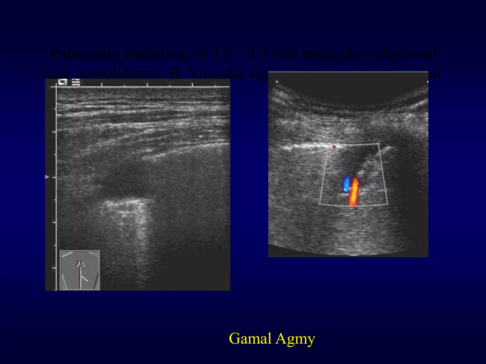


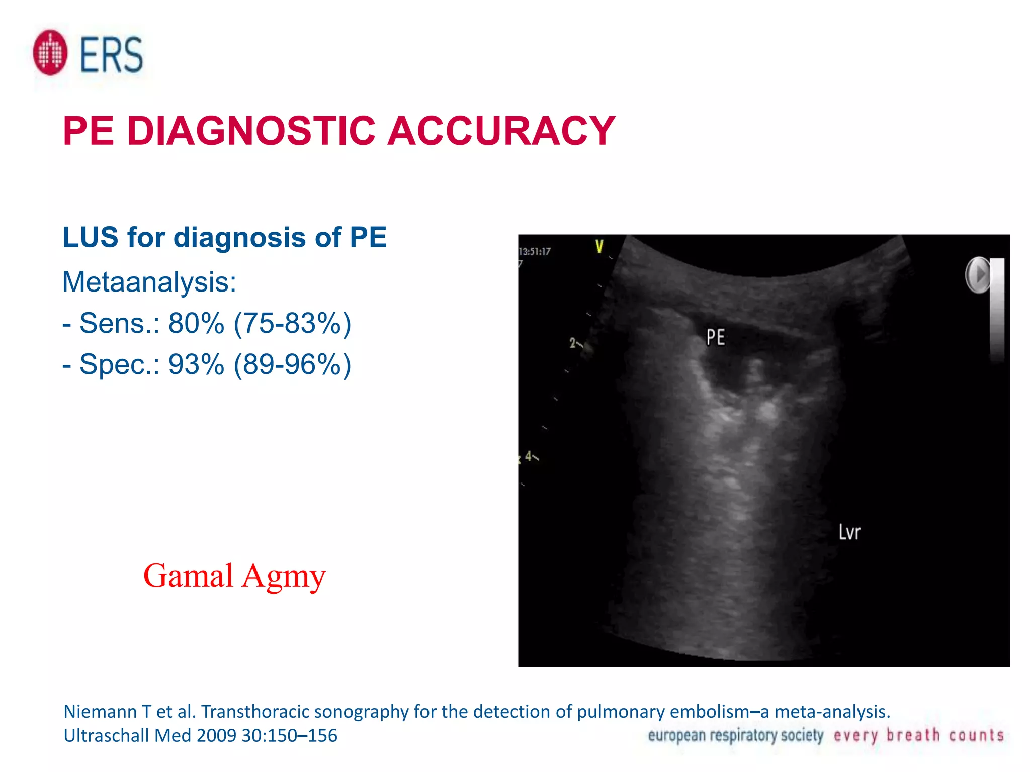


The document discusses pulmonary embolism (PE) and its diagnosis using lung ultrasound (LUS). It provides details on the diagnostic criteria for PE using LUS, including findings of typical lung lesions that indicate confirmed or probable PE. It also summarizes the sensitivity and specificity reported in studies on using LUS to diagnose PE. The document further discusses non-thrombotic causes of pulmonary embolism and the ultrasound appearance of various lung pathologies.


![Apporach to lung biopsy [Auto-saved].pptx latest](https://cdn.slidesharecdn.com/ss_thumbnails/apporachtolungbiopsyauto-saved-251211225655-93258539-thumbnail.jpg?width=640&height=640&fit=bounds)


