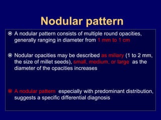
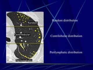
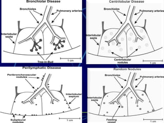
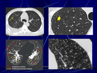
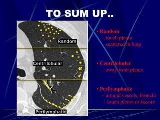
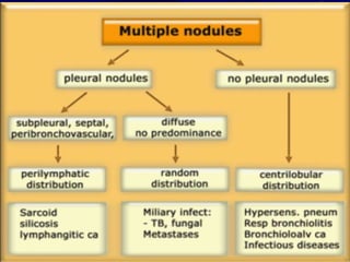
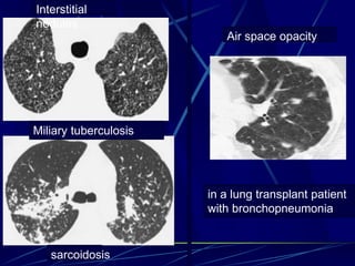
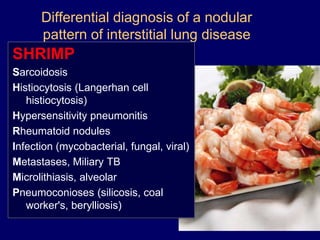
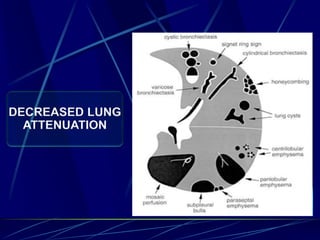
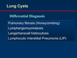
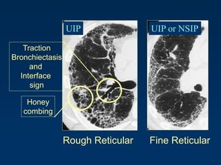
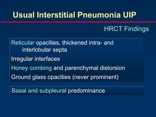
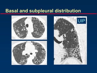
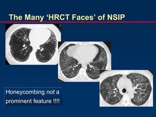
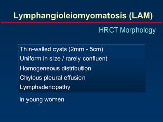
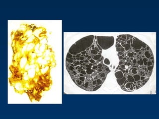
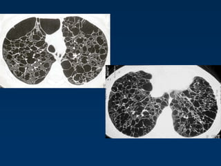
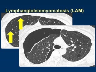
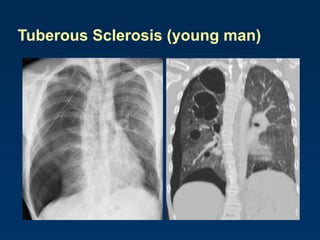
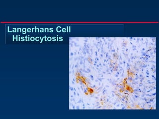
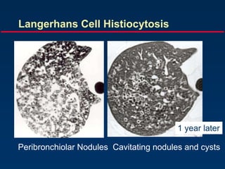
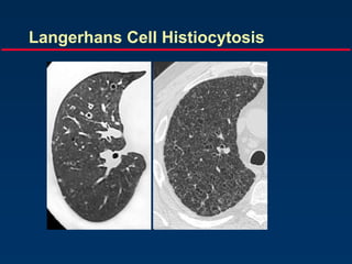
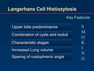
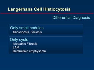
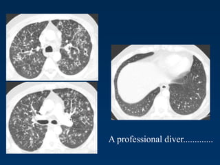
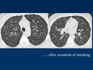
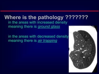
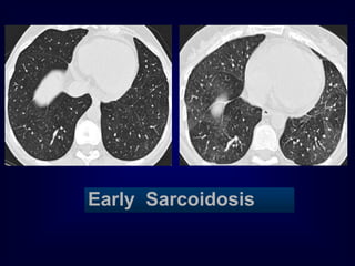
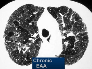
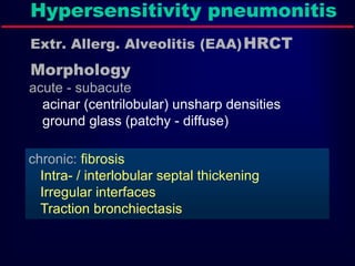
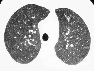
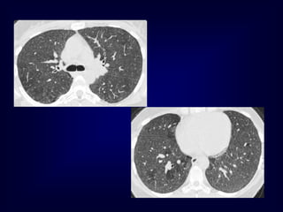
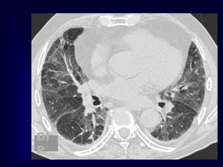
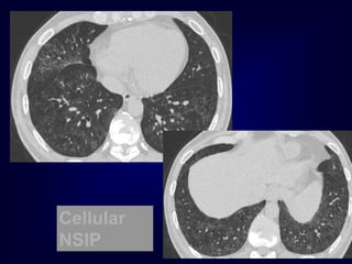
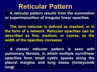
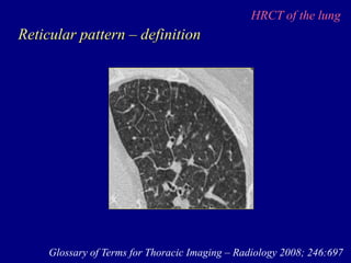
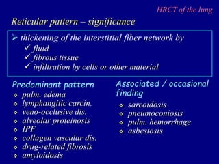
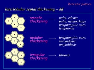
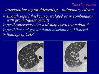
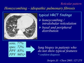
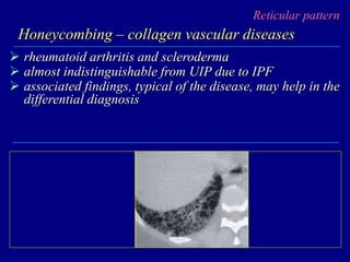
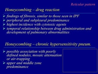
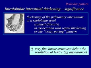
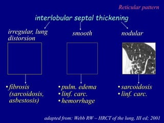
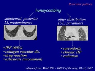
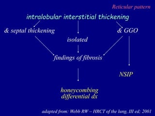
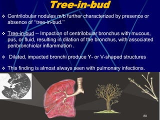
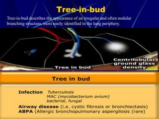
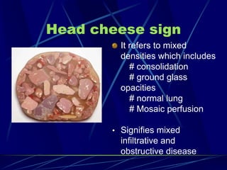
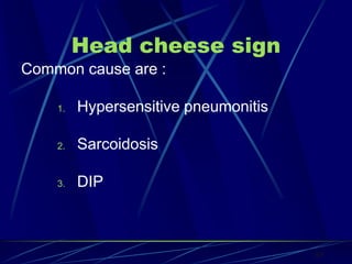
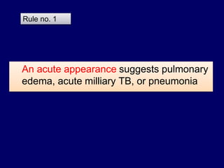
Snap Shots on HRCT
Patterns in Interstitial
Lung Diseases
Gamal Rabie Agmy, MD, FCCP
Professor of chest Diseases,
Assiut university
The interstitium of the lung is not normally visible radiographic-
ally; it becomes visible only when disease (e.g., edema,
fibrosis, tumor) increases its volume and attenuation.
The interstitial space is defined as continuum of loose
connective tissue throughout the lung composed of three
subdivisions:
(i) the bronchovascular (axial), surrounding the bronchi,
arteries, and veins from the lung root to the level of the
respiratory bronchiole
(ii) the parenchymal (acinar), situated between the alveolar
and capillary basement membranes
(iii) the subpleural, situated beneath the pleura, as well as in
the interlobular septae.
The Lung Interstitium
Secondary pulmonary lobular
anatomy
The terminal bronchiole in the center
divides into respiratory bronchioles with
acini that contain alveoli.
Lymphatics and veins run within the
interlobular septa
Centrilobular area in blue (left)
and perilymphatic area in yellow
(right)
Ideal ILD doctor
Radiologist
Pathologist
Pulmonologist
Nodular Patternitie
By
Gamal Rabie Agmy , MD , FCCP
Professor of Chest Diseases ,Assiut University
Nodular Pattern
Nodular pattern
 A nodular pattern consists of multiple round opacities,
generally ranging in diameter from 1 mm to 1 cm
 Nodular opacities may be described as miliary (1 to 2 mm,
the size of millet seeds), small, medium, or large, as the
diameter of the opacities increases
 A nodular pattern, especially with predominant distribution,
suggests a specific differential diagnosis
Perilymphatic distribution
Centrilobular distribution
Random distribution
ARE NODULES IN CONTACT
WITH PLEURA
NO
CENTRILOBULAR
YES
PERILYMPHATIC RANDOM
TO SUM UP..
• Random
– touch pleura
– scattered in lung
• Centrilobular
–away from pleura
• Perilymphatic
– around vessels, bronchi
– touch pleura or fissure
Interstitial
nodules
Air space opacity
Miliary tuberculosis
sarcoidosis
in a lung transplant patient
with bronchopneumonia
Differential diagnosis of a nodular
pattern of interstitial lung disease
SHRIMP
Sarcoidosis
Histiocytosis (Langerhan cell
histiocytosis)
Hypersensitivity pneumonitis
Rheumatoid nodules
Infection (mycobacterial, fungal, viral)
Metastases, Miliary TB
Microlithiasis, alveolar
Pneumoconioses (silicosis, coal
worker's, berylliosis)
Cystic Lung Lesions
By
Gamal Rabie Agmy , MD , FCCP
Professor of Chest Diseases ,Assiut University
DECREASED LUNG
ATTENUATION
Lung Cysts
Pulmonary fibrosis (Honeycombing)
Lymphangiomyomatosis
Langerhanscell histiocytosis
Lymphocytic Interstitial Pneumonia (LIP)
Differential Diagnosis
Rough Reticular Fine Reticular
Traction
Bronchiectasis
and
Interface
sign
Honey
combing
UIP UIP or NSIP
Usual Interstitial Pneumonia UIP
HRCT Findings
Reticular opacities, thickened intra- and
interlobular septa
Irregular interfaces
Honey combing and parenchymal distorsion
Ground glass opacities (never prominent)
Basal and subpleural predominance
Basal and subpleural distribution
UIP
The Many ‘HRCT Faces’ of NSIP
Honeycombing not a
prominent feature !!!!
Lymphangioleiomyomatosis (LAM)
HRCT Morphology
Thin-walled cysts (2mm - 5cm)
Uniform in size / rarely confluent
Homogeneous distribution
Chylous pleural effusion
Lymphadenopathy
in young women
Lymphangioleiomyomatosis (LAM)
Tuberous Sclerosis (young man)
Langerhans Cell
Histiocytosis
Langerhans Cell Histiocytosis
HRCT Findings
Small peribronchiolar nodules (1-5mm)
Thin-walled cysts (< 1cm),
Bizarre and confluent
Ground glass opacities
Late signs: irreversible / parenchymal fibrosis
Honey comb lung, septal thickening, bronchiectasis
1 year later
Peribronchiolar Nodules Cavitating nodules and cysts
Langerhans Cell Histiocytosis
Langerhans Cell Histiozytosis
Key Features
Upper lobe predominance
Combination of cysts and noduli
Characteristic stages
Increased Lung volume
Sparing of costophrenic angle
S
M
O
K
I
N
G
Langerhans Cell Histiocytosis
Differential Diagnosis
Only small nodules
Sarkoidosis, Silikosis
Only cysts
idiopathic Fibrosis
LAM
Destruktive emphysema
A professional diver.............
.......after cessation of smoking
Benign lymphoproliferative disorder
Diffuse interstitial infiltration of
mononuclear cells
Not limited to the air ways as
in follicular Bronchiolitis
LIP
= Lymphocytic Interstitial Pneumonia
Sjögren: LIP
LIP
= Lymphocytic Interstitial Pneumonia
Rarely idiopathic
In association with:
Sjögren’s syndrome
Immune deficiency syndromes, AIDS
Primary biliary cirrhosis
Multicentric Castlemean’s disease
Sjoegren disease
Dry eye and dry mouth
Fibrosis, bronchitis and bronchiolitis
LIP
Overlap
Sarcoid, DM/PM, MXCT
SLE, RA (pleural effusion)
Up to 40 x increased risk for lymphoma (mediastinal
adenopathy) and
2 x times increased risk for neoplasma
Mosiac pattern
Where is the pathology ???????
in the areas with increased density
meaning there is ground glass
in the areas with decreased density
meaning there is air trapping
Pathology in black areas
Airtrapping: Airway
Disease
Bronchiolitis obliterans (constrictive bronchiolitis)
idiopathic, connective tissue diseases, drug reaction,
after transplantation, after infection
Hypersensitivity pneumonitis
granulomatous inflammation of bronchiolar wall
Sarcoidosis
granulomatous inflammation of bronchiolar wall
Asthma / Bronchiectasis / Airway diseases
Airway Disease
what you see……
In inspiration
sharply demarcated areas of seemingly increased
density (normal) and decreased density
demarcation by interlobular septa
In expiration
‘black’ areas remain in volume and density
‘white’ areas decrease in volume and increase in
density
INCREASE IN CONTRAST
DIFFERENCES
AIRTRAPPING
Bronchiolitis
obliterans
Early Sarcoidosis
Chronic
EAA
Hypersensitivity pneumonitis
Extr. Allerg. Alveolitis (EAA)HRCT
Morphology
chronic: fibrosis
Intra- / interlobular septal thickening
Irregular interfaces
Traction bronchiectasis
acute - subacute
acinar (centrilobular) unsharp densities
ground glass (patchy - diffuse)
Pathology in white Areas
Alveolitis / Pneumonitis
Ground glass
desquamative intertitial pneumoinia (DIP)
nonspecific interstitial pneumonia (NSIP)
organizing pneumonia
In expiration
both areas (white and black) decrease in
volume and increase in density
DECREASE IN CONTRAST
DIFFERENCES
DI
P
Cellular
NSIP
Mosaic Perfusion
Chronic pulmonary embolism
LOOK FOR
Pulmonary hypertension
idiopathic, cardiac disease, pulmonary
disease
CTEPH =
Chronic thrombembolic
pulmonary hypertension
Reticular Pattern
Gamal Rabie Agmy, MD, FCCP
Professor of chest Diseases,
Assiut university
Reticular Pattern
A reticular pattern results from the summation
or superimposition of irregular linear opacities.
The term reticular is defined as meshed, or in
the form of a network. Reticular opacities can be
described as fine, medium, or coarse, as the
width of the opacities increases.
A classic reticular pattern is seen with
pulmonary fibrosis, in which multiple curvilinear
opacities form small cystic spaces along the
pleural margins and lung bases (honeycomb
lung)
HRCT of the lung
Reticular pattern – definition
Glossary of Terms for Thoracic Imaging – Radiology 2008; 246:697
HRCT of the lung
 thickening of the interstitial fiber network by
Reticular pattern – significance
 fluid
 fibrous tissue
 infiltration by cells or other material
 pulm. edema
 lymphangitic carcin.
 veno-occlusive dis.
 alveolar proteinosis
 IPF
 collagen vascular dis.
 drug-related fibrosis
 amyloidosis
Predominant pattern Associated / occasional
finding
 sarcoidosis
 pneumoconiosis
 pulm. hemorrhage
 asbestosis
HRCT of the lung
Reticular pattern – HRCT
numerous, clearly visible interlobular septa
outlining lobules of characteristic size and shape
interlobular septal thickening
very fine network of lines within visible lobules
intralobular interstitial thickening
several layers of air-filled cysts, 3-10 mm in diameter,
with thick walls (1-3 mm)
honeycombing
Reticular pattern
Interlobular septal thickening – dd
smooth
thickening
pulm. edema
pulm. hemorrhage
lymphangitic carc.
lymphoma
nodular
thickening
lymphangitic carc.
sarcoidosis
amyloidosis
irregular
thickening
fibrosis
Reticular pattern
Interlobular septal thickening – pulmonary edema
 smooth septal thickening, isolated or in combination
with ground-glass opacity
 peribronchovascular and subpleural interstitial th.
 perihilar and gravitational distribution, bilateral
 findings of CHF
Reticular pattern
Interlobular septal th. – lymphangitic carcinomatosis
 tumor filling of pulmonary vessels and lymphatics
 direct tumor infiltration of the interstitium
 vascular and lymphatic distension distally to tumor
emboli or obstruction
 breast ca.
 lung ca.
 stomach ca.
 pancreas ca.
Secondary to:
 prostate ca.
 adenoca. of
unknown origin
Reticular pattern
Interlobular septal th. – lymphangitic carcinomatosis
 smooth or nodular septal thickening
 smooth or nodular thickening of peribronchovascular
interstitium and fissures
 thickening of the intralobular axial interstitium
 focal or asymmetric distribution
Reticular pattern
Interlobular septal thickening – sarcoidosis
 reticulation is not a predominat finding
 distorsion of the lung architecture and secondary
lobule anatomy is common, especially when septal
thickening is present
 upper lobe predominance
Reticulation or not reticulation ……
“crazy paving”
Reticulation or not reticulation ……
alveolar proteinosis
Reticular pattern
Interlobular septal thickening – “crazy paving”
 scattered or diffuse ground-glass attenuation with
superimposed interlobular septal thickening and
intralobular lines
 described in a variety of infectious, neoplastic,
idiopathic, inhalation, and sanguineous disorders of
the lung
Rossi SE – Radiographics 2003; 23:1509
Reticular pattern
Honeycombing – significance
air-containing cystic spaces having thick,
fibrous walls lined by bronchiolar
epithelium

fibrosis is present
UIP is likely the histologic pattern
IPF is very likely, in the absence of a
known disease
Reticular pattern
Honeycombing – differential diagnosis
basal
distribution
middle/upper
distribution
chronic HP
sarcoidosis
IPF
collagen vasc. dis.
asbestosis
drugs
 honeycombing /
intralobular reticulation
 basal and peripheral
distribution
typical HRCT findings
sens. 77%
spec. 72%
PPV 85%
PPV 96%*
Swigris JJ – Chest 2005; 127:275
lung biopsy in patients who
do not show typical features
Reticular pattern
Honeycombing – idiopathic pulmonary fibrosis
* confident diagnosis
Reticular pattern
Honeycombing – collagen vascular diseases
 rheumatoid arthritis and scleroderma
 almost indistinguishable from UIP due to IPF
 associated findings, typical of the disease, may help in the
differential diagnosis
Reticular pattern
Honeycombing – drug reaction
 findings of fibrosis, similar to those seen in IPF
 peripheral and subpleural predominance
 highest incidence with cytotoxic agents
 temporal relationship between drug administration and
development of pulmonary abnormalities
Honeycombing – chronic hypersensitivity pneum.
 possible association with poorly
defined nodules, mosaic attenuation
or air-trapping
 upper and middle zone
predominance
Reticular pattern
Intralobular interstitial thickening – significance
thickening of the pulmonary interstitium
at a sublobular level
isolated (fibrosis)
in association with septal thickening
or the “crazy paving” pattern
very fine linear structures below the
resolution of HRCT (gg appearance)

Reticular pattern
adapted from: Webb RW – HRCT of the lung, III ed; 2001
interlobular septal thickening
irregular, lung
distorsion
nodular
smooth
•fibrosis
(sarcoidosis,
asbestosis)
•pulm. edema
•linf. carc.
•hemorrhage
•sarcoidosis
•linf. carc.
Reticular pattern
adapted from: Webb RW – HRCT of the lung, III ed; 2001
honeycombing
•IPF (60%)
•collagen vascular dis.
•drug reaction
•asbestosis (uncommon)
subpleural, posterior
LL predominance
•sarcoidosis
•chronic HP
•radiation
other distribution
(UL; parahilar)
Reticular pattern
adapted from: Webb RW – HRCT of the lung, III ed; 2001
intralobular interstitial thickening
& septal thickening & GGO
isolated
NSIP
findings of fibrosis
honeycombing
differential dx
Tree-in-bud
 Centrilobular nodules m/b further characterized by presence or
absence of ‘‘tree-in-bud.’’
 Tree-in-bud -- Impaction of centrilobular bronchus with mucous,
pus, or fluid, resulting in dilation of the bronchus, with associated
peribronchiolar inflammation .
 Dilated, impacted bronchi produce Y- or V-shaped structures
 This finding is almost always seen with pulmonary infections.
80
Tree-in-bud
Tree-in-bud describes the appearance of an irregular and often nodular
branching structure, most easily identified in the lung periphery.
Head cheese sign
It refers to mixed
densities which includes
# consolidation
# ground glass
opacities
# normal lung
# Mosaic perfusion
• Signifies mixed
infiltrative and
obstructive disease
Head cheese sign
Common cause are :
1. Hypersensitive pneumonitis
2. Sarcoidosis
3. DIP
83
How To Approach
a Practical
Diagnosis?
An acute appearance suggests pulmonary
edema, acute milliary TB, or pneumonia
Rule no. 1
Disseminated histoplasmosis and reticulonodular ILD.
A: PA chest radiograph, close-up of right upper lung, shows reticulonodular
ILD.
B: CT scan shows multiple circumscribed round pulmonary nodules, 2 to 3
mm in diameter.
Reticulonodular lower lung predominant
distribution with decreased lung volumes
suggests: (APC)
1. Asbestosis
2. Aspiration (chronic)
3. Pulmonary fibrosis (idiopathic)
4.Collagen vascular disease
Rule no. 2
Asbestos-related
pleural disease and
asbestosis
Pulmonary fibrosis and rheumatoid arthritis.
Systemic sclerosis.
A: PA chest radiograph shows a bibasilar and subpleural distribution of fine
reticular ILD. The presence of a dilated esophagus (arrows) provides a clue
to the correct diagnosis.
B: CT scan shows peripheral ILD and a dilated esophagus (arrow).
A middle or upper lung predominant distribution
suggests: (Mycobacterium Settle Superiorly in
Lung)
1. Mycobacterial or fungal disease
2. Silicosis
3. Sarcoidosis
4. Langerhans Cell Histiocytosis
Rule no. 3
Complicated silicosis. PA chest radiograph shows multiple
nodules involving the upper and middle lungs, with coalescence
of nodules in the left upper lobe resulting in early progressive
massive fibrosis
Sarcoidosis. CT scan shows nodular thickening of the bronchovascular
bundles (solid arrow) and subpleural nodules (dashed arrow), illustrating the
typical perilymphatic distribution of sarcoidosis.
Langerhan cell histiocytosis.
This 50-year-old man had a
30 pack-year history of
cigarette smoking.
A: PA chest radiograph
shows hyperinflation of the
lungs and fine bilateral
reticular ILD.
B: CT scan shows multiple
cysts (solid arrow) and
nodules (dashed arrow).
Associated lymphadenopathy suggests :
1.Sarcoidosis
2.neoplasm (lymphangitic carcinomatosis,
lymphoma, metastases)
3. infection (viral, mycobacterial, or fungal)
4. silicosis
Rule no. 4
Simple silicosis.
A: CT scan with lung windowing shows numerous
circumscribed pulmonary nodules, 2 to 3 mm in diameter
(arrows).
B: CT scan with mediastinal windowing shows densely
calcified hilar (solid arrows) and subcarinal (dashed arrow)
nodes.
Associated pleural thickening and/or
calcification suggest asbestosis.
Rule no. 5
Associated pleural effusion suggests :
1.pulmonary edema
2.lymphangitic carcinomatosis
3.lymphoma
4.collagen vascular disease
Rule no. 6
Cardiogenic pulmonary edema.
PA chest radiograph shows enlargement of the cardiac
silhouette, bilateral ILD, enlargement of the azygos vein
(solid arrow), and peribronchial cuffing (dashed arrow).
Lymphangitic carcinomatosis. This 53-year-old man
presented with chronic obstructive pulmonary disease and
large-cell bronchogenic carcinoma of the right lung.
CT scan shows unilateral nodular thickening (arrows) and a
malignant right pleural effusion.
Associated pneumothorax suggests
lymphangioleiomyomatosis or LCH.
Rule no. 7
Lymphangioleiomyomatosis
(LAM).
A: PA chest radiograph shows a
right basilar pneumothorax and
two right pleural drainage
catheters. The lung volumes are
increased, which is
characteristic of LAM, and there
is diffuse reticular ILD.
B: CT scan shows bilateral thin-
walled cysts and a loculated
right pneumothorax (P).
Snap Shots in ILDs.ppt

Snap Shots in ILDs.ppt

  • 2.
    Snap Shots onHRCT Patterns in Interstitial Lung Diseases Gamal Rabie Agmy, MD, FCCP Professor of chest Diseases, Assiut university
  • 3.
    The interstitium ofthe lung is not normally visible radiographic- ally; it becomes visible only when disease (e.g., edema, fibrosis, tumor) increases its volume and attenuation. The interstitial space is defined as continuum of loose connective tissue throughout the lung composed of three subdivisions: (i) the bronchovascular (axial), surrounding the bronchi, arteries, and veins from the lung root to the level of the respiratory bronchiole (ii) the parenchymal (acinar), situated between the alveolar and capillary basement membranes (iii) the subpleural, situated beneath the pleura, as well as in the interlobular septae. The Lung Interstitium
  • 4.
  • 5.
    The terminal bronchiolein the center divides into respiratory bronchioles with acini that contain alveoli. Lymphatics and veins run within the interlobular septa Centrilobular area in blue (left) and perilymphatic area in yellow (right)
  • 6.
  • 7.
    Nodular Patternitie By Gamal RabieAgmy , MD , FCCP Professor of Chest Diseases ,Assiut University
  • 8.
  • 9.
    Nodular pattern  Anodular pattern consists of multiple round opacities, generally ranging in diameter from 1 mm to 1 cm  Nodular opacities may be described as miliary (1 to 2 mm, the size of millet seeds), small, medium, or large, as the diameter of the opacities increases  A nodular pattern, especially with predominant distribution, suggests a specific differential diagnosis
  • 10.
  • 11.
    ARE NODULES INCONTACT WITH PLEURA NO CENTRILOBULAR YES PERILYMPHATIC RANDOM
  • 14.
    TO SUM UP.. •Random – touch pleura – scattered in lung • Centrilobular –away from pleura • Perilymphatic – around vessels, bronchi – touch pleura or fissure
  • 16.
    Interstitial nodules Air space opacity Miliarytuberculosis sarcoidosis in a lung transplant patient with bronchopneumonia
  • 17.
    Differential diagnosis ofa nodular pattern of interstitial lung disease SHRIMP Sarcoidosis Histiocytosis (Langerhan cell histiocytosis) Hypersensitivity pneumonitis Rheumatoid nodules Infection (mycobacterial, fungal, viral) Metastases, Miliary TB Microlithiasis, alveolar Pneumoconioses (silicosis, coal worker's, berylliosis)
  • 18.
    Cystic Lung Lesions By GamalRabie Agmy , MD , FCCP Professor of Chest Diseases ,Assiut University
  • 19.
  • 20.
    Lung Cysts Pulmonary fibrosis(Honeycombing) Lymphangiomyomatosis Langerhanscell histiocytosis Lymphocytic Interstitial Pneumonia (LIP) Differential Diagnosis
  • 21.
    Rough Reticular FineReticular Traction Bronchiectasis and Interface sign Honey combing UIP UIP or NSIP
  • 22.
    Usual Interstitial PneumoniaUIP HRCT Findings Reticular opacities, thickened intra- and interlobular septa Irregular interfaces Honey combing and parenchymal distorsion Ground glass opacities (never prominent) Basal and subpleural predominance
  • 23.
    Basal and subpleuraldistribution UIP
  • 24.
    The Many ‘HRCTFaces’ of NSIP Honeycombing not a prominent feature !!!!
  • 25.
    Lymphangioleiomyomatosis (LAM) HRCT Morphology Thin-walledcysts (2mm - 5cm) Uniform in size / rarely confluent Homogeneous distribution Chylous pleural effusion Lymphadenopathy in young women
  • 28.
  • 29.
  • 30.
  • 31.
    Langerhans Cell Histiocytosis HRCTFindings Small peribronchiolar nodules (1-5mm) Thin-walled cysts (< 1cm), Bizarre and confluent Ground glass opacities Late signs: irreversible / parenchymal fibrosis Honey comb lung, septal thickening, bronchiectasis
  • 32.
    1 year later PeribronchiolarNodules Cavitating nodules and cysts Langerhans Cell Histiocytosis
  • 33.
  • 34.
    Langerhans Cell Histiozytosis KeyFeatures Upper lobe predominance Combination of cysts and noduli Characteristic stages Increased Lung volume Sparing of costophrenic angle S M O K I N G
  • 35.
  • 36.
    Langerhans Cell Histiocytosis DifferentialDiagnosis Only small nodules Sarkoidosis, Silikosis Only cysts idiopathic Fibrosis LAM Destruktive emphysema
  • 37.
  • 38.
  • 39.
    Benign lymphoproliferative disorder Diffuseinterstitial infiltration of mononuclear cells Not limited to the air ways as in follicular Bronchiolitis LIP = Lymphocytic Interstitial Pneumonia
  • 40.
  • 41.
    LIP = Lymphocytic InterstitialPneumonia Rarely idiopathic In association with: Sjögren’s syndrome Immune deficiency syndromes, AIDS Primary biliary cirrhosis Multicentric Castlemean’s disease
  • 42.
    Sjoegren disease Dry eyeand dry mouth Fibrosis, bronchitis and bronchiolitis LIP Overlap Sarcoid, DM/PM, MXCT SLE, RA (pleural effusion) Up to 40 x increased risk for lymphoma (mediastinal adenopathy) and 2 x times increased risk for neoplasma
  • 43.
  • 44.
    Where is thepathology ??????? in the areas with increased density meaning there is ground glass in the areas with decreased density meaning there is air trapping
  • 45.
    Pathology in blackareas Airtrapping: Airway Disease Bronchiolitis obliterans (constrictive bronchiolitis) idiopathic, connective tissue diseases, drug reaction, after transplantation, after infection Hypersensitivity pneumonitis granulomatous inflammation of bronchiolar wall Sarcoidosis granulomatous inflammation of bronchiolar wall Asthma / Bronchiectasis / Airway diseases
  • 46.
    Airway Disease what yousee…… In inspiration sharply demarcated areas of seemingly increased density (normal) and decreased density demarcation by interlobular septa In expiration ‘black’ areas remain in volume and density ‘white’ areas decrease in volume and increase in density INCREASE IN CONTRAST DIFFERENCES AIRTRAPPING
  • 47.
  • 48.
  • 49.
  • 50.
    Hypersensitivity pneumonitis Extr. Allerg.Alveolitis (EAA)HRCT Morphology chronic: fibrosis Intra- / interlobular septal thickening Irregular interfaces Traction bronchiectasis acute - subacute acinar (centrilobular) unsharp densities ground glass (patchy - diffuse)
  • 53.
    Pathology in whiteAreas Alveolitis / Pneumonitis Ground glass desquamative intertitial pneumoinia (DIP) nonspecific interstitial pneumonia (NSIP) organizing pneumonia In expiration both areas (white and black) decrease in volume and increase in density DECREASE IN CONTRAST DIFFERENCES
  • 54.
  • 55.
  • 56.
    Mosaic Perfusion Chronic pulmonaryembolism LOOK FOR Pulmonary hypertension idiopathic, cardiac disease, pulmonary disease
  • 57.
  • 58.
    Reticular Pattern Gamal RabieAgmy, MD, FCCP Professor of chest Diseases, Assiut university
  • 59.
    Reticular Pattern A reticularpattern results from the summation or superimposition of irregular linear opacities. The term reticular is defined as meshed, or in the form of a network. Reticular opacities can be described as fine, medium, or coarse, as the width of the opacities increases. A classic reticular pattern is seen with pulmonary fibrosis, in which multiple curvilinear opacities form small cystic spaces along the pleural margins and lung bases (honeycomb lung)
  • 60.
    HRCT of thelung Reticular pattern – definition Glossary of Terms for Thoracic Imaging – Radiology 2008; 246:697
  • 61.
    HRCT of thelung  thickening of the interstitial fiber network by Reticular pattern – significance  fluid  fibrous tissue  infiltration by cells or other material  pulm. edema  lymphangitic carcin.  veno-occlusive dis.  alveolar proteinosis  IPF  collagen vascular dis.  drug-related fibrosis  amyloidosis Predominant pattern Associated / occasional finding  sarcoidosis  pneumoconiosis  pulm. hemorrhage  asbestosis
  • 62.
    HRCT of thelung Reticular pattern – HRCT numerous, clearly visible interlobular septa outlining lobules of characteristic size and shape interlobular septal thickening very fine network of lines within visible lobules intralobular interstitial thickening several layers of air-filled cysts, 3-10 mm in diameter, with thick walls (1-3 mm) honeycombing
  • 63.
    Reticular pattern Interlobular septalthickening – dd smooth thickening pulm. edema pulm. hemorrhage lymphangitic carc. lymphoma nodular thickening lymphangitic carc. sarcoidosis amyloidosis irregular thickening fibrosis
  • 64.
    Reticular pattern Interlobular septalthickening – pulmonary edema  smooth septal thickening, isolated or in combination with ground-glass opacity  peribronchovascular and subpleural interstitial th.  perihilar and gravitational distribution, bilateral  findings of CHF
  • 65.
    Reticular pattern Interlobular septalth. – lymphangitic carcinomatosis  tumor filling of pulmonary vessels and lymphatics  direct tumor infiltration of the interstitium  vascular and lymphatic distension distally to tumor emboli or obstruction  breast ca.  lung ca.  stomach ca.  pancreas ca. Secondary to:  prostate ca.  adenoca. of unknown origin
  • 66.
    Reticular pattern Interlobular septalth. – lymphangitic carcinomatosis  smooth or nodular septal thickening  smooth or nodular thickening of peribronchovascular interstitium and fissures  thickening of the intralobular axial interstitium  focal or asymmetric distribution
  • 67.
    Reticular pattern Interlobular septalthickening – sarcoidosis  reticulation is not a predominat finding  distorsion of the lung architecture and secondary lobule anatomy is common, especially when septal thickening is present  upper lobe predominance
  • 68.
    Reticulation or notreticulation …… “crazy paving”
  • 69.
    Reticulation or notreticulation …… alveolar proteinosis
  • 70.
    Reticular pattern Interlobular septalthickening – “crazy paving”  scattered or diffuse ground-glass attenuation with superimposed interlobular septal thickening and intralobular lines  described in a variety of infectious, neoplastic, idiopathic, inhalation, and sanguineous disorders of the lung Rossi SE – Radiographics 2003; 23:1509
  • 71.
    Reticular pattern Honeycombing –significance air-containing cystic spaces having thick, fibrous walls lined by bronchiolar epithelium  fibrosis is present UIP is likely the histologic pattern IPF is very likely, in the absence of a known disease
  • 72.
    Reticular pattern Honeycombing –differential diagnosis basal distribution middle/upper distribution chronic HP sarcoidosis IPF collagen vasc. dis. asbestosis drugs
  • 73.
     honeycombing / intralobularreticulation  basal and peripheral distribution typical HRCT findings sens. 77% spec. 72% PPV 85% PPV 96%* Swigris JJ – Chest 2005; 127:275 lung biopsy in patients who do not show typical features Reticular pattern Honeycombing – idiopathic pulmonary fibrosis * confident diagnosis
  • 74.
    Reticular pattern Honeycombing –collagen vascular diseases  rheumatoid arthritis and scleroderma  almost indistinguishable from UIP due to IPF  associated findings, typical of the disease, may help in the differential diagnosis
  • 75.
    Reticular pattern Honeycombing –drug reaction  findings of fibrosis, similar to those seen in IPF  peripheral and subpleural predominance  highest incidence with cytotoxic agents  temporal relationship between drug administration and development of pulmonary abnormalities Honeycombing – chronic hypersensitivity pneum.  possible association with poorly defined nodules, mosaic attenuation or air-trapping  upper and middle zone predominance
  • 76.
    Reticular pattern Intralobular interstitialthickening – significance thickening of the pulmonary interstitium at a sublobular level isolated (fibrosis) in association with septal thickening or the “crazy paving” pattern very fine linear structures below the resolution of HRCT (gg appearance) 
  • 77.
    Reticular pattern adapted from:Webb RW – HRCT of the lung, III ed; 2001 interlobular septal thickening irregular, lung distorsion nodular smooth •fibrosis (sarcoidosis, asbestosis) •pulm. edema •linf. carc. •hemorrhage •sarcoidosis •linf. carc.
  • 78.
    Reticular pattern adapted from:Webb RW – HRCT of the lung, III ed; 2001 honeycombing •IPF (60%) •collagen vascular dis. •drug reaction •asbestosis (uncommon) subpleural, posterior LL predominance •sarcoidosis •chronic HP •radiation other distribution (UL; parahilar)
  • 79.
    Reticular pattern adapted from:Webb RW – HRCT of the lung, III ed; 2001 intralobular interstitial thickening & septal thickening & GGO isolated NSIP findings of fibrosis honeycombing differential dx
  • 80.
    Tree-in-bud  Centrilobular nodulesm/b further characterized by presence or absence of ‘‘tree-in-bud.’’  Tree-in-bud -- Impaction of centrilobular bronchus with mucous, pus, or fluid, resulting in dilation of the bronchus, with associated peribronchiolar inflammation .  Dilated, impacted bronchi produce Y- or V-shaped structures  This finding is almost always seen with pulmonary infections. 80
  • 81.
    Tree-in-bud Tree-in-bud describes theappearance of an irregular and often nodular branching structure, most easily identified in the lung periphery.
  • 82.
    Head cheese sign Itrefers to mixed densities which includes # consolidation # ground glass opacities # normal lung # Mosaic perfusion • Signifies mixed infiltrative and obstructive disease
  • 83.
    Head cheese sign Commoncause are : 1. Hypersensitive pneumonitis 2. Sarcoidosis 3. DIP 83
  • 84.
    How To Approach aPractical Diagnosis?
  • 85.
    An acute appearancesuggests pulmonary edema, acute milliary TB, or pneumonia Rule no. 1
  • 86.
    Disseminated histoplasmosis andreticulonodular ILD. A: PA chest radiograph, close-up of right upper lung, shows reticulonodular ILD. B: CT scan shows multiple circumscribed round pulmonary nodules, 2 to 3 mm in diameter.
  • 87.
    Reticulonodular lower lungpredominant distribution with decreased lung volumes suggests: (APC) 1. Asbestosis 2. Aspiration (chronic) 3. Pulmonary fibrosis (idiopathic) 4.Collagen vascular disease Rule no. 2
  • 88.
  • 89.
    Pulmonary fibrosis andrheumatoid arthritis.
  • 90.
    Systemic sclerosis. A: PAchest radiograph shows a bibasilar and subpleural distribution of fine reticular ILD. The presence of a dilated esophagus (arrows) provides a clue to the correct diagnosis. B: CT scan shows peripheral ILD and a dilated esophagus (arrow).
  • 91.
    A middle orupper lung predominant distribution suggests: (Mycobacterium Settle Superiorly in Lung) 1. Mycobacterial or fungal disease 2. Silicosis 3. Sarcoidosis 4. Langerhans Cell Histiocytosis Rule no. 3
  • 92.
    Complicated silicosis. PAchest radiograph shows multiple nodules involving the upper and middle lungs, with coalescence of nodules in the left upper lobe resulting in early progressive massive fibrosis
  • 93.
    Sarcoidosis. CT scanshows nodular thickening of the bronchovascular bundles (solid arrow) and subpleural nodules (dashed arrow), illustrating the typical perilymphatic distribution of sarcoidosis.
  • 94.
    Langerhan cell histiocytosis. This50-year-old man had a 30 pack-year history of cigarette smoking. A: PA chest radiograph shows hyperinflation of the lungs and fine bilateral reticular ILD. B: CT scan shows multiple cysts (solid arrow) and nodules (dashed arrow).
  • 95.
    Associated lymphadenopathy suggests: 1.Sarcoidosis 2.neoplasm (lymphangitic carcinomatosis, lymphoma, metastases) 3. infection (viral, mycobacterial, or fungal) 4. silicosis Rule no. 4
  • 96.
    Simple silicosis. A: CTscan with lung windowing shows numerous circumscribed pulmonary nodules, 2 to 3 mm in diameter (arrows). B: CT scan with mediastinal windowing shows densely calcified hilar (solid arrows) and subcarinal (dashed arrow) nodes.
  • 97.
    Associated pleural thickeningand/or calcification suggest asbestosis. Rule no. 5
  • 98.
    Associated pleural effusionsuggests : 1.pulmonary edema 2.lymphangitic carcinomatosis 3.lymphoma 4.collagen vascular disease Rule no. 6
  • 99.
    Cardiogenic pulmonary edema. PAchest radiograph shows enlargement of the cardiac silhouette, bilateral ILD, enlargement of the azygos vein (solid arrow), and peribronchial cuffing (dashed arrow).
  • 100.
    Lymphangitic carcinomatosis. This53-year-old man presented with chronic obstructive pulmonary disease and large-cell bronchogenic carcinoma of the right lung. CT scan shows unilateral nodular thickening (arrows) and a malignant right pleural effusion.
  • 101.
  • 102.
    Lymphangioleiomyomatosis (LAM). A: PA chestradiograph shows a right basilar pneumothorax and two right pleural drainage catheters. The lung volumes are increased, which is characteristic of LAM, and there is diffuse reticular ILD. B: CT scan shows bilateral thin- walled cysts and a loculated right pneumothorax (P).