Diseases of the pulp:Part 1- Development, Physiology, Histology of Dental Pulp
•Download as PPTX, PDF•
44 likes•10,125 views

The development, physiology, histology of the dental pulp is briefly discussed. The features of the pulp as a connective tissue, its cells,fibers, innervation, vascularity are dealt with

Recommended
More Related Content
What's hot
What's hot (20)
Similar to Diseases of the pulp:Part 1- Development, Physiology, Histology of Dental Pulp
Similar to Diseases of the pulp:Part 1- Development, Physiology, Histology of Dental Pulp (20)
More from Deepthi P Ramachandran
More from Deepthi P Ramachandran (19)
Recently uploaded
Recently uploaded (20)
Diseases of the pulp:Part 1- Development, Physiology, Histology of Dental Pulp
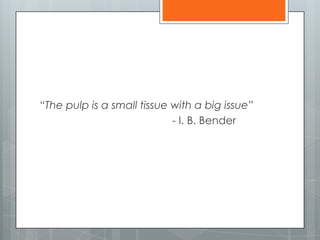
- 1. “The pulp is a small tissue with a big issue” - I. B. Bender
- 2. DISEASES OF THE DENTAL PULP- Part I DEEPTHI P.R. Ist YEAR MDS DEPT. OF CONSERVATIVE DENTISTRY AND ENDODONTICS
- 3. Introduction Development of pulp - disorders Pulp as a connective tissue - Cellular elements - Fibers - Ground substance - Vasculature - Nerve supply - Systemic factors affecting the pulp Dental pulp stem cells Conclusion
- 4. INTRODUCTION Unique tissue Soft tissue : mesenchymal origin Integral part of dentin – dentin pulp complex Rigid encasement: low compliance environment Incompressible: inflammation- increased tissue pressure External communication: apical foramen & lateral canals
- 5. DEFINITION ‘A richly vascularized and innervated specialized connective tissue of ectomesenchymal origin; contained in the central space of a tooth, surrounded by the dentin, with inductive, formative, nutritive, sensory and protective functions’ - Glossary of Endodontic terms
- 6. FUNCTIONS PRIMARY: Formative SECONDARY: tooth sensitivity, defense & hydration, nutrition Odontoblasts: Dentinogenesis Interaction with dental epithelium: Amelogenesis
- 7. DEVELOPMENT Downgrowths from dental lamina: Enamel organ Stages: Bud, Cap & Bell- deepening of invagination Tissue within the invagination: ‘ Dental Papilla’
- 8. DEVELOPMENT 8th week IUL: beginning of papilla Bell stage: inner layer of papilla- odontoblasts dentin Dental pulp: Cephalic neural crest cells
- 9. Blood supply Oval/ Circular reticulated plexus in alveolar bone (Saunders-1966 & Cutright- 1970) Series of blood vessels- dental papilla: future pulpal vessels Vessels in dental sac basal wall: course to papilla (Tobin 1972) Pulpal artery: plexus of vessels at pulpodentinal junction
- 10. Nerve supply Early development: few axons enter papilla- no peripheral nerve plexuses Eruptive stage: rapid development - plexus of Raschkow & terminals in odontoblastic layer Byers (1980)
- 11. Disorders: pulp development Vitamin D deficiency Down’s syndrome: Jaspers -1981 Dens invaginatus Pulpal dysplasia : Witkop- 1973 Regional odontodysplasia Hypophosphatasia: Houpt et al (1970) Beumer et al (1973) Hereditary hypophosphatemia: Archard & Witkop (1966) Hypophysectomy
- 12. Pulp as a Connective Tissue Cells, ground substance, fibers Cells: a fundamental matrix Site & precursor for the fiber complex Collagen & reticulin End product of the system
- 13. Cells of the Pulp Fibroblasts Odontoblasts Defense & other cells
- 14. Fibroblasts Basic cell type Baume: mesenchymal cells, pulpoblasts, or pulpocytes- progressive levels of maturation Active in collagen synthesis: fibers present on the cell body & processes
- 15. Fibroblasts Synthesize 6 Glycoproteins: fibronectin Fibronectin with Type III collagen: Reticulin fibers faint metachromasia, pho sphatase & ATP acitivity Galdames et al,Int. J. Morphol. vol.29 no.1 Temuco mar. 2011
- 16. Fibroblasts With age: more number & width of fibers & cells reduce More fibrous pulp: less defensive than young cellular pulp Responsible for increase in size of denticles
- 17. Odontoblasts Highly differentiated cell in pulp Main function: dentin production Uniformly stained hyperchromatic in tissue sections Cytoplasm: may or may not be evident
- 18. Morphologic variations: A, Pulp horn (pear shaped) B, coronal midpulp (spindle shape) C, coronal midroot level (elongated club shape) D, mid-third of root (short club shape) E, apical third of root (globules). Marion D et al, 1991
- 19. Electron Microscope Findings Large, closely aligned, multilayered sweet potato shaped cells 3 to 4 µm wide & 8 to 10 µm long Nucleus: • Ellipsoidal – chromatin & nucleolus • Double membrane covered • Granules attached to outer membrane
- 20. Electron Microscope Findings Nucleolus: One to four in number ( Ivanyi 1972) Ring shaped: fully developed- inactive RNA synthesis Compact: less developed- active RNA synthesis
- 21. Electron Microscope Findings Cytoplasm: Extensive rER & numerous transitional vesicles (Jesson-1968, Garant et al & Reith - 1968, Takuma & Nagai- 1971) Vesicles: fine fibrillar material Large Golgi apparatus : centre Membrane bound granules: lysosomes Secretory granules- abacus bodies: golgi complex
- 22. Electron Microscope Findings Mitochondria evenly distributed Centrioles present : rudimentary cilium Approx. 50 Ao diameter filaments 200 to 250 Ao diameter microtubules
- 23. Electron Microscope Findings Odontoblasts : 6-8 cells deep, palisade formation along predentin border Organelles: extend to terminal bar apparatus level Distal to this level: material constituting odontoblastic process
- 24. Electron Microscope Findings Odontoblastic process: Dentinal fibers/ Tomes’ fibers Traverses predentin, fills the lumen of dentinal tubule Coated vesicles: pinocytic & ingest material from predentin Numerous filaments: parallel to cell membrane- characteristic
- 26. Electron Microscope Findings Intercellular Junctions: Regions of plasma membranes between cells 3 types: Impermeable Adhering Communicating
- 27. Electron Microscope Findings Impermeable Junctions/ Tight Junctions: Helps: maintain a distinct internal environment Plasma membranes appear to fuse & offer a tight seal between cells
- 28. Electron Microscope Findings Adhering Junctions: Maintained by desmosomes: intercellular bridges 3 types: Belt, Spot & Hemidesmosomes Promote adhesion between cells
- 29. Electron Microscope Findings Communicating Junctions/ Gap Junctions: Mediate direct transfer of chemical messages between cells Exchange nutrients & signal molecules for coordination of function
- 30. Gap junction & Tight junction Desmosome like junction Sasaki T et al, 1982
- 31. Electron Microscope Findings Odontoblastic Junctional Complexes: Surface epithelial cells: terminal bars at apical extremities Consist of several components: junctional complexes Components: Zonula occludens, Zonula adherens & Macula adherens
- 32. Electron Microscope Findings Structures at border between odontoblastic process & cell bodies: small gap junctions, tight junctions & desmosome like junctions Tight adhesion between odontoblasts: not easily separated
- 33. Electron Microscope Findings Nerve endings: Presence of nerves in tubules: controversial Nerve endings in juxtaposition to odontoblastic processes: reported
- 34. Electron Microscope Findings Odontoblastic Communications: Odontoblastic nuclei: inner border of dentin Odontoblastic processes : adjacent processes through extensive lateral branch system (Kaye & Herold, 1966) Contact cells more centrally located: fine protoplasmic processes- fibronectin: cell to cell adhesion
- 35. Electron Microscope Findings Odontoblasts: mesenchymal syncytium- injury of one odontoblast affects others Continuity of cells lost: injury following operative procedures Cytoplasm stains for: RNA, lipids, ALP, ATPase, ACP, non specific esterases, protein carbohydrate complex : present
- 36. Electron Microscope Findings Cell free Zone/ Layer of Weil: under odontoblasts in coronal portion- nerve elements Not observed in middle & apical portions (Gotjamanos,1969) Cell rich Zone: Fibroblasts & undifferentiated mesenchyme cells
- 38. Defense cells Histiocytes and Macrophages: Pericytes : differentiate into fixed or wandering histiocytes under appropriate stimulation. Highly phagocytic: remove bacteria, foreign bodies, dead cells, debris. Pulpal macrophages & dendritic cells: Langerhans’ cells
- 39. Defense cells Polymorphonuclear Leukocytes: Commonest : pulpal inflammation Injury & cell death: rapidly migrate from nearby vessels Microabscess formation Bacteria & dead cells. Develop wider zones of inflammation. Silva et al, 2009
- 40. Defense cells Lymphocytes and Plasma Cells: Follows neutrophils. Injury & resultant immune responses Presence of a persistent irritant
- 41. Defense cells Mast Cells: Inflamed pulps Granules: histamine & heparin. Histamine: vasodilatation & increases vessel permeability
- 42. Reserve Cells Descendants of undifferentiated cells in the primitive dental papilla Multipotential cells : Fibroblast type Capable: dedifferentiate/redifferentiate- mature cell types. Cell-rich zone: concentrations of such cells.
- 43. Reserve Cells Produce little collagen: not mature fibroblasts (Frank- 1970) Cytoplasmic connections: odontoblasts & subjacent mesenchymal cells (Baume- 1980) Near vessels: other mature cell types Mast cells and odontoclasts: inflammation.
- 44. Reserve Cells Unique cells: calcified tissue - pulp cap/ pulpotomy[Ca(OH)2 ] Along the calcified tissue: base of tubules involved with caries, restorations, attrition, abrasion Not a true dentin; cells - not true odontoblasts
- 45. Fibers of the Pulp Reticular fibers: around blood vessels & odontoblasts Collagen- 640 Ao Type III collagen: 28% to 45%- histologically identified as reticulin Type I also
- 46. Fibers of the Pulp 2 types of filaments Rel. straight, approx 200 Ao diameter & 200 Ao periodicity Coiled, branched & irregularly beaded, 100 Ao diamter
- 47. Fibers of the Pulp von Korff fibers: Fine argyrophilic fibers Spirally twisted bundles- cork screw Unmineralised dentin/ predentin Fibrillar framework Bernick S
- 48. Fibers of the Pulp Collagen deposition Diffuse: no definite orientation Bundle: large, coarse bundles run parallel to nerves / independently (Stanley & Ranney, 1962) Apical portion: more fibrous than coronal (van Amerongen et al, 1983)
- 49. Fibers of the Pulp Coronal pulp: more bundle collagen Type III collagen & proteoglycans: arterial plexus & odontoblasts Extirpation of young cellular pulp: difficult Aged pulp: like absorbent paper point
- 50. Ground substance Structureless mass, gel-like in consistency: the bulk of the pulp Occupies the space between formed elements Influences: Spread of infection Metabolic changes Stability of crystalloids Effects of metabolic substances
- 51. Ground substance Proteins with glycoproteins, acid mucopolysaccharides GAGs: Hyaluronic acid (Engfeldt & Hjerpe, 1972) Water retention Ion Binding Electrolyte distribution during mineralization Collagen fibrillogenesis
- 52. Ground substance ‘Milieu interieur’: Engel (1958) Metabolites & breakdown products- exchange Hyaluronic acid: metabolite transport
- 53. Ground substance Pulp tissue hydroatatic pressure: 15 mm Hg increase- early stages of inflammation Depolymerization: microbial enzymes change in ground substance Hyaluronidases, chondroitin sulfatase Mucopolysaccharidase activity: resorbing deciduous teeth
- 54. Circulation of the Pulp Systemic circulation Microcirculation Lymphatics Control of blood flow Transcapillary fluid exchange Circulation in the inflamed pulp Clinical correlations
- 55. Arterial blood supply to teeth Right atrium Right ventricle Pulmonary artery Lungs Pulmonary vein (left ventricle) Aorta CCA ECA Internal Maxillary artery
- 56. Internal Maxillary artery pterygopalatinepterygoidmandibular Inferior alveolar Dental branch Lower Molars, premolars canines Incisive branch Lower Incisors Infraorbital artery ASA artery PSA artery Upper Incisors, canines Upper molars bicuspids
- 57. Venous drainage Nasopalatine, infraorbital, descending palatine, PSA, pharyngeal, Deep temporal, masseteric, Inferior alveolar, Middle meningeal Pterygoid venous plexus Internal maxillary vein with superficial temporal vein Retromandibular vein EJV/ IJV Innominate vein (right side) Superior venacava Heart (Right atrium)
- 58. Microcirculation Arterioles, capillaries & venules Arterioles: 50μ diameter: enter through apical foramen Branch : terminal arterioles capillary plexus – subodontoblastic zone Young teeth: extend into odontoblastic layer
- 59. Arteriovenous distribution of hemodynamics in rat dental pulp S. Kim et al, 1984
- 60. Takahashi et al- 1982
- 61. Microcirculation Capillaries: 8 to 10μ Coronal portion: capillary blood flow- twice that in the root Pulp horns: greatest blood flow
- 62. Microcirculation Fenestrations: rapid transport of fluid & metabolites Avg. capillary density: 1400/ mm3 : the greatest in the body Dr. K. Josephsen, Denmark
- 65. Arteriole distribution Main arteriole- 2 groups Coronally – pulp horn Between roof and floor of pulp chamber Takahashi et al, 1982
- 66. Microcirculation Pulpal venules: unusually thin walls, discontinuous muscular layer Diameter maximum: central region- 200μ Resting pulpal blood flow: 0.15 to 0.6 ml/ min/g tissue Blood volume: 3% pulpal wet weight
- 67. Microcirculation Changes measured: Laser Doppler flowmeters Detect revascularization: traumatized teeth Ideal : pulp vitality Limited: sensitivity, specificity, reproducibility & costs
- 68. Regulation of pulpal blood flow Neuronal, paracrine & endocrine mechanisms Vasodilatation: neighboring tissues- drop in pulpal blood flow & perfusion pressure Pulp: vulnerable in gingivitis/ periodontitis
- 69. Neuronal regulation Little/ no sympathetic vasoconstrictor tone Neuronal vasodilator tone: sensory neuropeptides Cervical sympathetic trunk: vasoconstriction Neuropeptide Y & norepinephrine
- 70. Neuronal regulation Blood flow sensory neuropeptides Vasodilatation : CGRP release Muscarinic receptors: ACh & VIP – vasodilatation (Yu CY et al- 2002) No parasympathetic vasodilatation: cat pulp (Sassano et al- 1995)
- 71. Local control Local tissue demands: regulate hemodynamics Endothelin-1 pulpal blood flow Prostacyclin, NO : endothelium Adenosine: ischemic & hypoxic tissue- low pulpal oxygen tension
- 72. Humoral control Angiotensin II : vasoconstrictive basal tone Receptors: AT1, AT2- rat pulp (Souza PP et al, 2007) DOPA, epinephrine: vasoconstriction ACh, Histamine, bradykinin : inhibit vasoconstriction
- 73. Lymphatics Drains filtered fluids & proteins: returns to blood Immune defense Lymphatic markers: extensive lymphatic system in pulp Capillaries- pulp horns; leave via apical foramen & lateral canals
- 74. From Berggreen E, Haug SR, Mkonyi LE, Bletsa A: Characterization of the dental lymphatic system and identification of cells immunopositive to specific lymphatic markers. Eur J Oral Sci 117(1):34–42, 2009
- 75. Lymphatics Arteriolar pulse pressure High interstitial pulsatile pressure Deformation of interstitial tissues Propulsion of lymph
- 76. Lymphatic drainage of teeth All maxillary teeth, Mandibular canines, premolars & molars Mandibular incisors Submaxillary glands Submental glands Superficial & deep cervical glands Thoracic duct (left) Jugular duct (right) Blood stream: junction of IJV & Subclavian veins
- 77. Transcapillary fluid exchange Regulated by : lymph flow & differences in colloidal osmotic & hydrostatic pressures Interstitial fluid volume: 0.6+ 0.03 ml/g Interstitial fluid pressure: 6- 10 mm Hg COP: rel. high- 83% plasma COP
- 78. Wiig H, Rubin K, Reed RK: New and active role of the interstitium in control of interstitial fluid pressure: potential therapeutic consequences. Acta Anaesthesiol Scand 47:111–121, 2003.
- 79. Circulation in the inflamed pulp Inflammation: vasodilatation & increased vascular permeability- interstitial fluid pressure Reabsorption of tissue fluid: pressure- disproves Pulpal strangulation theory (Heyeraas & Berggreen- 1999, Heyeraas & Kvinnsland- 1992)
- 80. Circulation in the inflamed pulp PGE2, Bradykinin, SP, Histamine: pulpal blood flow Serotonin: pulpal blood flow Acute inflammation: 200% of control flow & increased vascular permeability (Heyeraas & Kvinnsland- 1992, Heyeraas et al- 1996) LPS: circulatory dysfunction (Bletsa A et al, 2006)
- 81. Circulation in the inflamed pulp Endothelial perturbation: on exposure to endotoxin/ cytokines Reduced perfusion, VEGF down regulation & microvessel density : necrosis Lymphangiogenesis : inflamed pulps (Pimenta et al, 2003)
- 82. Vascular permeability: Inflamed pulp Vascular leakage: Prostaglandin, histamine, bradykinin, SP LPS, LTA, TNF-, IL-1: upregulate VEGF vascular permeability protein Transport COP
- 83. Circulation in the inflamed pulp: Clinical aspect Reduced distractions at night Pulpal blood flow : supine Further pulpal tissue pressure: activate sensitised nociceptors- spontaneous pain Throbbing : pulsations in the pulp - systole
- 84. Clinical correlations LOCAL ANESTHETICS: Blood flow infiltration : LA + epinephrine Pulp tissue pressure high conc. Vasoconstrictors (Van Hassel & Simard- Savoie et al 1973) No serious/ permanent damage
- 85. Clinical correlations GENERAL ANESTHETICS: Scott et al – 1972: rat study- pulpal blood flow velocity: zero in 30 seconds Effects: disappear in 1 hour AGING: Decreased circulation Atherosclerotic changes: calcification Cells atrophy & die; fibrosis
- 86. Clinical correlations TEMPERATURE CHANGES Elevation: 100C to 150C increase: intrapulpal pressure 2.5mm Hg/0C Irreversible changes: heating to 450C- prolonged (Van Hassel & Brown- 1969)
- 87. Clinical correlations Tooth preparation: affect pulpal blood flow Pulpal damage initiation: alteration in microvasculature No water spray: reduced blood flow- upto 1 hour (Kim et al, 1983)
- 88. Clinical correlations Reduction: Subfreezing temperatures: transient fall Intrapulpal pressure (Augsburger & Peters- 1981) < -20C: vascular engorgement & necrosis H2O2 & CO2 : reduce capillary blood flow
- 89. Clinical correlations ENDODONTIC THERAPY: Less hemorrhage: extirpation close to apex DEVELOPMENT: Blood vessel density increased coronally Subodontoblastic capillary plexus- larger : eruption Rich blood supply- floor of pulp chamber
- 90. Clinical correlations PERIODONTAL DISEASE: Reduction- circulation: degenerative changes Reparative processes diminished: older pulps: operative procedures- necrosis Excessive irradiation: necrosis Seltzer et al, 1963
- 91. Clinical correlations ANTERIOR OSTEOTOMY: Blood flow: maximum decrease immediate postop Apparently re established: normal response to stimuli (Pepersack- 1973, Theisen & Guernsey- 1976)
- 92. Nerve supply of the pulp Innervation of the teeth Theories of tooth pain perception Modulation of nerve impulses
- 93. Innervation of the teeth Vth N Ophthalmic Maxillary Mandibular PSA Infraorbital ASA Lingual Inferior alveolar Maxillary molars Maxillary premolars Maxillary anteriors Inferior dental Incisor Mandibular molars and premolars Mandibular cuspid and incisors
- 94. Convergence of sensory information : teeth to higher centres
- 95. Innervation Large no. of myelinated (A)& unmyelinated (C) fibers Premolar: 2000 Not all are nociceptors Afferent: sensory Efferent: Sympathetic: circulation & eruption
- 96. Characteristics of sensory fibers Fiber Myelination Location of Terminals Pain Characteristics Stimulation Threshold A-delta Yes Principally in region of pulp- dentin junction Sharp, pricking Relatively low C No Probably distributed throughout pulp Burning, aching, less bearable than A-delta fiber sensations Relatively high, usually associated with tissue injury
- 97. Sensory fibers Aδ: 1-5μ; 6-30 m/s C: 0.4-1μ; 0.5-2 m/s Pain localization: Single neuron innervation Low density propioceptors Electrical stimulation: A fibers
- 98. Fiber location within pulp
- 99. Sensory fibers Nerve bundles +blood vessels Dr. Inge Fristad, Department of Clinical Dentistry, University of Bergen
- 100. Plexus of Raschkow Mummery - 1919 Plexus of single nerve axons Develop: final stages of root formation Prolific branching: overlapping receptor fields A fibers: subodontoblastic plexus Terminal axons: free nerve endings
- 101. Types of nerve endings: Gunji T- 1982
- 102. Odontoblasts: receptor?? No anatomic communication: nerve fibers Low membrane potential: -24 to -30 mV Disruption of layer: no sensitivity Possibility: sodium channel activity/ factors release- neuromodulation Nerve fibers: resist necrosis Noxious stimuli: periapical tissues Pain in non vital teeth
- 103. Tissue injury & deafferentiation Deafferentiation: regeneration/ neuronal cell death V nuclei affected: pulp extirpation Phantom tooth pain Changes in gene expression: C-fos (Byers et al, 1993) A fibers: thermal & electric tests C fibers: pulp injured
- 104. Theories of tooth pain perception Dentinal nerve stimulation Dentinal receptor theory Hydrodynamic theory
- 105. Dentinal nerve stimulation Silver staining: controversial LM studies: variable penetration (Bernick- 1968) & termination (Rapp et al- 1957) EM studies: difficult interpretation No connection: nerves & odontoblasts (Fernehead – 1968)
- 106. Dentinal nerve stimulation Predentin: associated cells- origin questioned (Arwill- 1967) Arwill T
- 107. Dentinal nerve stimulation Axons: separated by narrow cleft (Byers et al) Nerves: beaded structures in SEM (Tidmarsh- 1981)
- 108. Dentinal nerve stimulation Frank et al- 1966 Nerve : concavity odontoblast(pic) ‘cork screw’ fibers Gap junctions: nerve cell processes & odontoblasts (Holland- 1975) Possible- no nerve connections Frank RM
- 109. Dentinal receptor theory Odontoblasts & processes: receptor Inconclusive Evidence: recording electrical activity Heat, cold, touch receptors (Scott & Tempel, Mumford- 1965) Electrical activity: nerves in pulp & not dentin (Matthews- 1970)
- 110. Dentinal receptor theory Intradentinal receptor: connections between odontoblastic process & nerve fiber (Frank- 1969) Transducer mechanism AChE: demonstrated in several studies (Avery and Rapp-1967); contrary too Adrenergic : pulpal blood vessel walls
- 111. Hydrodynamic theory Dentin pain & odontoblast displacement: related BrӓnstrӦm et al (1966, 1967, 1969, 1972) and Lilja (1980): hydrodynamic mechanism
- 112. Hydrodynamic theory Stimuli: expansion/ contraction – fluid Pulpward/ outward movement: nerve stimulation
- 113. Hydrodynamic theory Mechanisms - reduce fluid flow in dentin: Pashley et al- 1982 Plaque/ saliva bacteria Mineralized deposits- tubules Salivary/plasma proteins
- 114. Hydrodynamic theory- hypersensitive dentin 4 treatment modalities: Smear layer- burnishing root surface Oxalate compounds: insoluble ppts in tubules Tubule occlusion: pptd. Plasma proteins- HEMA + glutaraldehyde Dentin bonding agents application LASER : effects on pulp???
- 115. Pulpal tissue pressure & pain Blood flow, pressure changes, dental pain hydrostatic pressure: nerve fiber stimulation (Nӓhri- 1978) Pulp: mechanoreceptor- pain transmission
- 116. Polypeptides & Neurotransmitters PLASMA KININS: No pain: application to dentin ( Anderson and Naylor- 1972)0 SUBSTANCE P: Pulp: rich in SP Vasodilatation , increased capillary permeability (Pashley et al- 1982)
- 117. Polypeptides & Neurotransmitters PROSTAGLANDINS: Sensitize nociceptors: histamine, bradykinin, SP CGRP, Neuropeptide Y, NKA, VIP: painful pulps/ beneath caries Vasodilatation SP, CGRP: wound healing, inflammation CGRP release: vasoconstrictors
- 118. Systemic factors Vitamin deficiency Hormones Protein deficiency Systemic virus infection Hereditary diseases Tumor metastases
- 119. Vitamin deficiency Vitamin C - Fibroblasts - Odontoblasts: degenerate & lose morphology
- 120. Hormones & hormonal imbalance Steroids: Systemic corticosteroid Odontoblasts Inhibit reparative dentinogenesis Steroid :pulp therapy???
- 121. Hormones & hormonal imbalance Diabetes mellitus: Glucose concentration rise in dentinal pulp fluids Degenerative & inflammatory changes in pulp Dentinogenesis affected Atrophic pulp: non carious teeth Acute inflamed pulp: carious teeth Cohen et al, 1963
- 122. Hormones & hormonal imbalance Thyroid deficiency: Pulp vascularity Pulpal lumen Cellular elements
- 123. Protein deficiency No pulpal changes noted (Glickman & Shklar- 1954) Larger areas of periapical rarefaction ( Stahl et al -1958)
- 124. Systemic virus infection Odontoblasts injured: lymphocytic choriomeningitis (Hancock-1956) & Shope papilloma virus ( Fleming-1958) Degenerative changes & eventual necrosis: rats with Polyoma virus
- 125. Hereditary diseases Blood: Sickle cell anemia, leukemia Reticulo endothelial system: Hand- SchÜller- Christian disease Neurologic: Sturge- Weber disease Metachromatic leukodystrophy Krabbe’s leukodystrophy Fabry’s disease Niemann- Pick disease
- 126. Tumor transplantation Metastases: sparse reports Epitheliomas, sarcoma, Burkitt’s lymphomas- human dental pulps (Stanley- 1973)
- 127. Dental Pulp Stem Cells (DPSCs) Gronthos et al – 2000 Osteo/ odontogenic, adipogenic, neurogenic, chondrogenic, myogenic Tissue regeneration DPSCs: dentinal repair Appropriate carrier: dental implant Courtesy:
- 128. DPSCs DPSCs+ collagen + DMP1: pulp like tissue (Prescott et al, 2008) SHED: dental pulp tissue engg (Cordeiro et al,2008) Serum free medium + Insulin- transferrin- selenium- X & embryotrophic factor: suitable medium for culture (Hirata et al, 2010)
- 129. DPSCs Irreversible pulpitis: putative cells- stem cell properties (Wang et al, 2010) Regeneration in canine teeth – Gelfoam scaffold (Wang et al- 2013)
- 130. Conclusion Unique tissue Resembles embryonic connective tissue Dynamic response pattern
- 131. References Seltzer S, Bender J.B. Seltzer’s The Dental Pulp. Biological considerations in dental procedures. 3rd Edition Hargreaves KM, Cohen S. Cohen’s Pathways of the Pulp. 10th Edition Ingle JI, Bakland LK. Ingle’s Endodontics. 5th Edition
- 132. References Gronthos S, Mankani M, Brahim J, Gehron Roby P, Shi S. Postnatal human dental pulp stem cells (DPSCs) in vitro and in vivo. PNAS 2000; 97(25): 13625- 13630 In Vivo Generation of Dental Pulp-like Tissue by Using Dental Pulp Stem Cells, a Collagen Scaffold, and Dentin Matrix Protein 1 after SubcutaneousTransplantation in Mice. Prescott RS, Alsanea R, Fayad MI et al. J Endod 2008;34:421– 426
- 133. References Cordeiro MM, Dong Z, Kaneko T et al. Dental Pulp Tissue Engineering with Stem Cells from Exfoliated Deciduous Teeth. J Endod 2008;34:962–969 Wang Z, Pan J, Wright JT et al. Putative Stem Cells in Human Dental Pulp with Irreversible Pulpitis: An Exploratory Study. J Endod 2010;36:820–825)
- 134. References Hirata TM, Ishkitiev N, Yaeigaki K et al. Expression of Multiple Stem Cell Markers in Dental Pulp Cells Cultured in Serum-free Media. J Endod 2010;36:1139–1144 Wang Y, Zhao Y, Jia W, Yang J, Ge L. Preliminary Study on Dental Pulp Stem Cell–mediated Pulp Regeneration in Canine Immature Permanent Teeth. J Endod 2013;39:195–201
- 135. References Kim S, Lipowsky HH, Usami S, Chien S. Arteriovenous Distribution of Hemodynamic Parameters in the Rat Dental Pulp. Microvasc Res 27, 28-38 (1984)