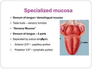
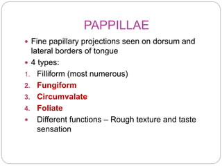
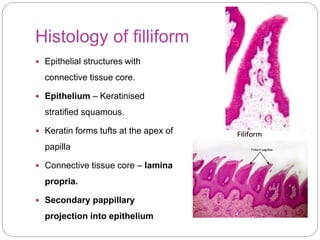
Dr. Meera K Pynadath
Dept. Oral Pathology &
Microbiology
Royal Dental College
Specialized Mucosa
Specialized mucosa
 Dorsum of tongue / dorsolingual mucosa
 Taste buds – sensory function
 “Sensory Mucosa”
 Dorsum of tongue – 2 parts
 Separated by sulcus terminalis
1. Anterior 2/3rd – papillary portion
2. Posterior 1/3rd – lymphatic portion
PAPPILLAE
 Fine papillary projections seen on dorsum and
lateral borders of tongue
 4 types:
1. Filliform (most numerous)
2. Fungiform
3. Circumvalate
4. Foliate
 Different functions – Rough texture and taste
sensation
Filliform papillae
 Most numerous
 Location: Anterior 2/3rd of
dorsolingual mucosa.
 Meaning : Filament like or thread
like
 Macroscopic structure:
 Numerous fine, pointed & cone
shaped projections
 “velvet appearance”
 No taste buds
 Functions:
1. Characteristic rough texture
2. Increase surface area & friction of
tongue – manipulate food bolus.
3. Sensation of touch
Histology of filliform
 Epithelial structures with
connective tissue core.
 Epithelium – Keratinised
stratified squamous.
 Keratin forms tufts at the apex of
papilla
 Connective tissue core – lamina
propria.
 Secondary pappillary
projection into epithelium
Fungiform papillae
 Location : Anterior 2/3rd
 Interspersed between filliform papillae
 Predominantly on tip and sides
 Meaning: “mushroom shape”
 Macroscopic structure:
 Round, reddish prominences
 Microscopic features
 Non keratinized
 Connective tissue core
 Rich vascular supply in lamina propria
 Dorsal portion – taste buds (1 to 3
nos)
Circumvalate papillae
 Also called valate papillae
 Location: Arranged anteriorly to sulcus
terminalis
 In a single row
 Number: 8 to 10
 Meaning : “surrounded by wall”
 Structure:
 Do not protrude to surface
 Dome shape
 Broader dorsal surface
 Narrow base
 Surrounded by deep circular
furrow/trough
 Trough surrounded elevated wall of
mucosa - vallum
Histology
 Stratified squamous epithelium
 Thin epithelium and larger CT core
 Epithelium shows secondary papillae of
connective tissue
 Superficial surface – keratinised
 Lateral surface – non keratinised
 Lateral aspects – taste buds
 Beneath the trough – von Ebner’s gland
open.
 Serous secretion clear the trough
 Contain salivary lipase.
Folliate Papillae
 Location: small vertical folds on lateral
border of posterior tongue
 Contain 4 to 11 parallel ridges.
 Meaning: Leaf like shape.
 Rudimentary in humans
 Contains taste buds
Taste buds
 Intraepithelial organs contain taste receptor cells or gustatory
cells
 Chemosensory - Responsible for taste sensation or gustation
 Detect 5 elements – sweet, sour, salt, bitter and umami
 Number: 4000 in oral cavity & upper ailmentary canal.
 Location: Seen papillae of tongue, soft palate, buccal mucosa,
epiglottis and esophagus.
 75% in oral cavity – 50% valate, 25% fungiform, and 25% folliate
Structure
 Shape: barrel or ovoid
 Size: 80µm high and 40µm thick
 Intraepithelial – from basal lamina to
the surface of the epithelium.
 Taste pore: outer opening of taste
(more than 1)
 Cells:
 Each tastebud has – 50 to 100
specialised cells.
1. Outer supporting cells –
 Outer longer cells -staves of barrel
 Inner shorter cells – spindle shape
2. Inner neuroepithelial cells –
receptor for taste stimuli (10 – 12
nos)
 Slender, dark staining cells, finger
like projections towards taste pore
Ultrastructural
findings
 4 cell types:
 Type I – dark cells –long cells- microvilli and
numerous vessicles related to gustatory nvs
 Type II– light cells- oval shape
 Type III- intermediate cells-light cells
 Type IV-rest on basement membrane,
precursor – undifferentiated cells.
Regions of taste
•Fungiform : sweet and salty
• Circum valate : bitter
•Foliate : sour
First concept
Second concept:
Taste cannot be broken down into areas
Taste is based on range of stimuli by chemical
substances in the food that fire up the gustatory nerves.
Taste is a continuum or total response of many gustatory
nerves
Posterior 1/3 rd of tongue
 Irregular studded cells with
numerous rounded or oval
prominence – Lingual
follicles
 Comprises of :
 lymph nodules with germinal
centers
 Lingual crypt on surface –
 lymphocytes migrate into
crypt through epithelium.
 Ducts of small posterior
lingual mucous glands opens
into crypt
 Lingual tonsil
Clinical significance
1. Depapillation
 Lingual papilla is lost
leaving a smooth, red
and painfull areas.
 Median rhomboid
glossitis and benign
migratory glossitis
(geographic tongue).
 Systemic diseases –iron
deficiency, pernicious
aneamia and vitamin
deficiency, vitamin B
complex deficiency – beefy
red tongue.
 Infections – scarlet fever
 Strawberry or raspberry
tongue
 Hairy tongue :
 Defective desquamation of
filliform papillae
 Hypertrophy of filliform
papillae.
 Normal papilla – 1mm
 Hairy tongue – 15 mm.
 Reasons:
 Lack mechanical debridement
and oral hygiene
 Tobacco use etc.
 Symptoms & signs:
 Tickling sensation esp palate
 Halitosis
 Burning sensation
Variation in other species
End session

Specialized Mucosa.pptx

  • 1.
    Dr. Meera KPynadath Dept. Oral Pathology & Microbiology Royal Dental College Specialized Mucosa
  • 2.
    Specialized mucosa  Dorsumof tongue / dorsolingual mucosa  Taste buds – sensory function  “Sensory Mucosa”  Dorsum of tongue – 2 parts  Separated by sulcus terminalis 1. Anterior 2/3rd – papillary portion 2. Posterior 1/3rd – lymphatic portion
  • 3.
    PAPPILLAE  Fine papillaryprojections seen on dorsum and lateral borders of tongue  4 types: 1. Filliform (most numerous) 2. Fungiform 3. Circumvalate 4. Foliate  Different functions – Rough texture and taste sensation
  • 4.
    Filliform papillae  Mostnumerous  Location: Anterior 2/3rd of dorsolingual mucosa.  Meaning : Filament like or thread like  Macroscopic structure:  Numerous fine, pointed & cone shaped projections  “velvet appearance”  No taste buds  Functions: 1. Characteristic rough texture 2. Increase surface area & friction of tongue – manipulate food bolus. 3. Sensation of touch
  • 5.
    Histology of filliform Epithelial structures with connective tissue core.  Epithelium – Keratinised stratified squamous.  Keratin forms tufts at the apex of papilla  Connective tissue core – lamina propria.  Secondary pappillary projection into epithelium
  • 6.
    Fungiform papillae  Location: Anterior 2/3rd  Interspersed between filliform papillae  Predominantly on tip and sides  Meaning: “mushroom shape”  Macroscopic structure:  Round, reddish prominences  Microscopic features  Non keratinized  Connective tissue core  Rich vascular supply in lamina propria  Dorsal portion – taste buds (1 to 3 nos)
  • 7.
    Circumvalate papillae  Alsocalled valate papillae  Location: Arranged anteriorly to sulcus terminalis  In a single row  Number: 8 to 10  Meaning : “surrounded by wall”  Structure:  Do not protrude to surface  Dome shape  Broader dorsal surface  Narrow base  Surrounded by deep circular furrow/trough  Trough surrounded elevated wall of mucosa - vallum
  • 8.
    Histology  Stratified squamousepithelium  Thin epithelium and larger CT core  Epithelium shows secondary papillae of connective tissue  Superficial surface – keratinised  Lateral surface – non keratinised  Lateral aspects – taste buds  Beneath the trough – von Ebner’s gland open.  Serous secretion clear the trough  Contain salivary lipase.
  • 9.
    Folliate Papillae  Location:small vertical folds on lateral border of posterior tongue  Contain 4 to 11 parallel ridges.  Meaning: Leaf like shape.  Rudimentary in humans  Contains taste buds
  • 10.
    Taste buds  Intraepithelialorgans contain taste receptor cells or gustatory cells  Chemosensory - Responsible for taste sensation or gustation  Detect 5 elements – sweet, sour, salt, bitter and umami  Number: 4000 in oral cavity & upper ailmentary canal.  Location: Seen papillae of tongue, soft palate, buccal mucosa, epiglottis and esophagus.  75% in oral cavity – 50% valate, 25% fungiform, and 25% folliate
  • 11.
    Structure  Shape: barrelor ovoid  Size: 80µm high and 40µm thick  Intraepithelial – from basal lamina to the surface of the epithelium.  Taste pore: outer opening of taste (more than 1)  Cells:  Each tastebud has – 50 to 100 specialised cells. 1. Outer supporting cells –  Outer longer cells -staves of barrel  Inner shorter cells – spindle shape 2. Inner neuroepithelial cells – receptor for taste stimuli (10 – 12 nos)  Slender, dark staining cells, finger like projections towards taste pore
  • 12.
    Ultrastructural findings  4 celltypes:  Type I – dark cells –long cells- microvilli and numerous vessicles related to gustatory nvs  Type II– light cells- oval shape  Type III- intermediate cells-light cells  Type IV-rest on basement membrane, precursor – undifferentiated cells.
  • 13.
    Regions of taste •Fungiform: sweet and salty • Circum valate : bitter •Foliate : sour First concept Second concept: Taste cannot be broken down into areas Taste is based on range of stimuli by chemical substances in the food that fire up the gustatory nerves. Taste is a continuum or total response of many gustatory nerves
  • 14.
    Posterior 1/3 rdof tongue  Irregular studded cells with numerous rounded or oval prominence – Lingual follicles  Comprises of :  lymph nodules with germinal centers  Lingual crypt on surface –  lymphocytes migrate into crypt through epithelium.  Ducts of small posterior lingual mucous glands opens into crypt  Lingual tonsil
  • 15.
    Clinical significance 1. Depapillation Lingual papilla is lost leaving a smooth, red and painfull areas.  Median rhomboid glossitis and benign migratory glossitis (geographic tongue).  Systemic diseases –iron deficiency, pernicious aneamia and vitamin deficiency, vitamin B complex deficiency – beefy red tongue.  Infections – scarlet fever  Strawberry or raspberry tongue
  • 16.
     Hairy tongue:  Defective desquamation of filliform papillae  Hypertrophy of filliform papillae.  Normal papilla – 1mm  Hairy tongue – 15 mm.  Reasons:  Lack mechanical debridement and oral hygiene  Tobacco use etc.  Symptoms & signs:  Tickling sensation esp palate  Halitosis  Burning sensation
  • 17.
  • 18.