Downloaded 694 times
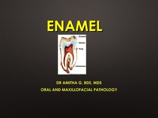


The document discusses the structure, composition, and development of dental enamel, the hardest biological tissue that covers the crown of teeth. It details the physical and chemical properties of enamel, stages of amelogenesis, and the life cycle of ameloblasts, including the significance of specific anatomical features. Multiple elements such as enamel rods, defects, and clinical implications are also addressed, providing a comprehensive overview of this dental tissue.


