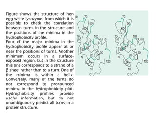
Protein Stability and
Folding
Dr.J. Vardhana
Assistant Professor, Dept of
Biotechnology
VISTAS
Protein Stability
-Native proteins are only marginally
stable under physiological conditions ->
high turnover
-Free energy required for denaturation is ~0.4
kJ/mol/ residue -> fully folded 100 residue
protein is ~40 kJ/mol more stable than its
unfolded form = energy of 2 hydrogen bonds
-But energy of all noncovalent interactions within
a protein is in the order of thousands of kJ/mol
=> native structure results from a delicate
balance of powerful counteracting forces
Proteins are stabilized by several
forces
-Protein structures are governed mainly by
hydrophobic effects and to lesser extend by
interactions between polar residues
-The hydrophobic effect causes nonpolar
substances to minimize their contact with water
(degree of order, entropy, of water is decreased
because water has not to form “cages” around
the hydrophobic groups)
-Relative hydropathy of residues: energy
required to solubilize a given amino acid in
water
Electrostatic interactions contribute to
protein stability
-Relatively week van der Waals forces are nevertheless
important to stabilize the protein in its native state
-But hydrogen bonds, which are a central feature of protein
structures, particularly secondary structures, make only a
minor contribution to the overall stability of a protein
-Because of extensive hydrogen bonding of surface residues
to water, difference between native and unfolded energy of
hydrogen bonding is ~-2 to 8 kJ/mol
- Ion pairing / salt bridges, i.e. between Lys+ and Asp-
75% of ionized residues are paired, mostly on surface, but
they have only a small stabilizing effect (paid by loss of
entropy and loss of solvation free energy) => poorly conserved
Examples of ion pairs in myoglobin
- Oppositely charged side chains from groups that are far
apart in sequence closely approach each other through
the formation of ion pairs
Disulfide bonds cross-link extracellular
proteins
-Intrachain or inter-chain disulfide bonds form during
folding of the protein
-While they are not necessary for the structure/function of
most proteins, they “lock in” a particular backbone fold
-Disulfide bonds are rarely in cytoplasmatic protein because
the cytosol is a reducing environment that would cleave the
disulfide bridges
-Most disulfide bridges occur in secreted proteins, i.e. they
are stable in the more oxidizing extracellular environment
Metal ions stabilize some small domains
-Metal ions, bound within a protein, can also serve to
stabilize the protein structure, through internal cross-linking
-For example zing fingers are motifs that frequently occur
in DNA binding proteins
-Zn2+ is tetrahedrally coordinated by side chain Cys, His and
occasionally Aps and Glu
-Zn2+ allows short stretches of polypeptides, 25-60 residues,
to fold into stable units
- Zinc fingers are not stable in the absence of Zn2+
- Zn2+ is stable and not oxidized/reduced (unlike Cu, or Fe)
A zing finger
motif
Coordinated
by Cys and
His
Protein Folding
-Protein folding is directed largely by residues that occupy
the interior of the folded protein
- How does a protein fold into its three dimensional
structure
-Does not occur through sampling of all possible
conformations ! This would take longer than the
universe exists
(n residues -> 2n torsion angles, each has 3 stable
conformations ->32n = ~10n possible conformations, 10-13 sec
for each conformation -> t = 10n/1013
=> for n=100 residues t = 1087 sec,
20 Mia years = 6 1017 sec)
A) Proteins follow folding
pathways
-Many proteins fold into their native
conformation in less than a few seconds
- They follow directed pathways, not
random
-Folding occurs locally by the formation of
secondary structures
-Followed by a hydrophobic collapse of the
structure to adopt a molten globule
= has most of the secondary structures
formed but not yet the proper
tertiary structure
- Proteins fold in a hierarchical manner
- Cooperative process
Protein disulfide isomerase acts
during protein folding
-Proteins fold more slowly in vitro than they fold in
vivo
-This is frequently due to the formation of non-
native disulfide bridges which are then slowly
exchanged to the native ones
-In vivo, disulfide bond formation is catalyzed by and
enzyme: protein disulfide isomerase (PDI)
-PDI binds a variety of unfolded proteins via a
hydrophobic patch to form a mixed disulfide
Mechanism of protein disulfide
isomerase
Mechanism of protein disulfide
isomerase
B) Molecular chaperones assist protein
folding
-Proteins begin to fold as they are synthesized and
grow on the ribosome
-In vivo, a peptide chain folds in the presence of a
very high concentration of other proteins
-Molecular chaperones are essential proteins that
help to fold newly synthesized or partially
unfolded proteins to re-fold correctly
-Many molecular chaperones were first described as
heat shock proteins (Hsp), their expression is
strongly induced upon heat treatment of cells
Chaperone activity requires ATP
Classes of molecular chaperones in prokaryotes and
eukaryotes
1.Hsp70 family, function as monomers with the
cochaperone Hsp40, folds newly made proteins
2. Chaperonins, large multisubunit proteins (see
below)
3. Hsp90 proteins, folding of proteins in signal
transduction such as steroid receptors
4. Trigger factor, associate with ribosome and
prevent improper folding of newly made
All of them operate by binding to solvent-
exposed hydrophobic surfaces and subsequent
release
All are ATPases
The GroEL/ES chaperonin forms closed
chambers in which proteins fold
The chaperonins in E. coli consist of two types
of subunits, GroEL and GroES
Structure: 14 identical 549-residue GroEL subunits
arranged in two stacked rings of seven subunits
each
Complex is capped at one end by domelike
heptameric ring of 97 Aa GroES subunits
Bullet-shaped complex with C7 symmetry
Central chamber of ~45 Å in which peptides
fold
X-raystructure of the GroEL-
GroES-(ADP)7
complex
GroES (cap)
GroEL
(cis)
GroEL
(trans)
Some diseases are caused by protein
misfolding
At least 20 – usually fatal – human diseases are
associated with extracellular deposition of normally
soluble proteins as insoluble fibrous aggregates =
amyloids (starch-like)
Amyloidoses: set of rare inherited diseases in which
mutant forms of normally soluble proteins, such as
lysozyme or fibrinogen, accumulate as amyloids
Symptoms usually become apparent only later in
life (30-70 years) progress over 5-15 years till
death
Applications of
hydrophobicity
Using a hydrophobicity scale that assigns a value to each amino acid we
can plot the variation of hydrophobicity along the sequence of a
protein. This is called a hydrophobicity profile.
Hydrophobicity profiles have been used to predict the positions of
turns between elements of secondary structure, exposed and buried
residues, membrane-spanning segments, and antigenic sites.
Use of hydrophobicity profiles to predict the positions turns
between helices
and strands of sheet
Figure shows the hydrophobicity profile of hen egg white lysozyme. It
has pronounced minima at the following residues: 17, 44, 70, 93, and
117.
Figure shows the structure of hen
egg white lysozyme, from which it is
possible to check the correlation
between turns in the structure and
the positions of the minima in the
hydrophobicity profile.
Four of the major minima in the
hydrophobicity profile appear at or
near the positions of turns. Another
minimum occurs in a surface-
exposed region, but in the structure
this one corresponds to a strand of a
β sheet rather than to a turn. One of
the minima is within a helix.
Conversely, many of the turns do
not correspond to pronounced
minima in the hydrophobicity plot.
Hydrophobicity profiles provide
useful information, but do not
unambiguously predict all turns in a
protein structure.
The helical wheel
O.B. Ptitsyn observed that α helices in globular proteins often have a
‘hydrophobic face’ turns inwards towards the protein interior, and a
‘hydrophilic face’ turned outwards towards the solvent. Each residue in
an α helix appears at a position 100° around the circumference of the
helix from its predecessor. Therefore, to achieve
Ptitsyn's effect, the sequence of residues should alternate between
hydrophobic and hydrophilic with a periodicity of approximately four.
To check this relationship, the residues can be projected onto a plane
perpendicular to a helix axis, a diagram called a helical wheel. This
example shows the sequence of an α helix of sperm whale myoglobin.
Charged and polar residues appear in green; others in black.
The helix has a hydrophobic face—
which points to the inside of the
structure, and a hydrophilic face—
which
points outside.
In this picture the hydrophilic face is at
the bottom of the diagram. From such
a pattern of hydrophobicity we can
predict whether a region of an amino
acid sequence is likely to form an α
helix in the native protein structure.
Superposition of
structures
• Some aspects of sequence analysis carry over fairly directly into
structural analysis, some must be generalized, and others have no
analogues at all.
• As in the case of sequences, a fundamental question in analysing
structures is to devise and compute a measure of similarity.
• If two molecules have very similar structures, we can imagine
• superposing them so that corresponding points are as close together as
possible.
• Then the average distance between corresponding points is a measure
of the structural similarity.
• In practice it is conventional to report the root-mean-square (r.m.s.)
deviation of the corresponding atoms:
where di is the distance between the ith pair of atoms after optimal
fitting, and n is the number of points. This assumes that we have
prespecified the correspondence between the points.
If the correspondence is not known, we must first determine it and
only then calculate the r.m.s. deviation of the alignable substructures.
Structural alignment of bovine γ-chymotrypsin and S.
aureus epidermolytic toxin A
Bovine chymotrypsin and S. aureus epidermolytic toxin A are both
members of the chymotrypsin family of proteinases. Figure shows a
structural superposition of PDB entries 8GCH (γ-chymotrypsin) (black)
and 1AGJ (S. aureus epidermolytic toxin A) (green). The molecules share
the common chymotrypsin-family serine
proteinase folding pattern, and the Ser-His-Asp catalytic triad (ball-and-
stick).
Structural superposition of γ-chymotrypsin [8GCH] (black) and S.
aureus epidermolytic toxin A [1AGJ] (green). The sidechains of the
catalytic triads are shown. Observe that the region around the
active site is the best-conserved part of the protein.

Protein Stability and folding.pptx/pdf..

  • 1.
    Protein Stability and Folding Dr.J.Vardhana Assistant Professor, Dept of Biotechnology VISTAS
  • 2.
    Protein Stability -Native proteinsare only marginally stable under physiological conditions -> high turnover -Free energy required for denaturation is ~0.4 kJ/mol/ residue -> fully folded 100 residue protein is ~40 kJ/mol more stable than its unfolded form = energy of 2 hydrogen bonds -But energy of all noncovalent interactions within a protein is in the order of thousands of kJ/mol => native structure results from a delicate balance of powerful counteracting forces
  • 3.
    Proteins are stabilizedby several forces -Protein structures are governed mainly by hydrophobic effects and to lesser extend by interactions between polar residues -The hydrophobic effect causes nonpolar substances to minimize their contact with water (degree of order, entropy, of water is decreased because water has not to form “cages” around the hydrophobic groups) -Relative hydropathy of residues: energy required to solubilize a given amino acid in water
  • 4.
    Electrostatic interactions contributeto protein stability -Relatively week van der Waals forces are nevertheless important to stabilize the protein in its native state -But hydrogen bonds, which are a central feature of protein structures, particularly secondary structures, make only a minor contribution to the overall stability of a protein -Because of extensive hydrogen bonding of surface residues to water, difference between native and unfolded energy of hydrogen bonding is ~-2 to 8 kJ/mol - Ion pairing / salt bridges, i.e. between Lys+ and Asp- 75% of ionized residues are paired, mostly on surface, but they have only a small stabilizing effect (paid by loss of entropy and loss of solvation free energy) => poorly conserved
  • 5.
    Examples of ionpairs in myoglobin - Oppositely charged side chains from groups that are far apart in sequence closely approach each other through the formation of ion pairs
  • 6.
    Disulfide bonds cross-linkextracellular proteins -Intrachain or inter-chain disulfide bonds form during folding of the protein -While they are not necessary for the structure/function of most proteins, they “lock in” a particular backbone fold -Disulfide bonds are rarely in cytoplasmatic protein because the cytosol is a reducing environment that would cleave the disulfide bridges -Most disulfide bridges occur in secreted proteins, i.e. they are stable in the more oxidizing extracellular environment
  • 7.
    Metal ions stabilizesome small domains -Metal ions, bound within a protein, can also serve to stabilize the protein structure, through internal cross-linking -For example zing fingers are motifs that frequently occur in DNA binding proteins -Zn2+ is tetrahedrally coordinated by side chain Cys, His and occasionally Aps and Glu -Zn2+ allows short stretches of polypeptides, 25-60 residues, to fold into stable units - Zinc fingers are not stable in the absence of Zn2+ - Zn2+ is stable and not oxidized/reduced (unlike Cu, or Fe)
  • 8.
  • 9.
    Protein Folding -Protein foldingis directed largely by residues that occupy the interior of the folded protein - How does a protein fold into its three dimensional structure -Does not occur through sampling of all possible conformations ! This would take longer than the universe exists (n residues -> 2n torsion angles, each has 3 stable conformations ->32n = ~10n possible conformations, 10-13 sec for each conformation -> t = 10n/1013 => for n=100 residues t = 1087 sec, 20 Mia years = 6 1017 sec)
  • 10.
    A) Proteins followfolding pathways -Many proteins fold into their native conformation in less than a few seconds - They follow directed pathways, not random -Folding occurs locally by the formation of secondary structures -Followed by a hydrophobic collapse of the structure to adopt a molten globule = has most of the secondary structures formed but not yet the proper tertiary structure - Proteins fold in a hierarchical manner - Cooperative process
  • 11.
    Protein disulfide isomeraseacts during protein folding -Proteins fold more slowly in vitro than they fold in vivo -This is frequently due to the formation of non- native disulfide bridges which are then slowly exchanged to the native ones -In vivo, disulfide bond formation is catalyzed by and enzyme: protein disulfide isomerase (PDI) -PDI binds a variety of unfolded proteins via a hydrophobic patch to form a mixed disulfide
  • 12.
    Mechanism of proteindisulfide isomerase
  • 13.
    Mechanism of proteindisulfide isomerase
  • 14.
    B) Molecular chaperonesassist protein folding -Proteins begin to fold as they are synthesized and grow on the ribosome -In vivo, a peptide chain folds in the presence of a very high concentration of other proteins -Molecular chaperones are essential proteins that help to fold newly synthesized or partially unfolded proteins to re-fold correctly -Many molecular chaperones were first described as heat shock proteins (Hsp), their expression is strongly induced upon heat treatment of cells
  • 15.
    Chaperone activity requiresATP Classes of molecular chaperones in prokaryotes and eukaryotes 1.Hsp70 family, function as monomers with the cochaperone Hsp40, folds newly made proteins 2. Chaperonins, large multisubunit proteins (see below) 3. Hsp90 proteins, folding of proteins in signal transduction such as steroid receptors 4. Trigger factor, associate with ribosome and prevent improper folding of newly made All of them operate by binding to solvent- exposed hydrophobic surfaces and subsequent release All are ATPases
  • 16.
    The GroEL/ES chaperoninforms closed chambers in which proteins fold The chaperonins in E. coli consist of two types of subunits, GroEL and GroES Structure: 14 identical 549-residue GroEL subunits arranged in two stacked rings of seven subunits each Complex is capped at one end by domelike heptameric ring of 97 Aa GroES subunits Bullet-shaped complex with C7 symmetry Central chamber of ~45 Å in which peptides fold
  • 17.
    X-raystructure of theGroEL- GroES-(ADP)7 complex GroES (cap) GroEL (cis) GroEL (trans)
  • 18.
    Some diseases arecaused by protein misfolding At least 20 – usually fatal – human diseases are associated with extracellular deposition of normally soluble proteins as insoluble fibrous aggregates = amyloids (starch-like) Amyloidoses: set of rare inherited diseases in which mutant forms of normally soluble proteins, such as lysozyme or fibrinogen, accumulate as amyloids Symptoms usually become apparent only later in life (30-70 years) progress over 5-15 years till death
  • 20.
    Applications of hydrophobicity Using ahydrophobicity scale that assigns a value to each amino acid we can plot the variation of hydrophobicity along the sequence of a protein. This is called a hydrophobicity profile. Hydrophobicity profiles have been used to predict the positions of turns between elements of secondary structure, exposed and buried residues, membrane-spanning segments, and antigenic sites. Use of hydrophobicity profiles to predict the positions turns between helices and strands of sheet Figure shows the hydrophobicity profile of hen egg white lysozyme. It has pronounced minima at the following residues: 17, 44, 70, 93, and 117.
  • 21.
    Figure shows thestructure of hen egg white lysozyme, from which it is possible to check the correlation between turns in the structure and the positions of the minima in the hydrophobicity profile. Four of the major minima in the hydrophobicity profile appear at or near the positions of turns. Another minimum occurs in a surface- exposed region, but in the structure this one corresponds to a strand of a β sheet rather than to a turn. One of the minima is within a helix. Conversely, many of the turns do not correspond to pronounced minima in the hydrophobicity plot. Hydrophobicity profiles provide useful information, but do not unambiguously predict all turns in a protein structure.
  • 22.
    The helical wheel O.B.Ptitsyn observed that α helices in globular proteins often have a ‘hydrophobic face’ turns inwards towards the protein interior, and a ‘hydrophilic face’ turned outwards towards the solvent. Each residue in an α helix appears at a position 100° around the circumference of the helix from its predecessor. Therefore, to achieve Ptitsyn's effect, the sequence of residues should alternate between hydrophobic and hydrophilic with a periodicity of approximately four. To check this relationship, the residues can be projected onto a plane perpendicular to a helix axis, a diagram called a helical wheel. This example shows the sequence of an α helix of sperm whale myoglobin. Charged and polar residues appear in green; others in black.
  • 23.
    The helix hasa hydrophobic face— which points to the inside of the structure, and a hydrophilic face— which points outside. In this picture the hydrophilic face is at the bottom of the diagram. From such a pattern of hydrophobicity we can predict whether a region of an amino acid sequence is likely to form an α helix in the native protein structure.
  • 24.
    Superposition of structures • Someaspects of sequence analysis carry over fairly directly into structural analysis, some must be generalized, and others have no analogues at all. • As in the case of sequences, a fundamental question in analysing structures is to devise and compute a measure of similarity. • If two molecules have very similar structures, we can imagine • superposing them so that corresponding points are as close together as possible. • Then the average distance between corresponding points is a measure of the structural similarity. • In practice it is conventional to report the root-mean-square (r.m.s.) deviation of the corresponding atoms: where di is the distance between the ith pair of atoms after optimal fitting, and n is the number of points. This assumes that we have prespecified the correspondence between the points. If the correspondence is not known, we must first determine it and only then calculate the r.m.s. deviation of the alignable substructures.
  • 25.
    Structural alignment ofbovine γ-chymotrypsin and S. aureus epidermolytic toxin A Bovine chymotrypsin and S. aureus epidermolytic toxin A are both members of the chymotrypsin family of proteinases. Figure shows a structural superposition of PDB entries 8GCH (γ-chymotrypsin) (black) and 1AGJ (S. aureus epidermolytic toxin A) (green). The molecules share the common chymotrypsin-family serine proteinase folding pattern, and the Ser-His-Asp catalytic triad (ball-and- stick). Structural superposition of γ-chymotrypsin [8GCH] (black) and S. aureus epidermolytic toxin A [1AGJ] (green). The sidechains of the catalytic triads are shown. Observe that the region around the active site is the best-conserved part of the protein.