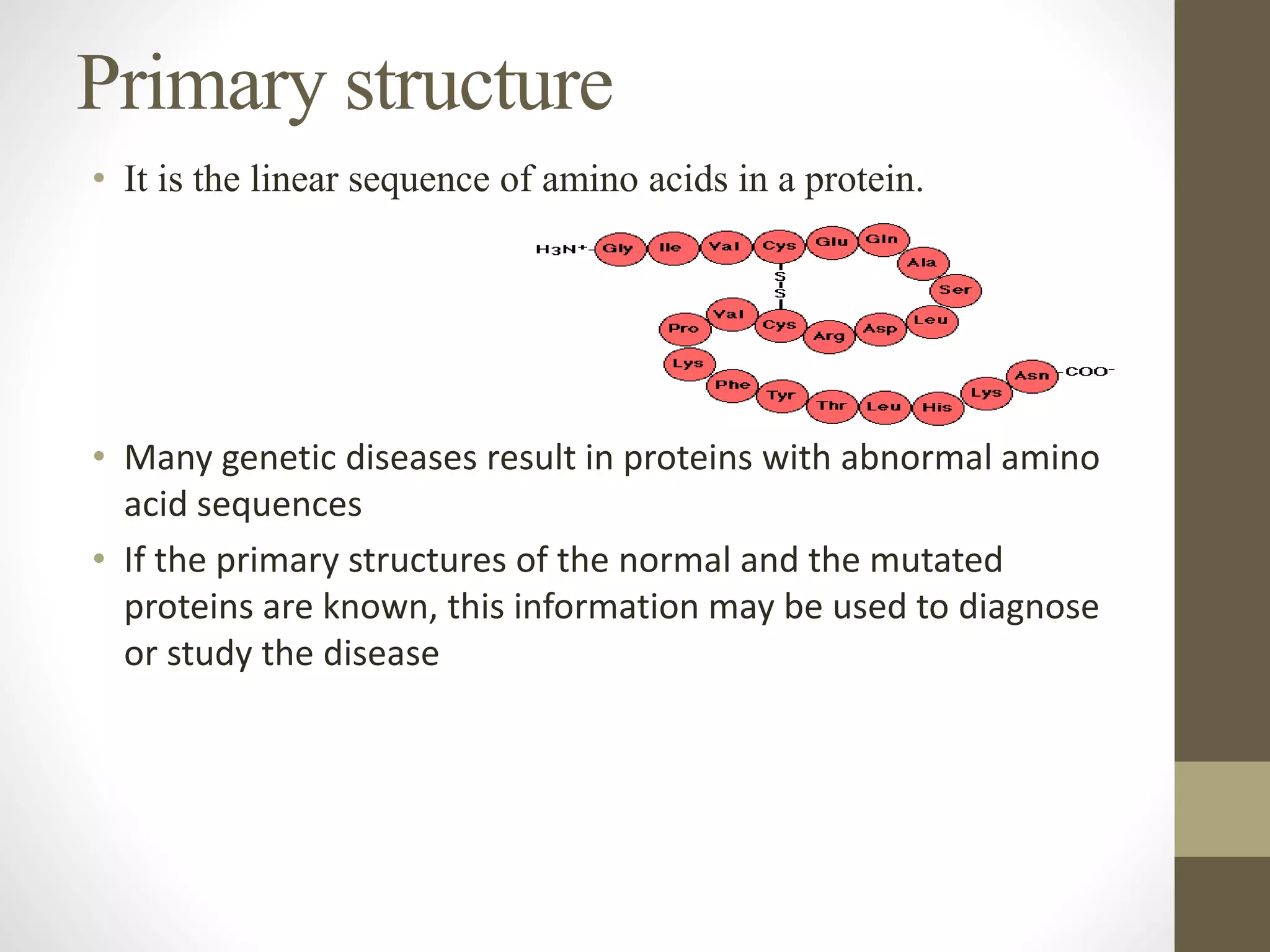
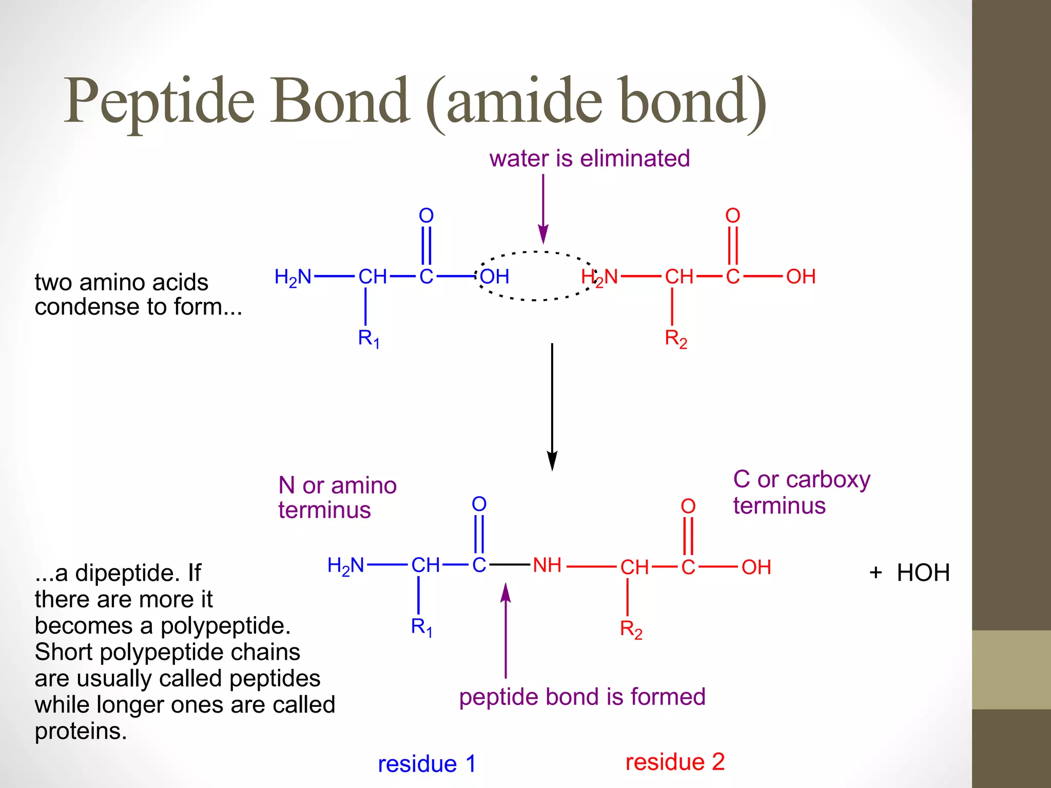
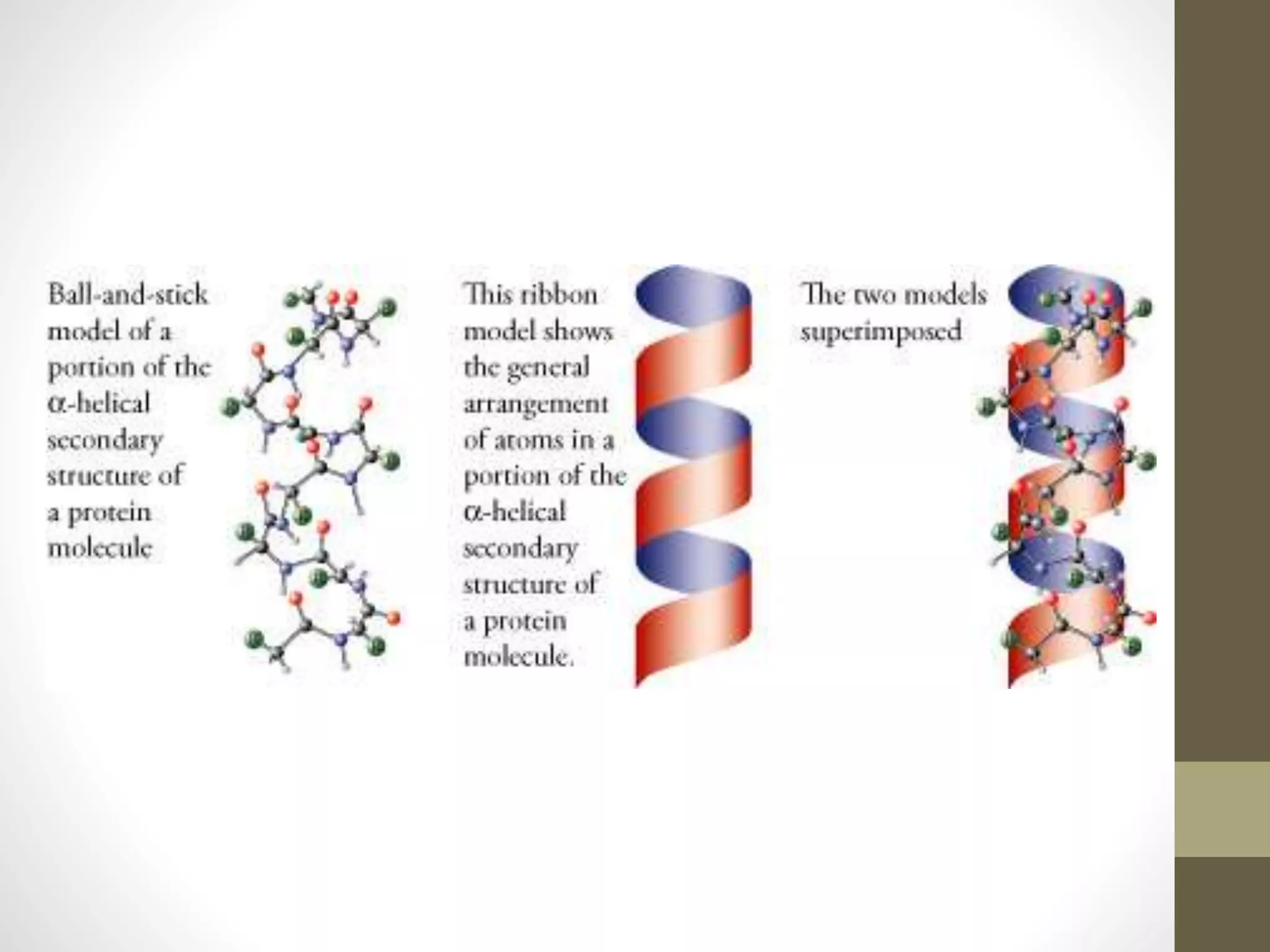
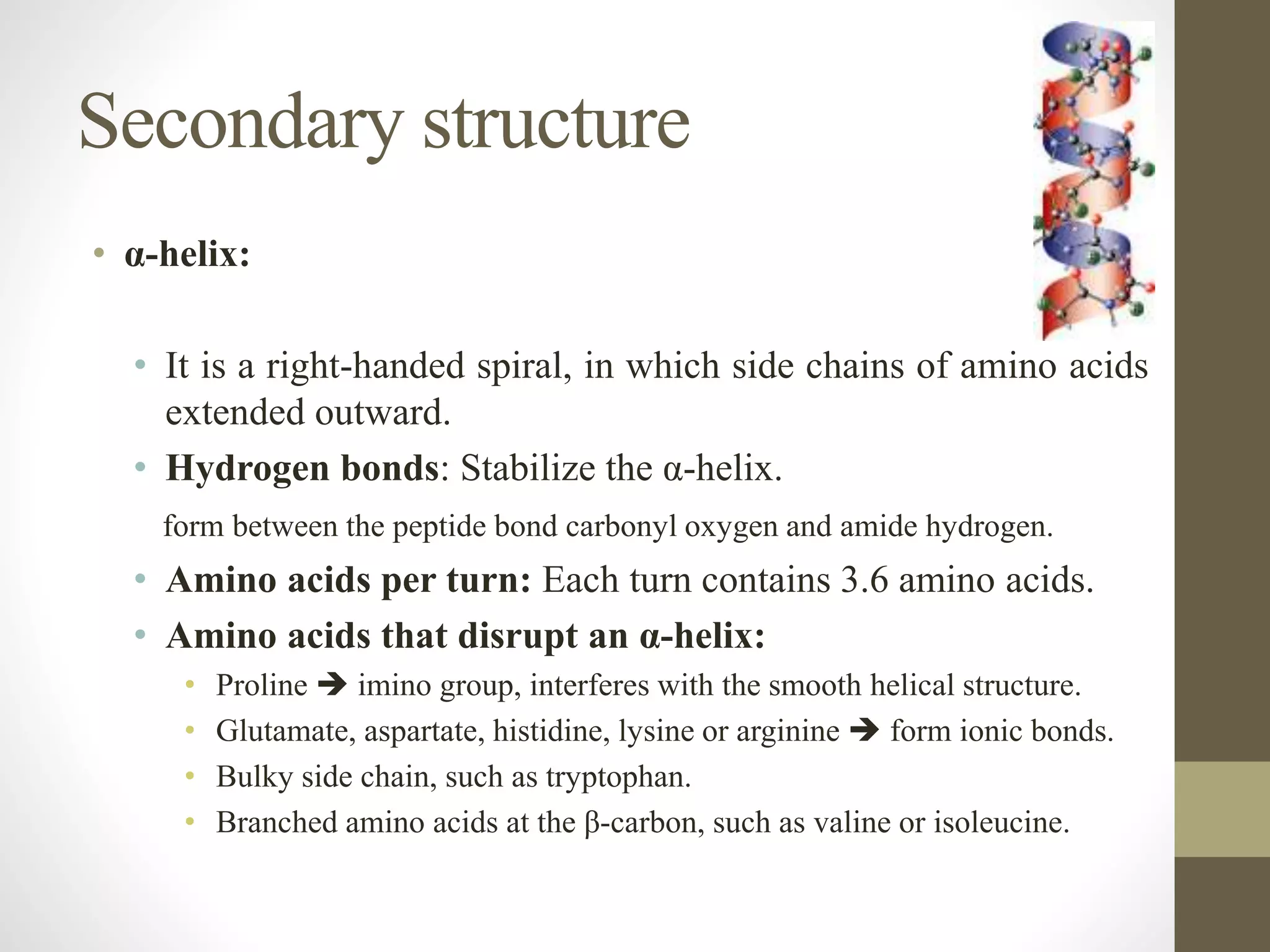
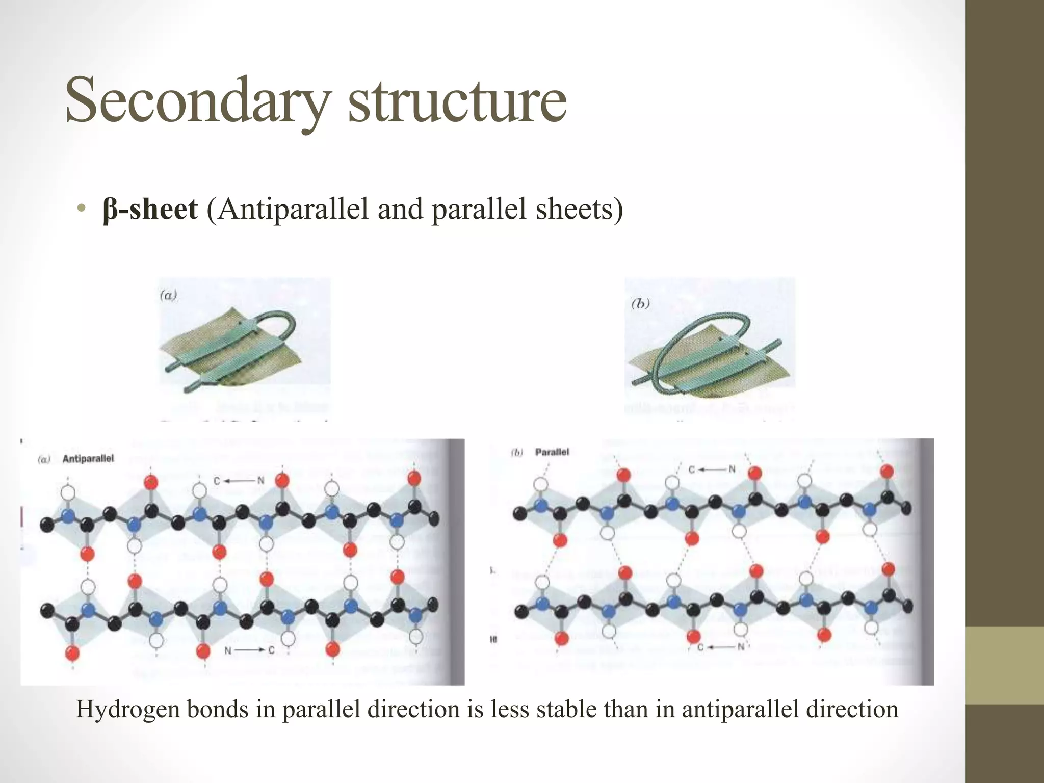
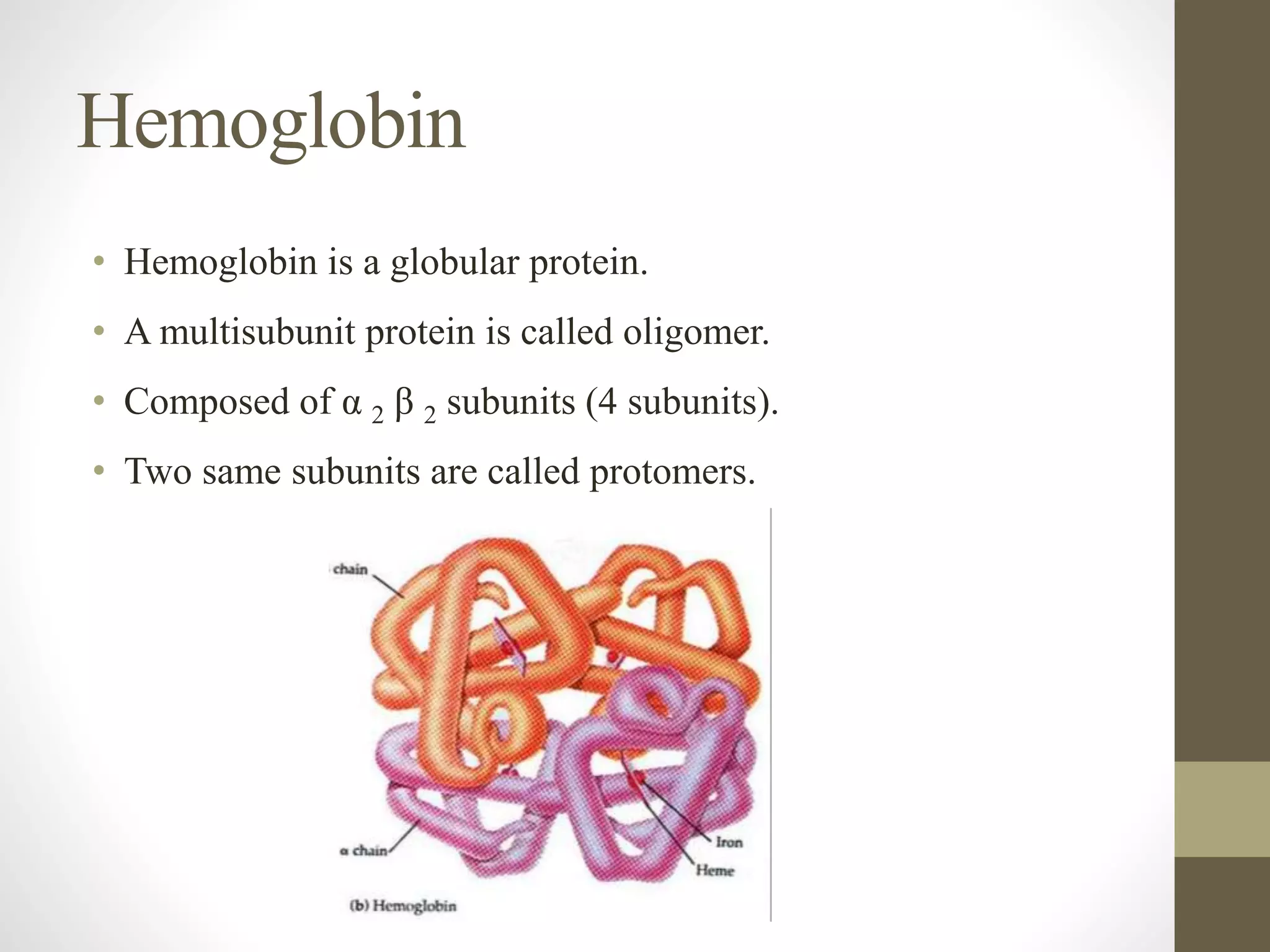
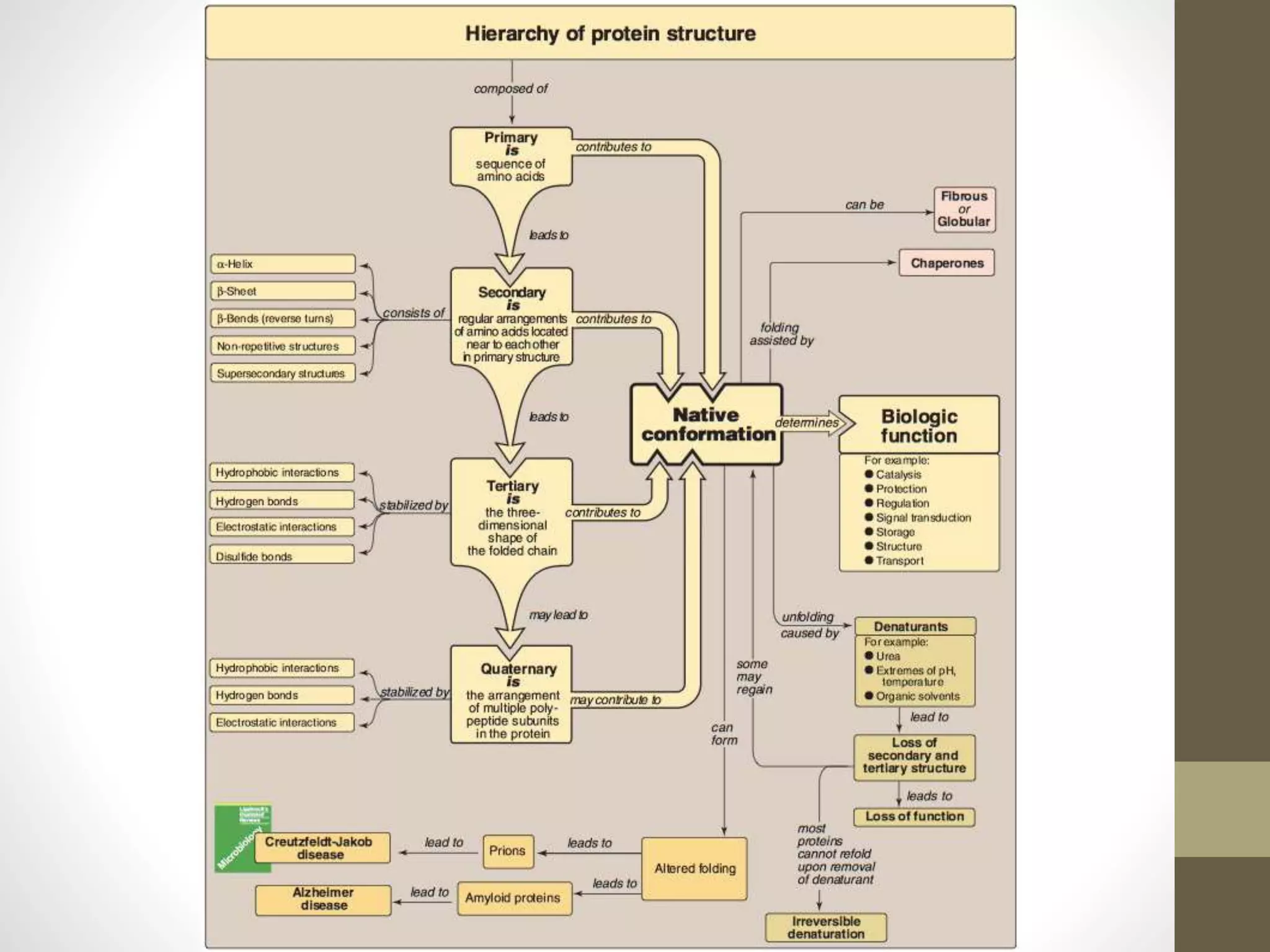
Protein structure
What are proteins?
• There are mainly 20 different types of amino acids that can be
combined to make a protein.
• The sequence of amino acids determines each protein’s unique
three-dimensional (3D) structure and its specific function.
• Proteins can be described according to their large range of
functions in the body e.g. antibody, enzyme, messenger,
structural component and transport/storage.
• It is the linear sequence of amino acids in a protein.
• Many genetic diseases result in proteins with abnormal amino
acid sequences
• If the primary structures of the normal and the mutated
proteins are known, this information may be used to diagnose
or study the disease
Primary structure
Peptide Bond (amide bond)
H2N CH C
R1
OH
O
H2N CH C
R2
OH
O
H2N CH C
R1
NH
O
CH C
R2
OH
O
peptide bond is formed
+ HOH
residue 1 residue 2
two amino acids
condense to form...
...a dipeptide. If
there are more it
becomes a polypeptide.
Short polypeptide chains
are usually called peptides
while longer ones are called
proteins.
water is eliminated
N or amino
terminus
C or carboxy
terminus
Peptides
• Amino acids can be polymerized to form chains:
• Two amino acids  dipeptide  one peptide bond.
• Three amino acids  tripeptide  two peptide bonds.
• Four amino acids  tetrapeptide  three peptide bonds.
• Few (2-20 amino acids)  oligopeptide.
• More (>20 amino acids)  polypeptide.
Peptide bond
• amino acids are joined covalently by peptide bonds, which are
amide linkages between the α-carboxyl group of one amino
acid and the α-amino group of another
• Peptide bonds are not broken by conditions that denature
proteins, such as heating or high concentrations of urea
• Prolonged exposure to a strong acid or base at elevated
temperatures is required to hydrolyze these bonds non
enzymatically
Naming the peptide
• the free amino end (N-terminal) of the peptide chain is
written to the left and the free carboxyl end(C-terminal) to the
right
• all amino acid sequences are read from the N- to the C-
terminal end of the peptide
• When a polypeptide is named, all amino acid residues have
their suffixes (-ine, -an, -ic, or -ate) changed to -yl, with the
exception of the C-terminal amino acid
Characteristics of a peptide
• The peptide bond has a partial double-bond character, that is,
it is shorter than a single bond and is rigid and planar
Polarity of a peptide
• Like all amide linkages, the – C=O and –NH groups of the
peptide bond are uncharged, and neither accept nor release
protons over the pH range of 2–12
• The – C=O and – NH groups of the peptide bond are polar, and
are involved in hydrogen bonds
Determination of the amino acid composition of a
polypeptide
I. identify and quantitate its constituent amino acids
II. polypeptide to be analyzed is first hydrolyzed by strong acid
at 110°C for 24 hours
III. individual amino acids, which can be separated by cation-
exchange chromatography
Sequencing of the peptide from its N-terminal end
• Sequencing is a stepwise process of identifying the specific
amino acid at each position in the peptide chain, beginning at
the N- terminal end
• Phenylisothiocyanate, known as Edman reagent, is used to
label the amino-terminal residue under mildly alkaline
conditions
• The resulting phenylthiohydantoin (PTH) derivative introduces
an instability in the N-terminal peptide bond that can be
selectively hydrolyzed without cleaving the other peptide
bonds
• Edman reagent can be applied repeatedly to the shortened
peptide
Cleavage of the polypeptide into smaller fragments
•primary structure composed of more than 100 amino acids. Such
molecules cannot be sequenced directly from end to end
•these large molecules can be cleaved at specific sites, and the
resulting fragments sequenced
•Enzymes that hydrolyze peptide bonds are termed peptidases
(proteases).
•Exopeptidases cut at the ends of proteins, and are divided into
aminopeptidases and carboxy peptidases. Carboxypeptidases are
used in determining the C-terminal amino acid. Endopeptidases
cleave within a protein
Determination of a protein’s primary structure by DNA Sequencing
The sequence of nucleotides in a protein-coding region of the DNA
specifies the amino acid sequence of a polypeptide. Therefore, if the
nucleotide sequence can be determined, it is possible, from knowledge
of the genetic code
Secondary structure
• It is regular arrangements of amino acids that are located near
to each other in the linear sequence.
• Excluding the conformations (3D arrangements) of its side
chains.
• α-helix, β-sheet and β-bend are examples of secondary
structures frequently found in proteins.
Secondary structure
• α-helix:
• It is a right-handed spiral, in which side chains of amino acids
extended outward.
• Hydrogen bonds: Stabilize the α-helix.
form between the peptide bond carbonyl oxygen and amide hydrogen.
• Amino acids per turn: Each turn contains 3.6 amino acids.
• Amino acids that disrupt an α-helix:
• Proline  imino group, interferes with the smooth helical structure.
• Glutamate, aspartate, histidine, lysine or arginine  form ionic bonds.
• Bulky side chain, such as tryptophan.
• Branched amino acids at the β-carbon, such as valine or isoleucine.
Secondary structure
• β-sheet (Composition of a β-sheet)
• Two or more polypeptide chains make hydrogen bonding
with each other.
• Also called pleated sheets because they appear as
folded structures with edges
• Comparison of a β-sheet and an α-helix:
• Unlike the α-helix, β-sheets are composed of two or more
peptide chains (β-strands),or segments of polypeptide chains,
which are almost fully extended
Secondary structure
• β-sheet (Antiparallel and parallel sheets)
Hydrogen bonds in parallel direction is less stable than in antiparallel direction
Secondary structure
• Other secondary structure examples:
• β-bends (reverse turns):
• Reverse the direction of a polypeptide chain.
• Usually found on the surface of the molecule and often include
charged residues.
• The name comes because they often connect successive strands of
antiparallel β-sheets.
• β-bends are generally composed of four amino acid residues, proline
or glycine are frequently found in β-bends.
• β-Bends are stabilized by the formation of hydrogen and ionic bonds.
• Nonrepetitive secondary structure:
e.g. loop or coil conformation.
Secondary structure
• Other secondary structure examples:
• Supersecondary structures (motifs):
A combination of secondary structural elements(α-helices, β-sheets,
nonrepetitive sequences).
α α motif: two α helices together
β α β motif: a helix connects two β sheets
β hairpin: reverse turns connect antiparallel β sheets
β barrels: rolls of β sheets
Tertiary structure
• It is the three-dimensional (3D) structure of an entire
polypeptide chain including side chains.
• The fundamental functional and 3D structural units of a
polypeptide known as domains, >200 amino acids fold into
two or more clusters.
• The core of a domain is built from combinations of
supersecondary structural elements (motifs) and their side
chains.
• Domains can be combined to form tertiary structure.
Tertiary structure
• Interactions stabilizing tertiary structure:
• Disulfide bonds.
• Hydrophobic interactions.
• Hydrogen bonds.
• Ionic interactions.
Tertiary structure
• Protein folding:
Tertiary structure
• Role of chaperons in protein folding:
• Chaperons are a specialized group of proteins, required for the
proper folding of many species of proteins.
• They also known as “heat chock” proteins.
• The interact with polypeptide at various stages during the folding
process.
Quaternary structure
• Some proteins contain two or more polypeptide chains that
may be structurally identical or totally unrelated.
• Each chain forms a 3D structure called subunit.
• According to the number of subunits: dimeric, trimeric, … or
multimeric.
• Subunits may either function independently of each other, or
work cooperatively, e.g. hemoglobin.
Hemoglobin
• Hemoglobin is a globular protein.
• A multisubunit protein is called oligomer.
• Composed of α 2 β 2 subunits (4 subunits).
• Two same subunits are called protomers.
Denaturation of proteins
• It results in the unfolding and disorganization of the protein’s
secondary and tertiary structures.
• Denaturating agents include:
• Heat.
• Organic solvents.
• Mechanical mixing.
• Strong acids or bases.
• Detergents.
• Ions of heavy metals (e.g. lead and mercury).
• Most proteins, once denatured, remain permanently disordered.
• Denatured proteins are often insoluble and, therefore,
precipitate from solution.
• Every protein must fold to achieve its normal conformation
and function.
• Abnormal folding of proteins leads to a number of diseases in
humans.
Protein misfolding
• Alzheimer’s disease:
• β amyloid protein is a misfolded protein.
• It forms fibrous deposits or plaques in the brains of
Alzheimer’s patients.
• Creutzfeldt-Jacob or prion disease:
• Prion protein is present in normal brain tissue.
• In diseased brains, the same protein is misfolded.
• It, therefore, forms insoluble fibrous aggregates that damage
brain cells.
Protein misfolding
Protein structure

Protein structure

  • 1.
  • 2.
    What are proteins? •There are mainly 20 different types of amino acids that can be combined to make a protein. • The sequence of amino acids determines each protein’s unique three-dimensional (3D) structure and its specific function. • Proteins can be described according to their large range of functions in the body e.g. antibody, enzyme, messenger, structural component and transport/storage.
  • 3.
    • It isthe linear sequence of amino acids in a protein. • Many genetic diseases result in proteins with abnormal amino acid sequences • If the primary structures of the normal and the mutated proteins are known, this information may be used to diagnose or study the disease Primary structure
  • 4.
    Peptide Bond (amidebond) H2N CH C R1 OH O H2N CH C R2 OH O H2N CH C R1 NH O CH C R2 OH O peptide bond is formed + HOH residue 1 residue 2 two amino acids condense to form... ...a dipeptide. If there are more it becomes a polypeptide. Short polypeptide chains are usually called peptides while longer ones are called proteins. water is eliminated N or amino terminus C or carboxy terminus
  • 5.
    Peptides • Amino acidscan be polymerized to form chains: • Two amino acids  dipeptide  one peptide bond. • Three amino acids  tripeptide  two peptide bonds. • Four amino acids  tetrapeptide  three peptide bonds. • Few (2-20 amino acids)  oligopeptide. • More (>20 amino acids)  polypeptide.
  • 6.
    Peptide bond • aminoacids are joined covalently by peptide bonds, which are amide linkages between the α-carboxyl group of one amino acid and the α-amino group of another • Peptide bonds are not broken by conditions that denature proteins, such as heating or high concentrations of urea • Prolonged exposure to a strong acid or base at elevated temperatures is required to hydrolyze these bonds non enzymatically Naming the peptide • the free amino end (N-terminal) of the peptide chain is written to the left and the free carboxyl end(C-terminal) to the right • all amino acid sequences are read from the N- to the C- terminal end of the peptide
  • 7.
    • When apolypeptide is named, all amino acid residues have their suffixes (-ine, -an, -ic, or -ate) changed to -yl, with the exception of the C-terminal amino acid
  • 8.
    Characteristics of apeptide • The peptide bond has a partial double-bond character, that is, it is shorter than a single bond and is rigid and planar Polarity of a peptide • Like all amide linkages, the – C=O and –NH groups of the peptide bond are uncharged, and neither accept nor release protons over the pH range of 2–12 • The – C=O and – NH groups of the peptide bond are polar, and are involved in hydrogen bonds Determination of the amino acid composition of a polypeptide I. identify and quantitate its constituent amino acids II. polypeptide to be analyzed is first hydrolyzed by strong acid at 110°C for 24 hours III. individual amino acids, which can be separated by cation- exchange chromatography
  • 9.
    Sequencing of thepeptide from its N-terminal end • Sequencing is a stepwise process of identifying the specific amino acid at each position in the peptide chain, beginning at the N- terminal end • Phenylisothiocyanate, known as Edman reagent, is used to label the amino-terminal residue under mildly alkaline conditions • The resulting phenylthiohydantoin (PTH) derivative introduces an instability in the N-terminal peptide bond that can be selectively hydrolyzed without cleaving the other peptide bonds • Edman reagent can be applied repeatedly to the shortened peptide
  • 10.
    Cleavage of thepolypeptide into smaller fragments •primary structure composed of more than 100 amino acids. Such molecules cannot be sequenced directly from end to end •these large molecules can be cleaved at specific sites, and the resulting fragments sequenced •Enzymes that hydrolyze peptide bonds are termed peptidases (proteases). •Exopeptidases cut at the ends of proteins, and are divided into aminopeptidases and carboxy peptidases. Carboxypeptidases are used in determining the C-terminal amino acid. Endopeptidases cleave within a protein Determination of a protein’s primary structure by DNA Sequencing The sequence of nucleotides in a protein-coding region of the DNA specifies the amino acid sequence of a polypeptide. Therefore, if the nucleotide sequence can be determined, it is possible, from knowledge of the genetic code
  • 11.
    Secondary structure • Itis regular arrangements of amino acids that are located near to each other in the linear sequence. • Excluding the conformations (3D arrangements) of its side chains. • α-helix, β-sheet and β-bend are examples of secondary structures frequently found in proteins.
  • 13.
    Secondary structure • α-helix: •It is a right-handed spiral, in which side chains of amino acids extended outward. • Hydrogen bonds: Stabilize the α-helix. form between the peptide bond carbonyl oxygen and amide hydrogen. • Amino acids per turn: Each turn contains 3.6 amino acids. • Amino acids that disrupt an α-helix: • Proline  imino group, interferes with the smooth helical structure. • Glutamate, aspartate, histidine, lysine or arginine  form ionic bonds. • Bulky side chain, such as tryptophan. • Branched amino acids at the β-carbon, such as valine or isoleucine.
  • 14.
    Secondary structure • β-sheet(Composition of a β-sheet) • Two or more polypeptide chains make hydrogen bonding with each other. • Also called pleated sheets because they appear as folded structures with edges • Comparison of a β-sheet and an α-helix: • Unlike the α-helix, β-sheets are composed of two or more peptide chains (β-strands),or segments of polypeptide chains, which are almost fully extended
  • 15.
    Secondary structure • β-sheet(Antiparallel and parallel sheets) Hydrogen bonds in parallel direction is less stable than in antiparallel direction
  • 16.
    Secondary structure • Othersecondary structure examples: • β-bends (reverse turns): • Reverse the direction of a polypeptide chain. • Usually found on the surface of the molecule and often include charged residues. • The name comes because they often connect successive strands of antiparallel β-sheets. • β-bends are generally composed of four amino acid residues, proline or glycine are frequently found in β-bends. • β-Bends are stabilized by the formation of hydrogen and ionic bonds. • Nonrepetitive secondary structure: e.g. loop or coil conformation.
  • 17.
    Secondary structure • Othersecondary structure examples: • Supersecondary structures (motifs): A combination of secondary structural elements(α-helices, β-sheets, nonrepetitive sequences). α α motif: two α helices together β α β motif: a helix connects two β sheets β hairpin: reverse turns connect antiparallel β sheets β barrels: rolls of β sheets
  • 18.
    Tertiary structure • Itis the three-dimensional (3D) structure of an entire polypeptide chain including side chains. • The fundamental functional and 3D structural units of a polypeptide known as domains, >200 amino acids fold into two or more clusters. • The core of a domain is built from combinations of supersecondary structural elements (motifs) and their side chains. • Domains can be combined to form tertiary structure.
  • 19.
    Tertiary structure • Interactionsstabilizing tertiary structure: • Disulfide bonds. • Hydrophobic interactions. • Hydrogen bonds. • Ionic interactions.
  • 20.
  • 21.
    Tertiary structure • Roleof chaperons in protein folding: • Chaperons are a specialized group of proteins, required for the proper folding of many species of proteins. • They also known as “heat chock” proteins. • The interact with polypeptide at various stages during the folding process.
  • 22.
    Quaternary structure • Someproteins contain two or more polypeptide chains that may be structurally identical or totally unrelated. • Each chain forms a 3D structure called subunit. • According to the number of subunits: dimeric, trimeric, … or multimeric. • Subunits may either function independently of each other, or work cooperatively, e.g. hemoglobin.
  • 23.
    Hemoglobin • Hemoglobin isa globular protein. • A multisubunit protein is called oligomer. • Composed of α 2 β 2 subunits (4 subunits). • Two same subunits are called protomers.
  • 24.
    Denaturation of proteins •It results in the unfolding and disorganization of the protein’s secondary and tertiary structures. • Denaturating agents include: • Heat. • Organic solvents. • Mechanical mixing. • Strong acids or bases. • Detergents. • Ions of heavy metals (e.g. lead and mercury). • Most proteins, once denatured, remain permanently disordered. • Denatured proteins are often insoluble and, therefore, precipitate from solution.
  • 25.
    • Every proteinmust fold to achieve its normal conformation and function. • Abnormal folding of proteins leads to a number of diseases in humans. Protein misfolding
  • 26.
    • Alzheimer’s disease: •β amyloid protein is a misfolded protein. • It forms fibrous deposits or plaques in the brains of Alzheimer’s patients. • Creutzfeldt-Jacob or prion disease: • Prion protein is present in normal brain tissue. • In diseased brains, the same protein is misfolded. • It, therefore, forms insoluble fibrous aggregates that damage brain cells. Protein misfolding