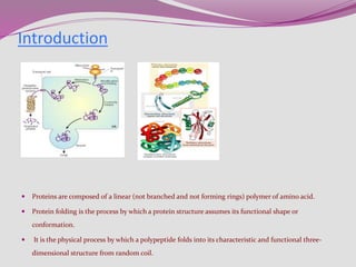
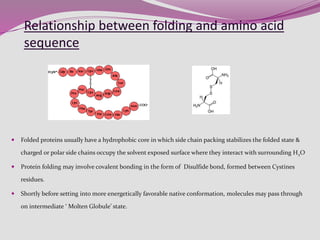
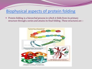
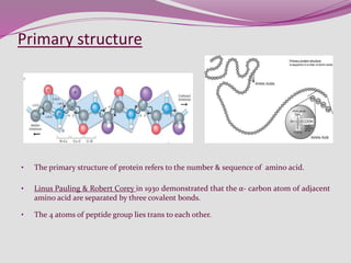
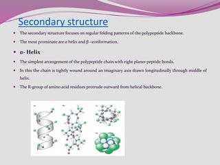
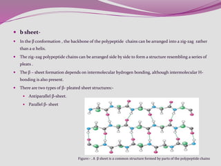
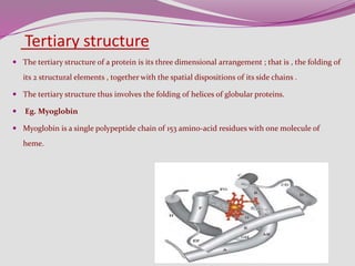
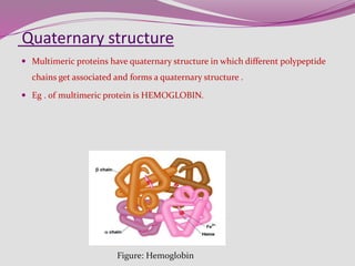
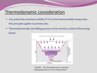
Protein folding
By
KAUSHAL KUMAR SAHU
Assistant Professor (Ad Hoc)
Department of Biotechnology
Govt. Digvijay Autonomous P. G. College
Raj-Nandgaon ( C. G. )
 Introduction-
 Levinthal paradox
 Biophysical aspects of protein folding
 Hierarchy in protein structure
 Thermodynamic stability
 Cellular aspects of protein folding
 Self assembly (folding) of protein
 Molecular assistance – Chaperons
 Enzymes involved in protein folding
 Conclusions
 References
Synopsis
 Proteins are composed of a linear (not branched and not forming rings) polymer of amino acid.
 Protein folding is the process by which a protein structure assumes its functional shape or
conformation.
 It is the physical process by which a polypeptide folds into its characteristic and functional three-
dimensional structure from random coil.
Introduction
• A newly synthesized polypeptide chain can theoretically assume high number of conformation.
• Let’s assume 10100 different conformation for a polypeptide and if each conformation were
sampled in every ~10-13 seconds, it would take about 1077 years to sample all possible
conformation.
• But experimentally it takes a few seconds or up to a minute for completion of process.
• Thus protein folding can not be a completely random & error process. There must be short
cuts.
• This was first pointed out by CYRUS LEVINTHAL in 1968, which is also called as
Levinthal’s Paradox.
Levinthal paradox
 Folded proteins usually have a hydrophobic core in which side chain packing stabilizes the folded state &
charged or polar side chains occupy the solvent exposed surface where they interact with surrounding H2O
 Protein folding may involve covalent bonding in the form of Disulfide bond, formed between Cystines
residues.
 Shortly before setting into more energetically favorable native conformation, molecules may pass through
on intermediate ‘ Molten Globule’ state.
Relationship between folding and amino acid
sequence
 Protein folding is a hierarchal process in which it folds from its primary
structure through a series and attains its final folding .These structures are :-
Biophysical aspects of protein folding
Primary structure
• The primary structure of protein refers to the number & sequence of amino acid.
• Linus Pauling & Robert Corey in 1930 demonstrated that the α- carbon atom of adjacent
amino acid are separated by three covalent bonds.
• The 4 atoms of peptide group lies trans to each other.
Secondary structure
 The secondary structure focuses on regular folding patterns of the polypeptide backbone.
 The most prominate are α helix and β –conformation.
 α- Helix
 The simplest arrangement of the polypeptide chain with right planer peptide bonds.
 In this the chain is tightly wound around an imaginary axis drawn longitudinally through middle of
helix.
 The R-group of amino acid residues protrude outward from helical backbone.
 b sheet-
 In the β conformation , the backbone of the polypeptide chains can be arranged into a zig-zag rather
than a α helix.
 The zig–zag polypeptide chains can be arranged side by side to form a structure resembling a series of
pleats .
 The β – sheet formation depends on intermolecular hydrogen bonding, although intermolecular H-
bonding is also present.
 There are two types of β- pleated sheet structures:-
 Antiparallel β-sheet.
 Parallel β- sheet
Figure:- . A β sheet is a common structure formed by parts of the polypeptide chains
Super secondary structure
 Super secondary structures, also called motifs or simply folds simply folds are particularly
stable arrangements of several elements of secondary structures and the connections
between them .
 Domains – polypeptides with more than a few hundred amino acid residues often folds
into two or more stable globular units called domains.
Figure:- Protein Domains. The cell-surface protein
CD4 consists of four similar domains.
Tertiary structure
 The tertiary structure of a protein is its three dimensional arrangement ; that is , the folding of
its 2 structural elements , together with the spatial dispositions of its side chains .
 The tertiary structure thus involves the folding of helices of globular proteins.
 Eg. Myoglobin
 Myoglobin is a single polypeptide chain of 153 amino-acid residues with one molecule of
heme.
Quaternary structure
 Multimeric proteins have quaternary structure in which different polypeptide
chains get associated and forms a quaternary structure .
 Eg . of multimeric protein is HEMOGLOBIN.
Figure: Hemoglobin
Thermodynamic consideration
 Any system has maximum stability if it is in the lowest possible energy state ,
this principles applies to proteins also .
 Thermodynamically, the folding process can be viewed as a kind of free energy
funnel
FIGURE: - The thermodynamics of protein
folding depicted as a Free-energy funnel.
(a) Self folding- e.g. Ribonuclease
(b) Molecular assistance-
Chaperons
Chaperonins
Cellular aspects of protein folding
By Chaperons
 Molecular chaperones are essential proteins that bind to unfolded and partially folded
polypeptide chains to prevent the improper association of exposed hydrophobic
segments that might lead to nonnative folding as well as polypeptide aggregation and
precipitation.
 1. Hsp 70 system-
 Identification of unfolded target protein
 Cycle of working of chaperons
Fig – Identification of unfolded target protein and Chaperones control protein folding interaction
Cycle of working of chaperons
Fig :- Cycle of binding and release of Hsp 40 – Hsp 70 .
By Chaperonins :- (Hsp60 / GroEL in E. Coli.)
 Chaperonin are the large structures
with hollow cavities often used for
handling the folding or degradation
of proteins
 The Hsp60 class of chaperones
forms a large apparatus that consist
of two types of subunit.
Fig :- Structure of Gro EL forming an oligomer of two ring
, each comprising a hollow cylinder made of 7 subunit.
Enzymes Involved In protein folding
 Protein disulfide isomerare ( PDI ) .
 Peptidyl prolylisomerase .
Fig :- Role of PDI in protein folding
Peptidyl prolyl isomerare
 This enzyme catalysis the isomerization of peptide bond .
 That involve protein reduce peptide bond between amino acid are almost always
in turns from .
Fig :- Isomers of protein
 Protein folding is the physical process by which a polypeptide folds into its
characteristics three dimensional structure.
 Failure to fold into native structure generally produces inactive proteins, but in
some instances misfolded proteins have modified or toxic functionality.
Conclusions-
 Books-
 Fundamental of biochemistry – Voet Voet Pratt – 4th edition
 Principle of biochemistry – David Nelson Michel M.Cox
 Websites-
 www.wekipidia.com
 www.KBiotech.com
 www.nature.com
References-

Protien folding by kk sahu 2

  • 1.
    Protein folding By KAUSHAL KUMARSAHU Assistant Professor (Ad Hoc) Department of Biotechnology Govt. Digvijay Autonomous P. G. College Raj-Nandgaon ( C. G. )
  • 2.
     Introduction-  Levinthalparadox  Biophysical aspects of protein folding  Hierarchy in protein structure  Thermodynamic stability  Cellular aspects of protein folding  Self assembly (folding) of protein  Molecular assistance – Chaperons  Enzymes involved in protein folding  Conclusions  References Synopsis
  • 3.
     Proteins arecomposed of a linear (not branched and not forming rings) polymer of amino acid.  Protein folding is the process by which a protein structure assumes its functional shape or conformation.  It is the physical process by which a polypeptide folds into its characteristic and functional three- dimensional structure from random coil. Introduction
  • 4.
    • A newlysynthesized polypeptide chain can theoretically assume high number of conformation. • Let’s assume 10100 different conformation for a polypeptide and if each conformation were sampled in every ~10-13 seconds, it would take about 1077 years to sample all possible conformation. • But experimentally it takes a few seconds or up to a minute for completion of process. • Thus protein folding can not be a completely random & error process. There must be short cuts. • This was first pointed out by CYRUS LEVINTHAL in 1968, which is also called as Levinthal’s Paradox. Levinthal paradox
  • 5.
     Folded proteinsusually have a hydrophobic core in which side chain packing stabilizes the folded state & charged or polar side chains occupy the solvent exposed surface where they interact with surrounding H2O  Protein folding may involve covalent bonding in the form of Disulfide bond, formed between Cystines residues.  Shortly before setting into more energetically favorable native conformation, molecules may pass through on intermediate ‘ Molten Globule’ state. Relationship between folding and amino acid sequence
  • 6.
     Protein foldingis a hierarchal process in which it folds from its primary structure through a series and attains its final folding .These structures are :- Biophysical aspects of protein folding
  • 7.
    Primary structure • Theprimary structure of protein refers to the number & sequence of amino acid. • Linus Pauling & Robert Corey in 1930 demonstrated that the α- carbon atom of adjacent amino acid are separated by three covalent bonds. • The 4 atoms of peptide group lies trans to each other.
  • 8.
    Secondary structure  Thesecondary structure focuses on regular folding patterns of the polypeptide backbone.  The most prominate are α helix and β –conformation.  α- Helix  The simplest arrangement of the polypeptide chain with right planer peptide bonds.  In this the chain is tightly wound around an imaginary axis drawn longitudinally through middle of helix.  The R-group of amino acid residues protrude outward from helical backbone.
  • 9.
     b sheet- In the β conformation , the backbone of the polypeptide chains can be arranged into a zig-zag rather than a α helix.  The zig–zag polypeptide chains can be arranged side by side to form a structure resembling a series of pleats .  The β – sheet formation depends on intermolecular hydrogen bonding, although intermolecular H- bonding is also present.  There are two types of β- pleated sheet structures:-  Antiparallel β-sheet.  Parallel β- sheet Figure:- . A β sheet is a common structure formed by parts of the polypeptide chains
  • 10.
    Super secondary structure Super secondary structures, also called motifs or simply folds simply folds are particularly stable arrangements of several elements of secondary structures and the connections between them .  Domains – polypeptides with more than a few hundred amino acid residues often folds into two or more stable globular units called domains. Figure:- Protein Domains. The cell-surface protein CD4 consists of four similar domains.
  • 11.
    Tertiary structure  Thetertiary structure of a protein is its three dimensional arrangement ; that is , the folding of its 2 structural elements , together with the spatial dispositions of its side chains .  The tertiary structure thus involves the folding of helices of globular proteins.  Eg. Myoglobin  Myoglobin is a single polypeptide chain of 153 amino-acid residues with one molecule of heme.
  • 12.
    Quaternary structure  Multimericproteins have quaternary structure in which different polypeptide chains get associated and forms a quaternary structure .  Eg . of multimeric protein is HEMOGLOBIN. Figure: Hemoglobin
  • 13.
    Thermodynamic consideration  Anysystem has maximum stability if it is in the lowest possible energy state , this principles applies to proteins also .  Thermodynamically, the folding process can be viewed as a kind of free energy funnel FIGURE: - The thermodynamics of protein folding depicted as a Free-energy funnel.
  • 14.
    (a) Self folding-e.g. Ribonuclease (b) Molecular assistance- Chaperons Chaperonins Cellular aspects of protein folding
  • 15.
    By Chaperons  Molecularchaperones are essential proteins that bind to unfolded and partially folded polypeptide chains to prevent the improper association of exposed hydrophobic segments that might lead to nonnative folding as well as polypeptide aggregation and precipitation.  1. Hsp 70 system-  Identification of unfolded target protein  Cycle of working of chaperons Fig – Identification of unfolded target protein and Chaperones control protein folding interaction
  • 16.
    Cycle of workingof chaperons Fig :- Cycle of binding and release of Hsp 40 – Hsp 70 .
  • 17.
    By Chaperonins :-(Hsp60 / GroEL in E. Coli.)  Chaperonin are the large structures with hollow cavities often used for handling the folding or degradation of proteins  The Hsp60 class of chaperones forms a large apparatus that consist of two types of subunit. Fig :- Structure of Gro EL forming an oligomer of two ring , each comprising a hollow cylinder made of 7 subunit.
  • 18.
    Enzymes Involved Inprotein folding  Protein disulfide isomerare ( PDI ) .  Peptidyl prolylisomerase . Fig :- Role of PDI in protein folding
  • 19.
    Peptidyl prolyl isomerare This enzyme catalysis the isomerization of peptide bond .  That involve protein reduce peptide bond between amino acid are almost always in turns from . Fig :- Isomers of protein
  • 20.
     Protein foldingis the physical process by which a polypeptide folds into its characteristics three dimensional structure.  Failure to fold into native structure generally produces inactive proteins, but in some instances misfolded proteins have modified or toxic functionality. Conclusions-
  • 21.
     Books-  Fundamentalof biochemistry – Voet Voet Pratt – 4th edition  Principle of biochemistry – David Nelson Michel M.Cox  Websites-  www.wekipidia.com  www.KBiotech.com  www.nature.com References-