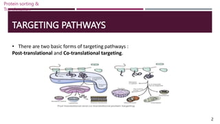
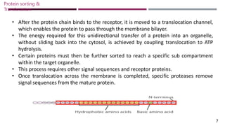
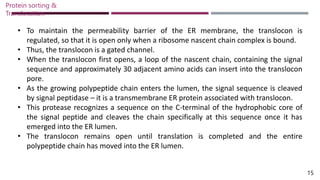
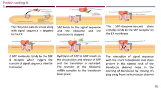
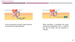
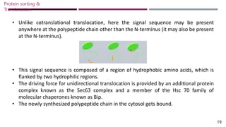
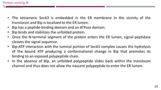
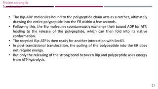
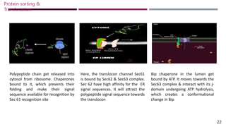
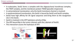
PROTEIN SORTING &
TRANSLOCATION
Submitted by,
A.T Milin Sera
Roll no : 1
1st M.Sc. Botany
ST. Teresa’s College, Ekm
Submitted to,
Ms. Nishitha I. K.
Assistant Professor
ST. Teresa’s College, Ekm
CONTENTS
• POST-TRANSLATIONAL TARGETING
• CO-TRANSLATIONAL TARGETING
• SIGNAL SEQUENCES
• SIGNAL RECOGNITION PARTICLE (SRP)
• TRANSLOCON
Protein sorting &
Translocation
INTRODUCTION
• In a mammalian cell, there are about 10,000 distinct varieties of proteins.
• Majority of these proteins are produced by cytosolic ribosomes and are kept in the
cytosol.
• Many of the proteins made by a cell are sent to a specific cell membrane or to the
cell surface for secretion.
• A cell cannot operate effectively unless all the proteins it produces find their
intended destinations.
• The delivery of newly synthesized proteins from the cytosol to their proper cellular
locations is referred to as “protein sorting, protein targeting or protein trafficking”.
1
Protein sorting &
Translocation
TARGETING PATHWAYS
• There are two basic forms of targeting pathways :
Post-translational and Co-translational targeting.
2
Protein sorting &
Translocation
POST-TRANSLATIONAL TARGETING
• It occurs soon after the synthesis of protein by translation at the ribosome.
• It occurs in the ribosomes that remain free in the cytosol.
• Such ribosomes synthesis polypeptides destined for the cytosol or for import into
the nucleus, mitochondria, chloroplasts or peroxisomes.
• When the polypeptide is complete, it is released from the ribosome.
• It is either remains in the cytosol or is transported into the appropriate organelle by
post-transitional import.
3
Protein sorting &
Translocation
CO-TRANSLATIONAL TARGETING
• Proteins as they are translated are targeted to the endoplasmic reticulum (ER) and
thereby enter the secretory pathway.
• These proteins are targeted to ER, Golgi apparatus, lysosomes, plasma membrane and
secreted proteins.
• It occurs in ribosomes that are attached to ER membranes.
• Such ribosomes synthesis polypeptides destined for the endomembrane system or for
export from the cell.
• Newly forming polypeptide is transferred across the ER membrane by co-translational
import.
• The completed polypeptide either remains in the ER or is transported via various
vesicles to another compartment.
4
Protein sorting &
Translocation
CO-TRANSLATIONAL TARGETING POST-TRANSLATIONAL TARGETING
Translocation takes place along with translation Translocation takes place after translation is
completed
Entire Ribosome-mRNA complex is translocated
translocated
Only the newly synthesized polypeptide chain
is translocated
Targeted to ER, Golgi bodies, lysosomes, plant
vacuoles, Plasma membrane etc.
Targeted to ER lumen, mitochondria,
peroxisomes, chloroplast etc.
Occurs in ribosomes that are associated with ER
(RER)
Occurs in free ribosomes
5
Protein sorting &
Translocation
SIGNAL SEQUENCES
• Signal sequences are the sequences that help in targeting proteins to their proper
cellular destinations.
• These sequences are present in the synthesized protein itself.
• They are about 20 – 50 amino acids long.
• Located at the N-terminus of the nascent chain.
• It includes a set of hydrophobic amino acids (6-12), which is preceded by a set of
basic amino acids.
• These signal sequences or uptake-targeting sequences are bounded by receptor
proteins.
• These govern the specificity of targeting.
6
Protein sorting &
Translocation
• After the protein chain binds to the receptor, it is moved to a translocation channel,
which enables the protein to pass through the membrane bilayer.
• The energy required for this unidirectional transfer of a protein into an organelle,
without sliding back into the cytosol, is achieved by coupling translocation to ATP
hydrolysis.
• Certain proteins must then be further sorted to reach a specific sub compartment
within the target organelle.
• This process requires other signal sequences and receptor proteins.
• Once translocation across the membrane is completed, specific proteases remove
signal sequences from the mature protein.
7
Protein sorting &
Translocation
TRANSLOCATION OF SECRETORY PROTEINS ACROSS ER
• The same secretory pathway is used by all eukaryotic cells for synthesizing and
sorting secreted proteins and soluble luminal proteins in the ER, Golgi apparatus and
lysosomes.
• These proteins are collectively referred to as “secretory proteins”.
• Despite the fact that all cells secrete a range of proteins, some cell are specialized to
release vast amounts of specific proteins.
• Pancreatic acinar cells are one example.
• They produce a lot of the digestive enzymes that are released into the ductules that
lead to the colon.
8
Protein sorting &
Translocation
SIGNAL SEQUENCES OF SECRETORY PROTEINS TO ER
• A 16 – 30 residue ER signal sequence is present in the nascent protein produced by
the ribosome.
• ER signal sequence directs the ribosome to the ER membrane.
• This initiates translocation of the growing polypeptide across the ER membrane.
• It is located at the N-terminus of the protein and form the first part of the protein.
• They contain one or more positively charged amino acids that are adjacent to a
continuous stretch of 6-12 hydrophobic residues.
• It is cleaved from the protein while it is still growing on the ribosome.
• Thus is usually not present in the mature proteins that are found in cells.
9
Protein sorting &
Translocation
SIGNAL RECOGNITION PARTICLE (SRP)
• SRPs are the key components in protein targeting.
• It is a cytosolic ribonucleoprotein particle.
• It transiently binds simultaneously to the ER signal sequence in a nascent protein.
• It also binds to the large ribosomal unit and to the SRP receptor that are present on
the membrane of the ER.
• Six discrete polypeptides and one small cytoplasmic RNA (7SL RNA) compose the
SRP.
• The six polypeptides are P54, P19, P68, P72, P9 & P14.
Protein sorting &
Translocation
10
• P54 & P19 are chemically cross linked to the ER signal sequences.
• The hydrophobic region of P54 contains a cleft which interacts with the
hydrophobic N-termini of nascent secretory proteins.
• This selectively targets them to the ER membrane.
• P68 & P72 binds to the SRP receptor.
• P9 & P14 binds to the smaller subunit of ribosome.
• Once the SRP get binds to the ribosome, translation is stopped and it restarts only
after the dissociation of the SRP.
Protein sorting &
Translocation
11
SRP RECEPTOR
• The SRP receptor is an integral membrane protein that is made up of two subunits α
and β.
• SRP receptor mediates the interaction of nascent secretory protein with the ER
membrane.
• It also permits the elongation and completion of the protein.
• The SRP and SRP receptor function to bring ribosomes that are synthesizing
secretory proteins to the ER membrane.
• The energy from GTP hydrolysis is used to release proteins lacking proper signal
sequences from SRP and SRP receptor complex.
• This prevents their mistargeting to the ER membrane.
Protein sorting &
Translocation
12
• The interaction of the SRP-nascent chain-ribosome complex with the SRP receptor is
promoted when GTP is bound by both P54 subunit of SRP and the α-subunit of SRP
receptor.
• This is followed by the transfer of the nascent chain and ribosome to a site on the ER
membrane where translocation can take place.
• Hydrolysis of the bound GTP takes place.
• After dissociating, SRP and its receptor release the bound GDP and recycle to the
cytosol.
• Then initiates another round of interaction between ribosomes synthesizing nascent
secretory proteins for their co-translational import to the ER.
Protein sorting &
Translocation
13
TRANSLOCATION INTO THE ER LUMEN
CO-TRANSLATIONAL TRANSLOCATION INTO ER
• After the targeting of the ribosome-synthesizing secretory protein to the ER
membrane, the ribosome and nascent chain are rapidly transferred to the
‘translocon’.
• Translocon is a protein-lined channel within the membrane.
• The process of translation continues and the elongating polypeptide passes directly
from the large ribosomal subunit into the central pore of the translocon.
• The 60S ribosomal subunit is aligned with the pore of the translocon.
• The growing chain is never exposed to the cytoplasm and does not fold until it
reaches the ER lumen.
Protein sorting &
Translocation
14
• To maintain the permeability barrier of the ER membrane, the translocon is
regulated, so that it is open only when a ribosome nascent chain complex is bound.
• Thus, the translocon is a gated channel.
• When the translocon first opens, a loop of the nascent chain, containing the signal
sequence and approximately 30 adjacent amino acids can insert into the translocon
pore.
• As the growing polypeptide chain enters the lumen, the signal sequence is cleaved
by signal peptidase – it is a transmembrane ER protein associated with translocon.
• This protease recognizes a sequence on the C-terminal of the hydrophobic core of
the signal peptide and cleaves the chain specifically at this sequence once it has
emerged into the ER lumen.
• The translocon remains open until translation is completed and the entire
polypeptide chain has moved into the ER lumen.
Protein sorting &
Translocation
15
The ribosome-nascent chain along
with signal sequence is targeted
to the ER
SRP binds to the signal sequence
and the ribosome and the
translation is stopped
This SRP-ribosome-nascent chain
complex binds to the SRP receptor on
the ER membrane
2 GTP molecules binds to the SRP
& receptor which triggers the
transfer of signal sequence into the
translocon
Hydrolysis of GTP to GDP results in
the dissociation and release of SRP
and the translation is restarted.
The transfer of the ribosome
mRNA complex to the translocon
takes place
The interaction of signal sequence
with the short hydrophobic side chain
present in the narrow neck of the
translocon channel helps in the
opening of translocon by moving it’s
plug away from the translocon channel
16
Protein sorting &
Translocation
As the translocation precedes, signal sequence
is cleaved by the signal peptidase
17
Protein sorting &
Translocation
When translation is completed, the newly
synthesized polypeptide chain is released
into the ER lumen and the ribosome is
released
POST-TRANSLATIONAL TRANSLOCATION INTO ER
• In most eukaryotes, secretory proteins enter the ER by co-translational translocation,
using the energy derived from translocation to pass through the membrane.
• In yeast, some secretory proteins enter the ER lumen after translation has been
completed, that is post-translational translocation.
• In this case, the translocating protein pass through the same translocon (Sec 61) used
in co-translational translocation.
• The SRP and SRP receptor are not involved in this case.
• In such cases, the direct interaction between the translocon and the signal sequence
is sufficient for targeting to the ER membrane.
Protein sorting &
Translocation
18
• Unlike cotranslational translocation, here the signal sequence may be present
anywhere at the polypeptide chain other than the N-terminus (it may also be present
at the N-terminus).
• This signal sequence is composed of a region of hydrophobic amino acids, which is
flanked by two hydrophilic regions.
• The driving force for unidirectional translocation is provided by an additional protein
complex known as the Sec63 complex and a member of the Hsc 70 family of
molecular chaperones known as Bip.
• The newly synthesized polypeptide chain in the cytosol gets bound.
Protein sorting &
Translocation
19
• The tetrameric Sec63 is embedded in the ER membrane in the vicinity of the
translocon and Bip is localized to the ER lumen.
• Bip has a peptide-binding domain and an ATPase domain.
• Bip binds and stabilizes the unfolded protein.
• Once the N-terminal segment of the protein enters the ER lumen, signal peptidase
cleaves the signal sequence.
• Bip-ATP interaction with the luminal portion of Sec63 complex causes the hydrolysis
of the bound ATP producing a conformational change in Bip that promotes its
binding to an exposed polypeptide chain.
• In the absence of Bip, an unfolded polypeptide slides back within the translocon
channel and thus does not allow the nascent polypeptide to enter the ER lumen.
20
Protein sorting &
Translocation
• The Bip-ADP molecules bound to the polypeptide chain acts as a ratchet, ultimately
drawing the entire polypeptide into the ER within a few seconds.
• Following this, the Bip molecules spontaneously exchange their bound ADP for ATP,
leading to the release of the polypeptide, which can then fold into its native
conformation.
• The recycled Bip-ATP is then ready for another interaction with Sec63.
• In post-translational translocation, the pulling of the polypeptide into the ER does
not require energy.
• But only the releasing of the strong bond between Bip and polypeptide uses energy
from ATP hydrolysis.
Protein sorting &
Translocation
21
Polypeptide chain get released into
cytosol from ribosome. Chaperones
bound to it, which prevents their
folding and make their signal
sequence available for recognition by
Sec 61 recognition site
Here, the translocon channel Sec61
is bound by Sec62 & Sec63 complex.
Sec 62 have high affinity for the ER
signal sequences. It will attract the
polypeptide signal sequence towards
the translocon
22
Protein sorting &
Translocation
Bip chaperone in the lumen get
bound by ATP. It moves towards the
Sec63 complex & interact with its j-
domain undergoing ATP hydrolysis,
which creates a conformational
change in Bip
When the polypeptide binds to the
translocon, a small part of it extends
to the ER lumen. ADP bound Bip
binds to this part and prevent its
slipping back out. Similarly, many
ADP-Bip molecules binds to the parts
of peptide chain that get exposed to
the lumen
Over time, the jostling of the
polypeptide and binding of Bip pull
the chain to the ER. This type of
transport system is called a ratchet
Polypeptide chain get released
from the Bip when Bip get bound
by ATP molecules and free
polypeptide chain remain in the ER
lumen
23
Protein sorting &
Translocation
TRANSLOCON
• The translocon (also known as a translocator or translocation channel) is a complex
of proteins associated with the translocation of polypeptides across membranes.
• In eukaryotes the term translocon most commonly refers to the complex that
transports nascent polypeptides with a targeting signal sequence into the interior
(cisternal or lumenal) space of the ER from the cytosol.
• This translocation process requires the protein to cross a hydrophobic lipid bilayer.
• The same complex is also used to integrate nascent proteins into the membrane
itself (membrane proteins).
• In prokaryotes, a similar protein complex transports polypeptides across the (inner)
plasma membrane or integrates membrane proteins.
Protein sorting &
Translocation
24
• In either case, the protein complex are formed from Sec proteins (Sec: secretory),
with the heterotrimeric Sec61 being the channel.
• In prokaryotes, the homologous channel complex is known as SecYEG.
• It consists of the subunits SecY, SecE, and SecG.
• SecY is the large pore subunit.
• In a side view, the channel has an hourglass shape, with a funnel on each side.
• The extracellular funnel has a little “plug” formed out of an alpha-helix.
• In the middle of the membrane is a construction, formed from a pore ring of six
hydrophobic amino acids that project their side chains inwards.
Protein sorting &
Translocation
25
• During protein translocation, the plug is moved out of the way, and a polypeptide
chain is moved from the cytoplasmic funnel, through the pore ring, the extracellular
funnel, into the extracellular space.
• Hydrophobic segments of membrane proteins exit sideways through the lateral gate
into the lipid phase and become membrane-spanning segments.
• In bacteria, SecYEG forms a complex with SecDF, YajC and YidC.
Protein sorting &
Translocation
26
Protein sorting &
Translocation
27
• In eukaryotes, Sec61 forms a complex with the oligosaccharyl transferase complex,
the TRAP complex, and the membrane protein TRAM (possible chaperone).
• In cotranslational translocation, only the Sec61 translocon channel is involved.
• In posttranslational translocation, the Sec61 is bound by Sec62 and Sec63 complex.
• Sec62 have high affinity for ER signal sequence and bring them to the recognition
site in the Sec61.
• Sec63 is involved in the ATP hydrolysis activity of Bip.
• The ATP bound Bip in the lumen interacts with the j-domain of Sec63.
• This interaction causes the hydrolysis of ATP.
REFERENCE
• Cooper, G., & Hausman, R. (2013). The Cell: A Molecular Approach (6th ed.).
Sunderland, MA: Sinauer Associates.
• Dalbey, R., & von Heijne, G. (Eds.). (2002). Protein targeting, transport, and
translocation. Elsevier.
• Malathi, V. (1899). Molecular Biology. Pearson Education India.
• Nagai, K., Oubridge, C., Kuglstatter, A., Menichelli, E., Isel, C., & Jovine, L. (2003).
Structure, function and evolution of the signal recognition particle. The EMBO journal,
22(14), 3479-3485.
• Segev, N., Fewell, S. W., & Brodsky, J. L. (2009). Entry into the endoplasmic reticulum:
protein translocation, folding and quality control. Trafficking Inside Cells: Pathways,
Mechanisms and Regulation, 119-142.
• https://www.cell.com/fulltext/S0092-8674(00)80780-1
28
Protein sorting &
Translocation
THANK YOU

Protein sorting and translocation. pptx

  • 1.
    PROTEIN SORTING & TRANSLOCATION Submittedby, A.T Milin Sera Roll no : 1 1st M.Sc. Botany ST. Teresa’s College, Ekm Submitted to, Ms. Nishitha I. K. Assistant Professor ST. Teresa’s College, Ekm
  • 2.
    CONTENTS • POST-TRANSLATIONAL TARGETING •CO-TRANSLATIONAL TARGETING • SIGNAL SEQUENCES • SIGNAL RECOGNITION PARTICLE (SRP) • TRANSLOCON Protein sorting & Translocation
  • 3.
    INTRODUCTION • In amammalian cell, there are about 10,000 distinct varieties of proteins. • Majority of these proteins are produced by cytosolic ribosomes and are kept in the cytosol. • Many of the proteins made by a cell are sent to a specific cell membrane or to the cell surface for secretion. • A cell cannot operate effectively unless all the proteins it produces find their intended destinations. • The delivery of newly synthesized proteins from the cytosol to their proper cellular locations is referred to as “protein sorting, protein targeting or protein trafficking”. 1 Protein sorting & Translocation
  • 4.
    TARGETING PATHWAYS • Thereare two basic forms of targeting pathways : Post-translational and Co-translational targeting. 2 Protein sorting & Translocation
  • 5.
    POST-TRANSLATIONAL TARGETING • Itoccurs soon after the synthesis of protein by translation at the ribosome. • It occurs in the ribosomes that remain free in the cytosol. • Such ribosomes synthesis polypeptides destined for the cytosol or for import into the nucleus, mitochondria, chloroplasts or peroxisomes. • When the polypeptide is complete, it is released from the ribosome. • It is either remains in the cytosol or is transported into the appropriate organelle by post-transitional import. 3 Protein sorting & Translocation
  • 6.
    CO-TRANSLATIONAL TARGETING • Proteinsas they are translated are targeted to the endoplasmic reticulum (ER) and thereby enter the secretory pathway. • These proteins are targeted to ER, Golgi apparatus, lysosomes, plasma membrane and secreted proteins. • It occurs in ribosomes that are attached to ER membranes. • Such ribosomes synthesis polypeptides destined for the endomembrane system or for export from the cell. • Newly forming polypeptide is transferred across the ER membrane by co-translational import. • The completed polypeptide either remains in the ER or is transported via various vesicles to another compartment. 4 Protein sorting & Translocation
  • 7.
    CO-TRANSLATIONAL TARGETING POST-TRANSLATIONALTARGETING Translocation takes place along with translation Translocation takes place after translation is completed Entire Ribosome-mRNA complex is translocated translocated Only the newly synthesized polypeptide chain is translocated Targeted to ER, Golgi bodies, lysosomes, plant vacuoles, Plasma membrane etc. Targeted to ER lumen, mitochondria, peroxisomes, chloroplast etc. Occurs in ribosomes that are associated with ER (RER) Occurs in free ribosomes 5 Protein sorting & Translocation
  • 8.
    SIGNAL SEQUENCES • Signalsequences are the sequences that help in targeting proteins to their proper cellular destinations. • These sequences are present in the synthesized protein itself. • They are about 20 – 50 amino acids long. • Located at the N-terminus of the nascent chain. • It includes a set of hydrophobic amino acids (6-12), which is preceded by a set of basic amino acids. • These signal sequences or uptake-targeting sequences are bounded by receptor proteins. • These govern the specificity of targeting. 6 Protein sorting & Translocation
  • 9.
    • After theprotein chain binds to the receptor, it is moved to a translocation channel, which enables the protein to pass through the membrane bilayer. • The energy required for this unidirectional transfer of a protein into an organelle, without sliding back into the cytosol, is achieved by coupling translocation to ATP hydrolysis. • Certain proteins must then be further sorted to reach a specific sub compartment within the target organelle. • This process requires other signal sequences and receptor proteins. • Once translocation across the membrane is completed, specific proteases remove signal sequences from the mature protein. 7 Protein sorting & Translocation
  • 10.
    TRANSLOCATION OF SECRETORYPROTEINS ACROSS ER • The same secretory pathway is used by all eukaryotic cells for synthesizing and sorting secreted proteins and soluble luminal proteins in the ER, Golgi apparatus and lysosomes. • These proteins are collectively referred to as “secretory proteins”. • Despite the fact that all cells secrete a range of proteins, some cell are specialized to release vast amounts of specific proteins. • Pancreatic acinar cells are one example. • They produce a lot of the digestive enzymes that are released into the ductules that lead to the colon. 8 Protein sorting & Translocation
  • 11.
    SIGNAL SEQUENCES OFSECRETORY PROTEINS TO ER • A 16 – 30 residue ER signal sequence is present in the nascent protein produced by the ribosome. • ER signal sequence directs the ribosome to the ER membrane. • This initiates translocation of the growing polypeptide across the ER membrane. • It is located at the N-terminus of the protein and form the first part of the protein. • They contain one or more positively charged amino acids that are adjacent to a continuous stretch of 6-12 hydrophobic residues. • It is cleaved from the protein while it is still growing on the ribosome. • Thus is usually not present in the mature proteins that are found in cells. 9 Protein sorting & Translocation
  • 12.
    SIGNAL RECOGNITION PARTICLE(SRP) • SRPs are the key components in protein targeting. • It is a cytosolic ribonucleoprotein particle. • It transiently binds simultaneously to the ER signal sequence in a nascent protein. • It also binds to the large ribosomal unit and to the SRP receptor that are present on the membrane of the ER. • Six discrete polypeptides and one small cytoplasmic RNA (7SL RNA) compose the SRP. • The six polypeptides are P54, P19, P68, P72, P9 & P14. Protein sorting & Translocation 10
  • 13.
    • P54 &P19 are chemically cross linked to the ER signal sequences. • The hydrophobic region of P54 contains a cleft which interacts with the hydrophobic N-termini of nascent secretory proteins. • This selectively targets them to the ER membrane. • P68 & P72 binds to the SRP receptor. • P9 & P14 binds to the smaller subunit of ribosome. • Once the SRP get binds to the ribosome, translation is stopped and it restarts only after the dissociation of the SRP. Protein sorting & Translocation 11
  • 14.
    SRP RECEPTOR • TheSRP receptor is an integral membrane protein that is made up of two subunits α and β. • SRP receptor mediates the interaction of nascent secretory protein with the ER membrane. • It also permits the elongation and completion of the protein. • The SRP and SRP receptor function to bring ribosomes that are synthesizing secretory proteins to the ER membrane. • The energy from GTP hydrolysis is used to release proteins lacking proper signal sequences from SRP and SRP receptor complex. • This prevents their mistargeting to the ER membrane. Protein sorting & Translocation 12
  • 15.
    • The interactionof the SRP-nascent chain-ribosome complex with the SRP receptor is promoted when GTP is bound by both P54 subunit of SRP and the α-subunit of SRP receptor. • This is followed by the transfer of the nascent chain and ribosome to a site on the ER membrane where translocation can take place. • Hydrolysis of the bound GTP takes place. • After dissociating, SRP and its receptor release the bound GDP and recycle to the cytosol. • Then initiates another round of interaction between ribosomes synthesizing nascent secretory proteins for their co-translational import to the ER. Protein sorting & Translocation 13
  • 16.
    TRANSLOCATION INTO THEER LUMEN CO-TRANSLATIONAL TRANSLOCATION INTO ER • After the targeting of the ribosome-synthesizing secretory protein to the ER membrane, the ribosome and nascent chain are rapidly transferred to the ‘translocon’. • Translocon is a protein-lined channel within the membrane. • The process of translation continues and the elongating polypeptide passes directly from the large ribosomal subunit into the central pore of the translocon. • The 60S ribosomal subunit is aligned with the pore of the translocon. • The growing chain is never exposed to the cytoplasm and does not fold until it reaches the ER lumen. Protein sorting & Translocation 14
  • 17.
    • To maintainthe permeability barrier of the ER membrane, the translocon is regulated, so that it is open only when a ribosome nascent chain complex is bound. • Thus, the translocon is a gated channel. • When the translocon first opens, a loop of the nascent chain, containing the signal sequence and approximately 30 adjacent amino acids can insert into the translocon pore. • As the growing polypeptide chain enters the lumen, the signal sequence is cleaved by signal peptidase – it is a transmembrane ER protein associated with translocon. • This protease recognizes a sequence on the C-terminal of the hydrophobic core of the signal peptide and cleaves the chain specifically at this sequence once it has emerged into the ER lumen. • The translocon remains open until translation is completed and the entire polypeptide chain has moved into the ER lumen. Protein sorting & Translocation 15
  • 18.
    The ribosome-nascent chainalong with signal sequence is targeted to the ER SRP binds to the signal sequence and the ribosome and the translation is stopped This SRP-ribosome-nascent chain complex binds to the SRP receptor on the ER membrane 2 GTP molecules binds to the SRP & receptor which triggers the transfer of signal sequence into the translocon Hydrolysis of GTP to GDP results in the dissociation and release of SRP and the translation is restarted. The transfer of the ribosome mRNA complex to the translocon takes place The interaction of signal sequence with the short hydrophobic side chain present in the narrow neck of the translocon channel helps in the opening of translocon by moving it’s plug away from the translocon channel 16 Protein sorting & Translocation
  • 19.
    As the translocationprecedes, signal sequence is cleaved by the signal peptidase 17 Protein sorting & Translocation When translation is completed, the newly synthesized polypeptide chain is released into the ER lumen and the ribosome is released
  • 20.
    POST-TRANSLATIONAL TRANSLOCATION INTOER • In most eukaryotes, secretory proteins enter the ER by co-translational translocation, using the energy derived from translocation to pass through the membrane. • In yeast, some secretory proteins enter the ER lumen after translation has been completed, that is post-translational translocation. • In this case, the translocating protein pass through the same translocon (Sec 61) used in co-translational translocation. • The SRP and SRP receptor are not involved in this case. • In such cases, the direct interaction between the translocon and the signal sequence is sufficient for targeting to the ER membrane. Protein sorting & Translocation 18
  • 21.
    • Unlike cotranslationaltranslocation, here the signal sequence may be present anywhere at the polypeptide chain other than the N-terminus (it may also be present at the N-terminus). • This signal sequence is composed of a region of hydrophobic amino acids, which is flanked by two hydrophilic regions. • The driving force for unidirectional translocation is provided by an additional protein complex known as the Sec63 complex and a member of the Hsc 70 family of molecular chaperones known as Bip. • The newly synthesized polypeptide chain in the cytosol gets bound. Protein sorting & Translocation 19
  • 22.
    • The tetramericSec63 is embedded in the ER membrane in the vicinity of the translocon and Bip is localized to the ER lumen. • Bip has a peptide-binding domain and an ATPase domain. • Bip binds and stabilizes the unfolded protein. • Once the N-terminal segment of the protein enters the ER lumen, signal peptidase cleaves the signal sequence. • Bip-ATP interaction with the luminal portion of Sec63 complex causes the hydrolysis of the bound ATP producing a conformational change in Bip that promotes its binding to an exposed polypeptide chain. • In the absence of Bip, an unfolded polypeptide slides back within the translocon channel and thus does not allow the nascent polypeptide to enter the ER lumen. 20 Protein sorting & Translocation
  • 23.
    • The Bip-ADPmolecules bound to the polypeptide chain acts as a ratchet, ultimately drawing the entire polypeptide into the ER within a few seconds. • Following this, the Bip molecules spontaneously exchange their bound ADP for ATP, leading to the release of the polypeptide, which can then fold into its native conformation. • The recycled Bip-ATP is then ready for another interaction with Sec63. • In post-translational translocation, the pulling of the polypeptide into the ER does not require energy. • But only the releasing of the strong bond between Bip and polypeptide uses energy from ATP hydrolysis. Protein sorting & Translocation 21
  • 24.
    Polypeptide chain getreleased into cytosol from ribosome. Chaperones bound to it, which prevents their folding and make their signal sequence available for recognition by Sec 61 recognition site Here, the translocon channel Sec61 is bound by Sec62 & Sec63 complex. Sec 62 have high affinity for the ER signal sequences. It will attract the polypeptide signal sequence towards the translocon 22 Protein sorting & Translocation Bip chaperone in the lumen get bound by ATP. It moves towards the Sec63 complex & interact with its j- domain undergoing ATP hydrolysis, which creates a conformational change in Bip
  • 25.
    When the polypeptidebinds to the translocon, a small part of it extends to the ER lumen. ADP bound Bip binds to this part and prevent its slipping back out. Similarly, many ADP-Bip molecules binds to the parts of peptide chain that get exposed to the lumen Over time, the jostling of the polypeptide and binding of Bip pull the chain to the ER. This type of transport system is called a ratchet Polypeptide chain get released from the Bip when Bip get bound by ATP molecules and free polypeptide chain remain in the ER lumen 23 Protein sorting & Translocation
  • 26.
    TRANSLOCON • The translocon(also known as a translocator or translocation channel) is a complex of proteins associated with the translocation of polypeptides across membranes. • In eukaryotes the term translocon most commonly refers to the complex that transports nascent polypeptides with a targeting signal sequence into the interior (cisternal or lumenal) space of the ER from the cytosol. • This translocation process requires the protein to cross a hydrophobic lipid bilayer. • The same complex is also used to integrate nascent proteins into the membrane itself (membrane proteins). • In prokaryotes, a similar protein complex transports polypeptides across the (inner) plasma membrane or integrates membrane proteins. Protein sorting & Translocation 24
  • 27.
    • In eithercase, the protein complex are formed from Sec proteins (Sec: secretory), with the heterotrimeric Sec61 being the channel. • In prokaryotes, the homologous channel complex is known as SecYEG. • It consists of the subunits SecY, SecE, and SecG. • SecY is the large pore subunit. • In a side view, the channel has an hourglass shape, with a funnel on each side. • The extracellular funnel has a little “plug” formed out of an alpha-helix. • In the middle of the membrane is a construction, formed from a pore ring of six hydrophobic amino acids that project their side chains inwards. Protein sorting & Translocation 25
  • 28.
    • During proteintranslocation, the plug is moved out of the way, and a polypeptide chain is moved from the cytoplasmic funnel, through the pore ring, the extracellular funnel, into the extracellular space. • Hydrophobic segments of membrane proteins exit sideways through the lateral gate into the lipid phase and become membrane-spanning segments. • In bacteria, SecYEG forms a complex with SecDF, YajC and YidC. Protein sorting & Translocation 26
  • 29.
    Protein sorting & Translocation 27 •In eukaryotes, Sec61 forms a complex with the oligosaccharyl transferase complex, the TRAP complex, and the membrane protein TRAM (possible chaperone). • In cotranslational translocation, only the Sec61 translocon channel is involved. • In posttranslational translocation, the Sec61 is bound by Sec62 and Sec63 complex. • Sec62 have high affinity for ER signal sequence and bring them to the recognition site in the Sec61. • Sec63 is involved in the ATP hydrolysis activity of Bip. • The ATP bound Bip in the lumen interacts with the j-domain of Sec63. • This interaction causes the hydrolysis of ATP.
  • 30.
    REFERENCE • Cooper, G.,& Hausman, R. (2013). The Cell: A Molecular Approach (6th ed.). Sunderland, MA: Sinauer Associates. • Dalbey, R., & von Heijne, G. (Eds.). (2002). Protein targeting, transport, and translocation. Elsevier. • Malathi, V. (1899). Molecular Biology. Pearson Education India. • Nagai, K., Oubridge, C., Kuglstatter, A., Menichelli, E., Isel, C., & Jovine, L. (2003). Structure, function and evolution of the signal recognition particle. The EMBO journal, 22(14), 3479-3485. • Segev, N., Fewell, S. W., & Brodsky, J. L. (2009). Entry into the endoplasmic reticulum: protein translocation, folding and quality control. Trafficking Inside Cells: Pathways, Mechanisms and Regulation, 119-142. • https://www.cell.com/fulltext/S0092-8674(00)80780-1 28 Protein sorting & Translocation
  • 31.