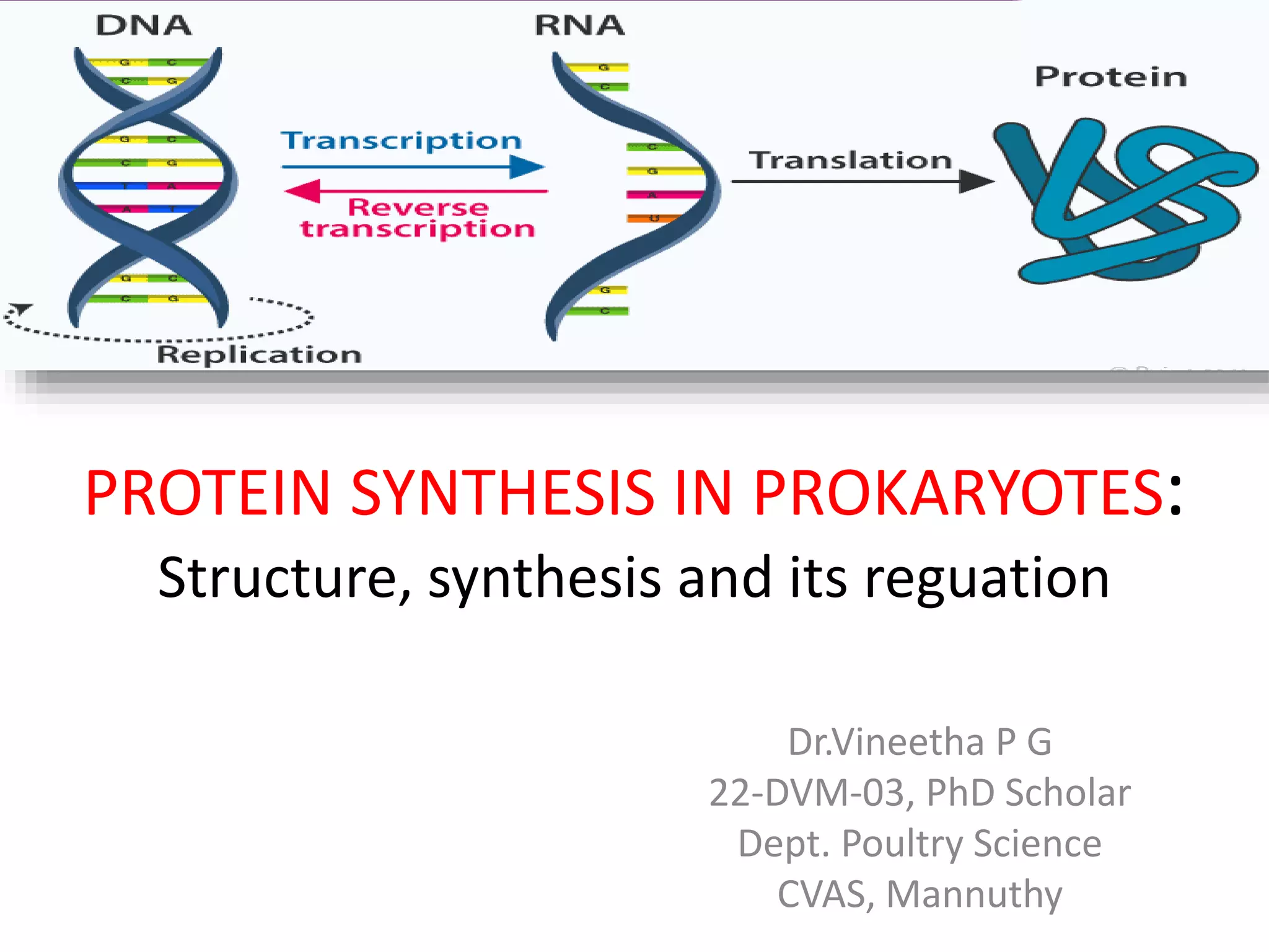
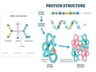
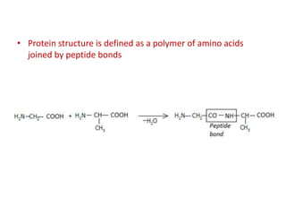
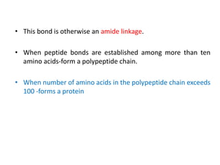
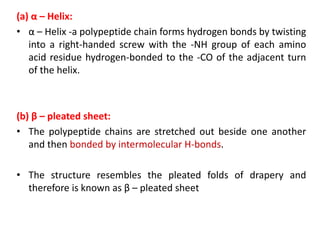
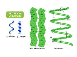
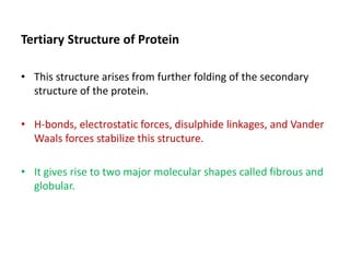
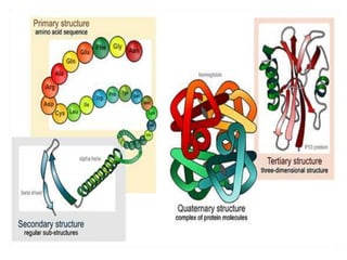
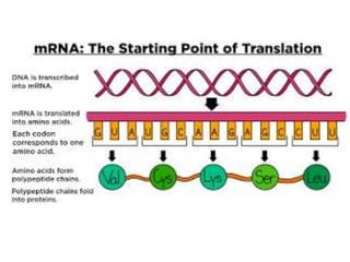
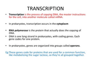
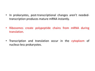
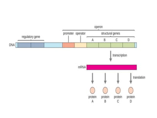
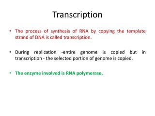
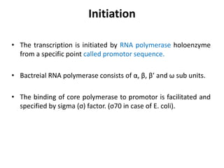
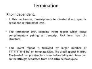
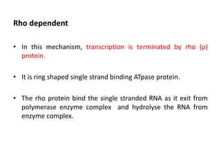
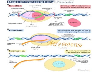
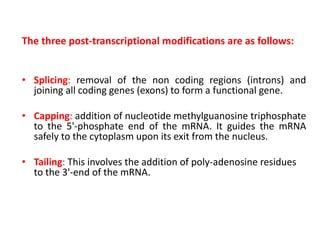
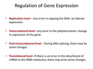
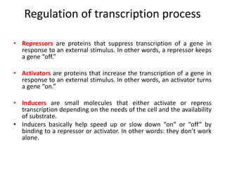
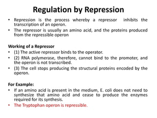
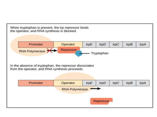
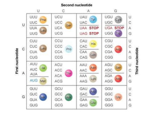
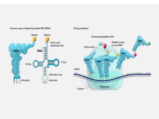
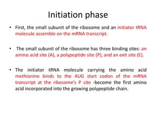
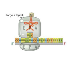
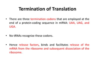
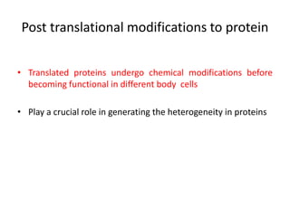
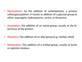
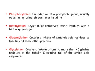
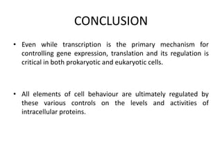
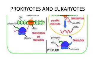
PROTEIN SYNTHESIS IN PROKARYOTES:
Structure, synthesis and its reguation
Dr.Vineetha P G
22-DVM-03, PhD Scholar
Dept. Poultry Science
CVAS, Mannuthy
• Protein structure is defined as a polymer of amino acids
joined by peptide bonds
• This bond is otherwise an amide linkage.
• When peptide bonds are established among more than ten
amino acids-form a polypeptide chain.
• When number of amino acids in the polypeptide chain exceeds
100 -forms a protein
Classification of Proteins
• Based on the molecular shape, proteins can be classified into
two types.
1. Fibrous Proteins:
• When the polypeptide chains run parallel and are held
together by hydrogen and disulfide bonds, then the fiber-like
structure is formed.
• These are water-insoluble proteins.
Example – keratin (present in hair, wool, and silk) and myosin
(present in muscles), etc.
2. Globular Proteins:
• This structure results when the chains of polypeptides coil
around to give a spherical shape.
• These are usually soluble in water.
Example – Insulin and albumins are common examples of
globular proteins.
Levels of Protein Structure
Linderstrom-Lang (1952) -suggested a hierarchy of protein
structure with four levels: primary, secondary, tertiary , and
quaternary.
Primary Structure of Protein
• The Primary structure of proteins is the exact ordering of
amino acids forming their chains.
• Covalent, peptide bonds which connect the amino acids
together maintain the primary structure of a protein.
Secondary Structure of Protein
• These polypeptide chains fold due to the interaction between
the amine and carboxyl group of the peptide link.
• They are found to exist in two different types of structures α –
helix and β – pleated sheet structures.
• This structure arises due to the regular folding of the
backbone of the polypeptide chain due to hydrogen bonding
between -CO group and -NH groups of the peptide bond.
(a) α – Helix:
• α – Helix -a polypeptide chain forms hydrogen bonds by twisting
into a right-handed screw with the -NH group of each amino
acid residue hydrogen-bonded to the -CO of the adjacent turn
of the helix.
(b) β – pleated sheet:
• The polypeptide chains are stretched out beside one another
and then bonded by intermolecular H-bonds.
• The structure resembles the pleated folds of drapery and
therefore is known as β – pleated sheet
Tertiary Structure of Protein
• This structure arises from further folding of the secondary
structure of the protein.
• H-bonds, electrostatic forces, disulphide linkages, and Vander
Waals forces stabilize this structure.
• It gives rise to two major molecular shapes called fibrous and
globular.
Quaternary Structure of Protein
• Some of the proteins are composed of two or more
polypeptide chains referred to as sub-units.
• The spatial arrangement of these subunits with respect to
each other is known as quaternary structure.
• A protein’s shape is determined by its primary structure (the
amino acid sequence).
TRANSCRIPTION
• Transcription is the process of copying DNA, the master instructions
for the cell, into another molecule called mRNA.
• In prokaryotes, transcription occurs in the cytoplasm
• RNA polymerase is the protein that actually does the copying of
DNA.
• DNA is one long strand in prokaryotes, with coding genes. Each
gene codes for one protein.
• In prokaryotes, genes are organized into groups called operons.
Eg:These genes code for proteins that are used for a common function,
like metabolizing the sugar lactose, so they're all grouped together.
• In prokaryotes, post-transcriptional changes aren’t needed-
transcription produces mature mRNA instantly.
• Ribosomes create polypeptide chains from mRNA during
translation.
• Transcription and translation occur in the cytoplasm of
nucleus-less prokaryotes.
• Proteins are the active players in most cell processes,
implementing the myriad tasks that are directed by the
information encoded in genomic DNA.
• Protein synthesis is thus the final stage of gene expression.
• The polypeptide chain fold into the three-dimensional
conformation undergo various processing steps before being
converted to its active form.
Operon
• François Jacob and Jacques Monod - showed the organization
of bacterial genes into operons-studies on the lac operon of E.
coli.
• In E. coli, all structural genes encode enzymes needed to use
lactose lie next to each other in the lactose (or lac) operon
under the control of a single promoter, the lac promoter.
• They won the Nobel Prize in Physiology or Medicine in 1965.
• In prokaryotes, operons whose gene products are required
consistently – expression is unregulated-transcribed and
translated to the protein products.
• Such genes encode enzymes in housekeeping functions -
cellular maintenance, including DNA replication, repair, and
expression, enzymes involved in core metabolism.
• Prokaryotic operons that are expressed only when needed
and are regulated by repressors, activators, and inducers
• Each operon includes DNA sequences that influence its own
transcription; these are located in a region called the
regulatory region.
• The regulatory region includes the promoter, to
which transcription factors can bind.
• Transcription factors influence the binding of RNA
polymerase to the promoter - to transcribe structural genes.
Transcription
• The process of synthesis of RNA by copying the template
strand of DNA is called transcription.
• During replication -entire genome is copied but in
transcription - the selected portion of genome is copied.
• The enzyme involved is RNA polymerase.
THREE steps involved
• Initiation
– Closed complex formation
– Open complex formation
– Tertiary complex formation
• Elongation
• Termination
Initiation
• The transcription is initiated by RNA polymerase holoenzyme
from a specific point called promotor sequence.
• Bactreial RNA polymerase consists of α, β, β’ and ω sub units.
• The binding of core polymerase to promotor is facilitated and
specified by sigma (σ) factor. (σ70 in case of E. coli).
• The core polymerase along with σ-factor is called Holo-
enzyme ie. RNA polymerase holoenzyme.
• In case of e. coli, promotor consists of two conserved
sequences 5’-TTGACA-3’ at -35 element and 5’-TATAAT-3’ at -
10 element.
• Binding of holoenzyme to two conserve sequence of
promotor form close complex.
• After formation of closed complex, the RNA polymerase
holoenzyme separates 10-14 bases called melting. So that
open complex is formed.
• This changing from closed complex to open complex is
called isomerization.
• RNA polymerase starts synthesizing nucleotide. It does not
require the help of primase.
Elongation
• After synthesis of RNA more than 10 bp long, the σ-factor is
ejected and the enzyme move along 5’-3’ direction
continuously synthesizing RNA.
• The synthesized RNA exit from RNA exit channel.
• The synthesized RNA is proof reads by Hydrolytic editing. For
this the polymerase back track by one or more nucleotide and
cleave the RNA removing the error and synthesize the correct
one. The Gre factor enhance this proof reading process.
• Pyrophospholytic editing another mechanism of removing
altered nucleotide
Termination
Rho independent:
• In this mechanism, transcription is terminated due to specific
sequence in terminator DNA.
• The terminator DNA contains invert repeat which cause
complimentary pairing as transcript RNA form hair pin
structure.
• This invert repeat is followed by larger number of
TTTTTTTT(~8 bp) on template DNA. The uracil appear in RNA.
The load of hair pin structure is not tolerated by A=U base pair
so the RNA get separated from RNA-DNA heteroduplex.
Rho dependent
• In this mechanism, transcription is terminated by rho (ρ)
protein.
• It is ring shaped single strand binding ATpase protein.
• The rho protein bind the single stranded RNA as it exit from
polymerase enzyme complex and hydrolyse the RNA from
enzyme complex.
The three post-transcriptional modifications are as follows:
• Splicing: removal of the non coding regions (introns) and
joining all coding genes (exons) to form a functional gene.
• Capping: addition of nucleotide methylguanosine triphosphate
to the 5'-phosphate end of the mRNA. It guides the mRNA
safely to the cytoplasm upon its exit from the nucleus.
• Tailing: This involves the addition of poly-adenosine residues
to the 3'-end of the mRNA.
Regulation of Gene Expression
• Replication level – Any error in copying the DNA -an altered
expression.
• Transcriptional level –any error in the polymerization -change
in expression of the gene.
• Post-transcriptional level – During RNA splicing, there may be
some changes.
• Translational level –if there is an error in the attachment of
mRNA to the tRNA molecules, there may arise some changes.
Regulation of transcription process
• Repressors are proteins that suppress transcription of a gene in
response to an external stimulus. In other words, a repressor keeps
a gene “off.”
• Activators are proteins that increase the transcription of a gene in
response to an external stimulus. In other words, an activator turns
a gene “on.”
• Inducers are small molecules that either activate or repress
transcription depending on the needs of the cell and the availability
of substrate.
• Inducers basically help speed up or slow down “on” or “off” by
binding to a repressor or activator. In other words: they don’t work
alone.
Regulation by Repression
• Repression is the process whereby a repressor inhibits the
transcription of an operon.
• The repressor is usually an amino acid, and the proteins produced
from the repressible operon
Working of a Repressor
• (1) The active repressor binds to the operator.
• (2) RNA polymerase, therefore, cannot bind to the promoter, and
the operon is not transcribed.
• (3) The cell stops producing the structural proteins encoded by the
operon.
For Example:
• If an amino acid is present in the medium, E. coli does not need to
synthesize that amino acid and cease to produce the enzymes
required for its synthesis.
• The Tryptophan operon is repressible.
Regulation by Catabolite Repression
• Some operons (e.g., lac and ara) are not expressed when glucose is
present in the medium. These operons require cAMP for their
expression.
Working:
• (1) Glucose causes cAMP levels in the cells to decrease.
• (2) When glucose decreases, cAMP levels rise.
• (3) cAMP binds to the catabolite-activator protein (CAP).
• (4) The cAMP–protein complex binds to a site near the promoter of
the operon and facilitates binding of RNA polymerase to the
promoter.
Example:
• The lac operon exhibits catabolite repression. In the presence of
lactose and the absence of glucose, the lac repressor is inactivated,
and the high levels of cAMP facilitate the binding of RNA
polymerase to the promoter. The operon is transcribed, and the
proteins that allow the cells to utilize lactose are produced.
Regulation by Induction
• Induction is the process whereby an inducer (a small molecule) stimulates
the transcription of an operon.
Working of an Inducer:
• (1) The inducer binds to the repressor, inactivating it.
• (2) The inactive repressor does not bind to the operator.
• (3) RNA polymerase, therefore, can bind to the promoter and transcribe
the operon.
• (4) The structural proteins encoded by the operon are produced.
Example:
• If glucose is not present in the provided medium but another sugar is
available, E. coli produces the enzymes to utilize that sugar.
• The Lac operon is inducible.
TRANSLATION
• Following transcription is translation-production of proteins
based on mRNA blueprints.
• Components-Ribosomes, initiation factors, elongation factors,
amino acids, tRNAs, and aminoacyl-tRNA synthetase, peptidyl
transferases.
• Three steps : initiation, elongation, and termination.
• Posttranslational modifications happen
Initiation phase
• First, the small subunit of the ribosome and an initiator tRNA
molecule assemble on the mRNA transcript.
• The small subunit of the ribosome has three binding sites: an
amino acid site (A), a polypeptide site (P), and an exit site (E).
• The initiator tRNA molecule carrying the amino acid
methionine binds to the AUG start codon of the mRNA
transcript at the ribosome’s P site -become the first amino
acid incorporated into the growing polypeptide chain.
Elongation phase
• First, the ribosome moves along the mRNA in the 5'-to-3'direction,
which requires the elongation factor G, in a process
called translocation.
• The tRNA that corresponds to the second codon can then bind to
the A site, a step that requires elongation factors (in E. coli, these are
called EF-Tu and EF-Ts), as well as guanosine triphosphate (GTP) as
an energy source for the process.
• Upon binding of the tRNA-amino acid complex in the A site, GTP is
cleaved to form guanosine diphosphate (GDP), then released along
with EF-Tu to be recycled by EF-Ts for the next round.
• Next, peptide bonds between the now-adjacent first and second
amino acids are formed through a peptidyl transferase activity.
• After the peptide bond is formed, the ribosome shifts, or
translocates, again, thus causing the tRNA to occupy the E
site.
• The tRNA is then released to the cytoplasm to pick up another
amino acid.
• In addition, the A site is now empty and ready to receive the
tRNA for the next codon.
• This process is repeated until all the codons in the mRNA have
been read by tRNA molecules, and the nascent protein must
be released from the mRNA and ribosome.
Termination of Translation
• There are three termination codons that are employed at the
end of a protein-coding sequence in mRNA: UAA, UAG, and
UGA.
• No tRNAs recognize these codons.
• Hence release factors, binds and facilitates release of the
mRNA from the ribosome and subsequent dissociation of the
ribosome.
Post translational modifications to protein
• Translated proteins undergo chemical modifications before
becoming functional in different body cells
• Play a crucial role in generating the heterogeneity in proteins
• Glycosylation: by the addition of carbohydrates, a process
called glycosylation. It results in addition of a glycosyl group to
either asparagine, hydroxylysine, serine, or threonine.
• Acetylation: the addition of an acetyl group, usually at the N-
terminus of the protein.
• Alkylation: The addition of an alkyl group (e.g. methyl, ethyl)
• Methylation: The addition of a methyl group, usually at lysine
or arginine residues.
• Phosphorylation: the addition of a phosphate group, usually
to serine, tyrosine, threonine or histidine
• Biotinylation: Acylation of conserved lysine residues with a
biotin appendage.
• Glutamylation: Covalent linkage of glutamic acid residues to
tubulin and some other proteins.
• Glycylation: Covalent linkage of one to more than 40 glycine
residues to the tubulin C-terminal tail of the amino acid
sequence.
• Isoprenylation: The addition of an isoprenoid group (e.g.
farnesol and geranylgeraniol).
• Lipoylation: The attachment of a lipoate functionality.
• Phosphopantetheinylation: The addition of a 4'-
phosphopantetheinyl moiety to acyl carrier proteins
• Sulfation: The addition of a sulfate group to a tyrosine.
CONCLUSION
• Even while transcription is the primary mechanism for
controlling gene expression, translation and its regulation is
critical in both prokaryotic and eukaryotic cells.
• All elements of cell behaviour are ultimately regulated by
these various controls on the levels and activities of
intracellular proteins.
• DNA mutations modify the mRNA sequence, which modifies
the amino acid sequence.
• Mutations can shorten polypeptide chains by causing early
translation termination.
• Alternatively, a mutation in the mRNA sequence affects the
encoded amino acid.
• This amino acid variation affect protein function or folding.
PROKRYOTES AND EUKARYOTES
References
• Smith, C. M., Marks, A. D., Lieberman, M. A., Marks, D. B., &
Marks, D. B. (2005). Marks’ basic medical biochemistry: A
clinical approach. Philadelphia: Lippincott Williams & Wilkins.
• David Hames and Nigel Hooper (2005). Biochemistry and
Genetics. Third ed. Taylor & Francis Group: New York.
• Sastry A.S. & Bhat S.K. (2016). Essentials of Medical
Microbiology. New Delhi: Jaypee Brothers Medical Publishers
• https://byjus.com/biology/lac-operon-regulation-gene-
expression
• https://www.khanacademy.org
THANK YOU

protein synthesis in prokaryotes.pptx

  • 1.
    PROTEIN SYNTHESIS INPROKARYOTES: Structure, synthesis and its reguation Dr.Vineetha P G 22-DVM-03, PhD Scholar Dept. Poultry Science CVAS, Mannuthy
  • 3.
    • Protein structureis defined as a polymer of amino acids joined by peptide bonds
  • 4.
    • This bondis otherwise an amide linkage. • When peptide bonds are established among more than ten amino acids-form a polypeptide chain. • When number of amino acids in the polypeptide chain exceeds 100 -forms a protein
  • 5.
    Classification of Proteins •Based on the molecular shape, proteins can be classified into two types. 1. Fibrous Proteins: • When the polypeptide chains run parallel and are held together by hydrogen and disulfide bonds, then the fiber-like structure is formed. • These are water-insoluble proteins. Example – keratin (present in hair, wool, and silk) and myosin (present in muscles), etc.
  • 6.
    2. Globular Proteins: •This structure results when the chains of polypeptides coil around to give a spherical shape. • These are usually soluble in water. Example – Insulin and albumins are common examples of globular proteins.
  • 7.
    Levels of ProteinStructure Linderstrom-Lang (1952) -suggested a hierarchy of protein structure with four levels: primary, secondary, tertiary , and quaternary. Primary Structure of Protein • The Primary structure of proteins is the exact ordering of amino acids forming their chains. • Covalent, peptide bonds which connect the amino acids together maintain the primary structure of a protein.
  • 8.
    Secondary Structure ofProtein • These polypeptide chains fold due to the interaction between the amine and carboxyl group of the peptide link. • They are found to exist in two different types of structures α – helix and β – pleated sheet structures. • This structure arises due to the regular folding of the backbone of the polypeptide chain due to hydrogen bonding between -CO group and -NH groups of the peptide bond.
  • 9.
    (a) α –Helix: • α – Helix -a polypeptide chain forms hydrogen bonds by twisting into a right-handed screw with the -NH group of each amino acid residue hydrogen-bonded to the -CO of the adjacent turn of the helix. (b) β – pleated sheet: • The polypeptide chains are stretched out beside one another and then bonded by intermolecular H-bonds. • The structure resembles the pleated folds of drapery and therefore is known as β – pleated sheet
  • 11.
    Tertiary Structure ofProtein • This structure arises from further folding of the secondary structure of the protein. • H-bonds, electrostatic forces, disulphide linkages, and Vander Waals forces stabilize this structure. • It gives rise to two major molecular shapes called fibrous and globular.
  • 12.
    Quaternary Structure ofProtein • Some of the proteins are composed of two or more polypeptide chains referred to as sub-units. • The spatial arrangement of these subunits with respect to each other is known as quaternary structure. • A protein’s shape is determined by its primary structure (the amino acid sequence).
  • 15.
    TRANSCRIPTION • Transcription isthe process of copying DNA, the master instructions for the cell, into another molecule called mRNA. • In prokaryotes, transcription occurs in the cytoplasm • RNA polymerase is the protein that actually does the copying of DNA. • DNA is one long strand in prokaryotes, with coding genes. Each gene codes for one protein. • In prokaryotes, genes are organized into groups called operons. Eg:These genes code for proteins that are used for a common function, like metabolizing the sugar lactose, so they're all grouped together.
  • 16.
    • In prokaryotes,post-transcriptional changes aren’t needed- transcription produces mature mRNA instantly. • Ribosomes create polypeptide chains from mRNA during translation. • Transcription and translation occur in the cytoplasm of nucleus-less prokaryotes.
  • 17.
    • Proteins arethe active players in most cell processes, implementing the myriad tasks that are directed by the information encoded in genomic DNA. • Protein synthesis is thus the final stage of gene expression. • The polypeptide chain fold into the three-dimensional conformation undergo various processing steps before being converted to its active form.
  • 18.
    Operon • François Jacoband Jacques Monod - showed the organization of bacterial genes into operons-studies on the lac operon of E. coli. • In E. coli, all structural genes encode enzymes needed to use lactose lie next to each other in the lactose (or lac) operon under the control of a single promoter, the lac promoter. • They won the Nobel Prize in Physiology or Medicine in 1965.
  • 19.
    • In prokaryotes,operons whose gene products are required consistently – expression is unregulated-transcribed and translated to the protein products. • Such genes encode enzymes in housekeeping functions - cellular maintenance, including DNA replication, repair, and expression, enzymes involved in core metabolism. • Prokaryotic operons that are expressed only when needed and are regulated by repressors, activators, and inducers
  • 20.
    • Each operonincludes DNA sequences that influence its own transcription; these are located in a region called the regulatory region. • The regulatory region includes the promoter, to which transcription factors can bind. • Transcription factors influence the binding of RNA polymerase to the promoter - to transcribe structural genes.
  • 22.
    Transcription • The processof synthesis of RNA by copying the template strand of DNA is called transcription. • During replication -entire genome is copied but in transcription - the selected portion of genome is copied. • The enzyme involved is RNA polymerase.
  • 23.
    THREE steps involved •Initiation – Closed complex formation – Open complex formation – Tertiary complex formation • Elongation • Termination
  • 24.
    Initiation • The transcriptionis initiated by RNA polymerase holoenzyme from a specific point called promotor sequence. • Bactreial RNA polymerase consists of α, β, β’ and ω sub units. • The binding of core polymerase to promotor is facilitated and specified by sigma (σ) factor. (σ70 in case of E. coli).
  • 25.
    • The corepolymerase along with σ-factor is called Holo- enzyme ie. RNA polymerase holoenzyme. • In case of e. coli, promotor consists of two conserved sequences 5’-TTGACA-3’ at -35 element and 5’-TATAAT-3’ at - 10 element. • Binding of holoenzyme to two conserve sequence of promotor form close complex.
  • 27.
    • After formationof closed complex, the RNA polymerase holoenzyme separates 10-14 bases called melting. So that open complex is formed. • This changing from closed complex to open complex is called isomerization. • RNA polymerase starts synthesizing nucleotide. It does not require the help of primase.
  • 28.
    Elongation • After synthesisof RNA more than 10 bp long, the σ-factor is ejected and the enzyme move along 5’-3’ direction continuously synthesizing RNA. • The synthesized RNA exit from RNA exit channel. • The synthesized RNA is proof reads by Hydrolytic editing. For this the polymerase back track by one or more nucleotide and cleave the RNA removing the error and synthesize the correct one. The Gre factor enhance this proof reading process. • Pyrophospholytic editing another mechanism of removing altered nucleotide
  • 29.
    Termination Rho independent: • Inthis mechanism, transcription is terminated due to specific sequence in terminator DNA. • The terminator DNA contains invert repeat which cause complimentary pairing as transcript RNA form hair pin structure. • This invert repeat is followed by larger number of TTTTTTTT(~8 bp) on template DNA. The uracil appear in RNA. The load of hair pin structure is not tolerated by A=U base pair so the RNA get separated from RNA-DNA heteroduplex.
  • 31.
    Rho dependent • Inthis mechanism, transcription is terminated by rho (ρ) protein. • It is ring shaped single strand binding ATpase protein. • The rho protein bind the single stranded RNA as it exit from polymerase enzyme complex and hydrolyse the RNA from enzyme complex.
  • 33.
    The three post-transcriptionalmodifications are as follows: • Splicing: removal of the non coding regions (introns) and joining all coding genes (exons) to form a functional gene. • Capping: addition of nucleotide methylguanosine triphosphate to the 5'-phosphate end of the mRNA. It guides the mRNA safely to the cytoplasm upon its exit from the nucleus. • Tailing: This involves the addition of poly-adenosine residues to the 3'-end of the mRNA.
  • 34.
    Regulation of GeneExpression • Replication level – Any error in copying the DNA -an altered expression. • Transcriptional level –any error in the polymerization -change in expression of the gene. • Post-transcriptional level – During RNA splicing, there may be some changes. • Translational level –if there is an error in the attachment of mRNA to the tRNA molecules, there may arise some changes.
  • 35.
    Regulation of transcriptionprocess • Repressors are proteins that suppress transcription of a gene in response to an external stimulus. In other words, a repressor keeps a gene “off.” • Activators are proteins that increase the transcription of a gene in response to an external stimulus. In other words, an activator turns a gene “on.” • Inducers are small molecules that either activate or repress transcription depending on the needs of the cell and the availability of substrate. • Inducers basically help speed up or slow down “on” or “off” by binding to a repressor or activator. In other words: they don’t work alone.
  • 36.
    Regulation by Repression •Repression is the process whereby a repressor inhibits the transcription of an operon. • The repressor is usually an amino acid, and the proteins produced from the repressible operon Working of a Repressor • (1) The active repressor binds to the operator. • (2) RNA polymerase, therefore, cannot bind to the promoter, and the operon is not transcribed. • (3) The cell stops producing the structural proteins encoded by the operon. For Example: • If an amino acid is present in the medium, E. coli does not need to synthesize that amino acid and cease to produce the enzymes required for its synthesis. • The Tryptophan operon is repressible.
  • 38.
    Regulation by CataboliteRepression • Some operons (e.g., lac and ara) are not expressed when glucose is present in the medium. These operons require cAMP for their expression. Working: • (1) Glucose causes cAMP levels in the cells to decrease. • (2) When glucose decreases, cAMP levels rise. • (3) cAMP binds to the catabolite-activator protein (CAP). • (4) The cAMP–protein complex binds to a site near the promoter of the operon and facilitates binding of RNA polymerase to the promoter. Example: • The lac operon exhibits catabolite repression. In the presence of lactose and the absence of glucose, the lac repressor is inactivated, and the high levels of cAMP facilitate the binding of RNA polymerase to the promoter. The operon is transcribed, and the proteins that allow the cells to utilize lactose are produced.
  • 40.
    Regulation by Induction •Induction is the process whereby an inducer (a small molecule) stimulates the transcription of an operon. Working of an Inducer: • (1) The inducer binds to the repressor, inactivating it. • (2) The inactive repressor does not bind to the operator. • (3) RNA polymerase, therefore, can bind to the promoter and transcribe the operon. • (4) The structural proteins encoded by the operon are produced. Example: • If glucose is not present in the provided medium but another sugar is available, E. coli produces the enzymes to utilize that sugar. • The Lac operon is inducible.
  • 42.
  • 43.
    • Following transcriptionis translation-production of proteins based on mRNA blueprints. • Components-Ribosomes, initiation factors, elongation factors, amino acids, tRNAs, and aminoacyl-tRNA synthetase, peptidyl transferases. • Three steps : initiation, elongation, and termination. • Posttranslational modifications happen
  • 46.
    Initiation phase • First,the small subunit of the ribosome and an initiator tRNA molecule assemble on the mRNA transcript. • The small subunit of the ribosome has three binding sites: an amino acid site (A), a polypeptide site (P), and an exit site (E). • The initiator tRNA molecule carrying the amino acid methionine binds to the AUG start codon of the mRNA transcript at the ribosome’s P site -become the first amino acid incorporated into the growing polypeptide chain.
  • 48.
    Elongation phase • First,the ribosome moves along the mRNA in the 5'-to-3'direction, which requires the elongation factor G, in a process called translocation. • The tRNA that corresponds to the second codon can then bind to the A site, a step that requires elongation factors (in E. coli, these are called EF-Tu and EF-Ts), as well as guanosine triphosphate (GTP) as an energy source for the process. • Upon binding of the tRNA-amino acid complex in the A site, GTP is cleaved to form guanosine diphosphate (GDP), then released along with EF-Tu to be recycled by EF-Ts for the next round. • Next, peptide bonds between the now-adjacent first and second amino acids are formed through a peptidyl transferase activity.
  • 49.
    • After thepeptide bond is formed, the ribosome shifts, or translocates, again, thus causing the tRNA to occupy the E site. • The tRNA is then released to the cytoplasm to pick up another amino acid. • In addition, the A site is now empty and ready to receive the tRNA for the next codon. • This process is repeated until all the codons in the mRNA have been read by tRNA molecules, and the nascent protein must be released from the mRNA and ribosome.
  • 51.
    Termination of Translation •There are three termination codons that are employed at the end of a protein-coding sequence in mRNA: UAA, UAG, and UGA. • No tRNAs recognize these codons. • Hence release factors, binds and facilitates release of the mRNA from the ribosome and subsequent dissociation of the ribosome.
  • 52.
    Post translational modificationsto protein • Translated proteins undergo chemical modifications before becoming functional in different body cells • Play a crucial role in generating the heterogeneity in proteins
  • 53.
    • Glycosylation: bythe addition of carbohydrates, a process called glycosylation. It results in addition of a glycosyl group to either asparagine, hydroxylysine, serine, or threonine. • Acetylation: the addition of an acetyl group, usually at the N- terminus of the protein. • Alkylation: The addition of an alkyl group (e.g. methyl, ethyl) • Methylation: The addition of a methyl group, usually at lysine or arginine residues.
  • 54.
    • Phosphorylation: theaddition of a phosphate group, usually to serine, tyrosine, threonine or histidine • Biotinylation: Acylation of conserved lysine residues with a biotin appendage. • Glutamylation: Covalent linkage of glutamic acid residues to tubulin and some other proteins. • Glycylation: Covalent linkage of one to more than 40 glycine residues to the tubulin C-terminal tail of the amino acid sequence.
  • 55.
    • Isoprenylation: Theaddition of an isoprenoid group (e.g. farnesol and geranylgeraniol). • Lipoylation: The attachment of a lipoate functionality. • Phosphopantetheinylation: The addition of a 4'- phosphopantetheinyl moiety to acyl carrier proteins • Sulfation: The addition of a sulfate group to a tyrosine.
  • 57.
    CONCLUSION • Even whiletranscription is the primary mechanism for controlling gene expression, translation and its regulation is critical in both prokaryotic and eukaryotic cells. • All elements of cell behaviour are ultimately regulated by these various controls on the levels and activities of intracellular proteins.
  • 58.
    • DNA mutationsmodify the mRNA sequence, which modifies the amino acid sequence. • Mutations can shorten polypeptide chains by causing early translation termination. • Alternatively, a mutation in the mRNA sequence affects the encoded amino acid. • This amino acid variation affect protein function or folding.
  • 59.
  • 60.
    References • Smith, C.M., Marks, A. D., Lieberman, M. A., Marks, D. B., & Marks, D. B. (2005). Marks’ basic medical biochemistry: A clinical approach. Philadelphia: Lippincott Williams & Wilkins. • David Hames and Nigel Hooper (2005). Biochemistry and Genetics. Third ed. Taylor & Francis Group: New York. • Sastry A.S. & Bhat S.K. (2016). Essentials of Medical Microbiology. New Delhi: Jaypee Brothers Medical Publishers
  • 61.
  • 62.