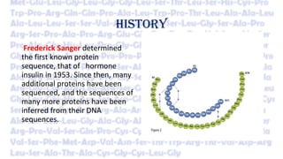
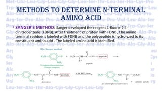
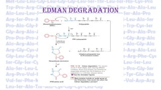
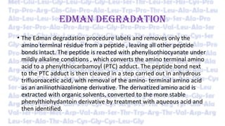
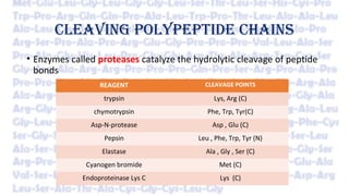
AMINO ACID SEQUENCING
MADE BY:MADE BY:
VAISHALI JAINVAISHALI JAIN
ROLL NO: 06ROLL NO: 06
DEFINITION
Peptide sequence, or amino acid sequence, is the order in
which amino acid residues, connected by peptide bonds, lie in
the chain in peptides and proteins. The sequence is generally
reported from the N-terminal end containing free amino
group to the C-terminal end containing free carboxyl group.
Shorter peptides are sequenced with automated procedures
but larger proteins must be sequenced in smaller segments.
IMPORTANCE OF AMINO ACID SEQUENCING
• Knowledge of a protein’s amino acid sequence is prerequisite for
determining its three-dimensional structure and is essential for
understanding its molecular mechanism of action.
• Many inherited diseases are caused by mutations that result in an
amino acid change in a protein. Amino acid sequence analysis can
assist in the development of diagnostic tests and effective therapies.
• Sequence comparisons among analogous proteins from different
species yield insights into protein function and reveal evolutionary
relationships among the proteins and the organisms that produce
them.
HISTORY
Frederick Sanger determined
the first known protein
sequence, that of hormone
insulin in 1953. Since then, many
additional proteins have been
sequenced, and the sequences of
many more proteins have been
inferred from their DNA
sequences.
N-TERMINAL ANALYSIS
Each polypeptide chain (if it is not chemically blocked) has an N-
terminal residue. Identifying this “end group” can establish the
number of chemically distinct polypeptides in a protein. For
example, insulin has equal amounts of the N-terminal residues Gly
and Phe, which indicates that it has equal numbers of two
nonidentical polypeptide chains.
METHODS TO DETERMINE N-TERMINAL
AMINO ACID
• SANGER’S METHOD: Sanger developed the reagent 1-fluoro-2,4-
dinitrobenzene (FDNB). After treatment of protein with FDNB , the amino
terminal residue is labeled with FDNB and the polypeptide is hydrolyzed to its
constituent amino acid . The labeled amino acid is identified.
DANSYL CHLORIDE METHOD
DISADVANTAGE:
•The hydrolysis stage destroys the polypeptide , these procedures
cannot be used to sequence a polypeptide beyond its amino-terminal
residue .
ADVANTAGE:
•It can help to determine the number of chemically distinct
polypeptides in a protein , provided each has a different amino
terminal residue.
EDMAN DEGRADATION
EDMAN DEGRADATION
• The Edman degradation procedure labels and removes only the
amino terminal residue from a peptide , leaving all other peptide
bonds intact. The peptide is reacted with phenylisothiocyanate under
mildly alkaline conditions , which converts the amino terminal amino
acid to a phenylthiocarbamoyl (PTC) adduct. The peptide bond next
to the PTC adduct is then cleaved in a step carried out in anhydrous
trifluoroacetic acid, with removal of the amino- terminal amino acid
as an anilinothiazolinone derivative. The derivatized amino acid is
extracted with organic solvents, converted to the more stable
phenylthiohydantoin derivative by treatment with aqueous acid and
then identified.
EDMAN DEGRADATION
• After removal and identification of the amino terminal residue, the
new amino-terminal residue so exposed can be labeled, removed,
and identified through the same series of reactions .This procedure is
repeated until the entire sequence is determined.
• The Edman degradation is carried out in a machine, called a
sequenator, that mixes reagents in the proper proportions, separates
the products, identifies them, and records the results.
• These methods are extremely sensitive. Often, the complete amino
acid sequence can be determined starting with only a few
micrograms of protein.
SEQUENCING OF LARGE PROTEINSSEQUENCING OF LARGE PROTEINS
BREAKING DISULFIDE BONDS
CLEAVING POLYPEPTIDE CHAINS
• Enzymes called proteases catalyze the hydrolytic cleavage of peptide
bonds
REAGENT CLEAVAGE POINTS
trypsin Lys, Arg (C)
chymotrypsin Phe, Trp, Tyr(C)
Asp-N-protease Asp , Glu (C)
Pepsin Leu , Phe, Trp, Tyr (N)
Elastase Ala , Gly , Ser (C)
Cyanogen bromide Met (C)
Endoproteinase Lys C Lys (C)
SEQUENCING THE PEPTIDE
• Each peptide fragment resulting from the action of protease is
sequenced separately by the Edman procedure.
ORDERING THE PEPTIDE FRAGMENTS
• Another sample of the intact polypeptide is cleaved into fragments
using a different enzyme or reagent , one that cleaves peptide bonds
at points other than those cleaved by first protease .
• The amino acid sequences of each fragment obtained by the two
cleavage procedures are examined.
• Overlapping fragments obtained from the second fragmentation yield
the correct order of the peptide fragments produced in the first.
QUESTION
• TRYPSIN TREATMENT- CHYMOTRYPSIN TREATMENT
Leu–Glu Gln–Ala–Phe
Gly–Tyr–Asn–Arg Asn-Arg-Leu-Glu
Gln–Ala–Phe–Val–Lys Val-Lys-Gly-Tyr
ANSWER
Gln–Ala–Phe
Gln-Ala-Phe-Val-Lys
Val-Lys-Gly-Tyr
Gly-Tyr-Asn-Arg
Asn-Arg-Leu-Glu
Leu-Glu
Gln-Ala-Phe-Val-Lys-Gly-Tyr-Asn-Arg-Leu-Glu
MASS SPECTROMETRY
• Mass spectrometry has emerged as an important technique for
characterizing and sequencing polypeptides.
• Mass spectrometry accurately measures the mass to-charge (m/z)
ratio for ions in the gas phase (where m is the ion’s mass and z is its
charge).
TANDEM MASS SPECTROMETRY
• Used to sequence short stretches of polypeptide
• A solution containing the protein under investigation is first treated with a
protease to hydrolyze it to a mixture of shorter peptides and the mixture is then
injected into a device that is essentially two mass spectrometers in tandem.
• The first mass spectrometer functions to select and separate the peptide ion of
interest from peptide ions of different masses as well as any contaminants that
may be present.
• The selected peptide ion is then passed into a collision cell, where it collides with
chemically inert atoms such as helium. The energy thereby imparted to the
peptide ion causes it to fragment predominantly at only one of its several
peptide bonds, thereby yielding one or two charged fragments per original ion.
• The molecular masses of the numerous charged fragments so produced are then
determined by the second mass spectrometer.
• By comparing the molecular masses of successively larger members of
a family of fragments, the molecular masses and therefore the
identities of the corresponding amino acid residues can be
determined.
• The sequence of an entire polypeptide can thus be elucidated
• Computerization of the mass-comparison process has reduced the
time required to sequence a short polypeptide to only a few minutes
• Mass spectrometry can also be used to sequence peptides with
chemically blocked N-termini (which prevents Edman degradation)
and to characterize other posttranslational modifications such as the
addition of phosphate or carbohydrate groups
Amino acid sequencing

Amino acid sequencing

  • 1.
    AMINO ACID SEQUENCING MADEBY:MADE BY: VAISHALI JAINVAISHALI JAIN ROLL NO: 06ROLL NO: 06
  • 2.
    DEFINITION Peptide sequence, oramino acid sequence, is the order in which amino acid residues, connected by peptide bonds, lie in the chain in peptides and proteins. The sequence is generally reported from the N-terminal end containing free amino group to the C-terminal end containing free carboxyl group. Shorter peptides are sequenced with automated procedures but larger proteins must be sequenced in smaller segments.
  • 3.
    IMPORTANCE OF AMINOACID SEQUENCING • Knowledge of a protein’s amino acid sequence is prerequisite for determining its three-dimensional structure and is essential for understanding its molecular mechanism of action. • Many inherited diseases are caused by mutations that result in an amino acid change in a protein. Amino acid sequence analysis can assist in the development of diagnostic tests and effective therapies. • Sequence comparisons among analogous proteins from different species yield insights into protein function and reveal evolutionary relationships among the proteins and the organisms that produce them.
  • 4.
    HISTORY Frederick Sanger determined thefirst known protein sequence, that of hormone insulin in 1953. Since then, many additional proteins have been sequenced, and the sequences of many more proteins have been inferred from their DNA sequences.
  • 5.
    N-TERMINAL ANALYSIS Each polypeptidechain (if it is not chemically blocked) has an N- terminal residue. Identifying this “end group” can establish the number of chemically distinct polypeptides in a protein. For example, insulin has equal amounts of the N-terminal residues Gly and Phe, which indicates that it has equal numbers of two nonidentical polypeptide chains.
  • 6.
    METHODS TO DETERMINEN-TERMINAL AMINO ACID • SANGER’S METHOD: Sanger developed the reagent 1-fluoro-2,4- dinitrobenzene (FDNB). After treatment of protein with FDNB , the amino terminal residue is labeled with FDNB and the polypeptide is hydrolyzed to its constituent amino acid . The labeled amino acid is identified.
  • 7.
  • 8.
    DISADVANTAGE: •The hydrolysis stagedestroys the polypeptide , these procedures cannot be used to sequence a polypeptide beyond its amino-terminal residue . ADVANTAGE: •It can help to determine the number of chemically distinct polypeptides in a protein , provided each has a different amino terminal residue.
  • 9.
  • 10.
    EDMAN DEGRADATION • TheEdman degradation procedure labels and removes only the amino terminal residue from a peptide , leaving all other peptide bonds intact. The peptide is reacted with phenylisothiocyanate under mildly alkaline conditions , which converts the amino terminal amino acid to a phenylthiocarbamoyl (PTC) adduct. The peptide bond next to the PTC adduct is then cleaved in a step carried out in anhydrous trifluoroacetic acid, with removal of the amino- terminal amino acid as an anilinothiazolinone derivative. The derivatized amino acid is extracted with organic solvents, converted to the more stable phenylthiohydantoin derivative by treatment with aqueous acid and then identified.
  • 11.
    EDMAN DEGRADATION • Afterremoval and identification of the amino terminal residue, the new amino-terminal residue so exposed can be labeled, removed, and identified through the same series of reactions .This procedure is repeated until the entire sequence is determined. • The Edman degradation is carried out in a machine, called a sequenator, that mixes reagents in the proper proportions, separates the products, identifies them, and records the results. • These methods are extremely sensitive. Often, the complete amino acid sequence can be determined starting with only a few micrograms of protein.
  • 12.
    SEQUENCING OF LARGEPROTEINSSEQUENCING OF LARGE PROTEINS
  • 13.
  • 14.
    CLEAVING POLYPEPTIDE CHAINS •Enzymes called proteases catalyze the hydrolytic cleavage of peptide bonds REAGENT CLEAVAGE POINTS trypsin Lys, Arg (C) chymotrypsin Phe, Trp, Tyr(C) Asp-N-protease Asp , Glu (C) Pepsin Leu , Phe, Trp, Tyr (N) Elastase Ala , Gly , Ser (C) Cyanogen bromide Met (C) Endoproteinase Lys C Lys (C)
  • 15.
    SEQUENCING THE PEPTIDE •Each peptide fragment resulting from the action of protease is sequenced separately by the Edman procedure.
  • 16.
    ORDERING THE PEPTIDEFRAGMENTS • Another sample of the intact polypeptide is cleaved into fragments using a different enzyme or reagent , one that cleaves peptide bonds at points other than those cleaved by first protease . • The amino acid sequences of each fragment obtained by the two cleavage procedures are examined. • Overlapping fragments obtained from the second fragmentation yield the correct order of the peptide fragments produced in the first.
  • 17.
    QUESTION • TRYPSIN TREATMENT-CHYMOTRYPSIN TREATMENT Leu–Glu Gln–Ala–Phe Gly–Tyr–Asn–Arg Asn-Arg-Leu-Glu Gln–Ala–Phe–Val–Lys Val-Lys-Gly-Tyr ANSWER Gln–Ala–Phe Gln-Ala-Phe-Val-Lys Val-Lys-Gly-Tyr Gly-Tyr-Asn-Arg Asn-Arg-Leu-Glu Leu-Glu Gln-Ala-Phe-Val-Lys-Gly-Tyr-Asn-Arg-Leu-Glu
  • 18.
    MASS SPECTROMETRY • Massspectrometry has emerged as an important technique for characterizing and sequencing polypeptides. • Mass spectrometry accurately measures the mass to-charge (m/z) ratio for ions in the gas phase (where m is the ion’s mass and z is its charge).
  • 19.
    TANDEM MASS SPECTROMETRY •Used to sequence short stretches of polypeptide • A solution containing the protein under investigation is first treated with a protease to hydrolyze it to a mixture of shorter peptides and the mixture is then injected into a device that is essentially two mass spectrometers in tandem. • The first mass spectrometer functions to select and separate the peptide ion of interest from peptide ions of different masses as well as any contaminants that may be present. • The selected peptide ion is then passed into a collision cell, where it collides with chemically inert atoms such as helium. The energy thereby imparted to the peptide ion causes it to fragment predominantly at only one of its several peptide bonds, thereby yielding one or two charged fragments per original ion. • The molecular masses of the numerous charged fragments so produced are then determined by the second mass spectrometer.
  • 20.
    • By comparingthe molecular masses of successively larger members of a family of fragments, the molecular masses and therefore the identities of the corresponding amino acid residues can be determined. • The sequence of an entire polypeptide can thus be elucidated • Computerization of the mass-comparison process has reduced the time required to sequence a short polypeptide to only a few minutes • Mass spectrometry can also be used to sequence peptides with chemically blocked N-termini (which prevents Edman degradation) and to characterize other posttranslational modifications such as the addition of phosphate or carbohydrate groups